Protocols for Human Immune Monitoring

Protocols for Human Immune Monitoring

Protocols for Human Immune Monitoring

Flow cytometry and the more recently developed mass cytometry—or cytometry by time-of-flight (CyTOF)— are key technologies broadly used by researchers to study the immune system. Since its invention in the late 1960s, fluorescence-based flow cytometry has advanced considerably and now allows the simultaneous detection of 18-20 proteins expressed by individual cells. Fluorescence-based flow cytometry has enable researchers to better understand the complexity of the immune system. Yet, the phenotypic and functional heterogeneity of immune cells has trigger the need for even more proteins to be detected at once. To overcome the limitation imposed by the spectral overlap of fluorochromes, CyTOF technology uses elemental isotopes instead of fluorochromes to label antibodies. CyTOF enables simultaneous detection of 30-50 proteins and could, in theory, detect up to 100 markers. Using these technologies, immunologists have developed a broad range of staining procedures, gating strategies, and functional assays to better understand the immune system.

Our special issue on human immune monitoring presents a comprehensive collection of detailed and peer reviewed protocols focused on human sample processing for flow cytometry analysis (section 3), immune assays to assess the composition, phenotype and functionality of human immune cells by fluorescence-based flow cytometry (section 2) and by CyTOF technology (section 1). Bio-protocols’ uniquely interactive platform supports communication between scientists – through feedback, Q&A, and protocol updates sections – and will allow you to set up cytometry based technologies for your research. Bio-protocol is a living platform and our Protocols for Human Immune Monitoring special issue will grow with the cytometry field, giving you access to the latest developments.


Editorial Committee Protocol List

Managing Editor

Associate Editor

Reviewer

Determination of Cellular Uptake and Endocytic Pathways

Determination of Cellular Uptake and Endocytic Pathways

7 Favorites
10562 Views
Feb 20, 2019

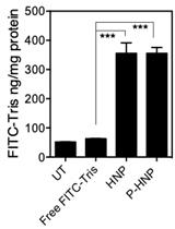
Efficiency of drug and gene delivery via nonviral vehicles is contingent on proper cellular uptake and intracellular release. Further, various cargos, such as nucleases for gene editing or inhibitors for endosomal receptors, require transport to specific compartments of the cell. Hence, characterization of cellular uptake and endocytic pathways is crucial for the optimization of any nanoparticle-mediated intracellular delivery system. Previous work on endocytic pathways looks at the effect of various pathway inhibitors on the uptake efficiency of nanoparticles carrying fluorescently-labeled cargo. While this helps attribute particle uptake to specific pathways like caveolae-mediated or clathrin-mediated endocytosis, this does not provide a holistic picture of the delivery process. Here, we provide a general protocol that combines systematic studies of inhibitor effects on efficiency with quantification of particle-induced cell membrane permeability. By applying this methodology to a nucleic acid delivery system, for example a helical polypeptide-based nanoparticle for plasmid and guide RNA delivery, we gain understanding of the endocytic mechanisms and cell uptake for intelligent design of intracellular delivery.

Isolation of PBMCs Using Vacutainer<sup>®</sup> Cellular Preparation Tubes (CPT<sup>TM</sup>)

Isolation of PBMCs Using Vacutainer® Cellular Preparation Tubes (CPTTM)

12 Favorites
37394 Views
Jan 20, 2017

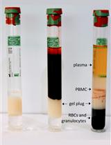
Peripheral blood mononuclear cell (PBMC) isolation is commonly done via density gradient centrifugation over Ficoll-Hypaque, a labor-intensive procedure that requires skilled technicians and can contribute to sample variability. Cellular Preparation Tubes (CPTs) are Vacutainer blood draw tubes that contain Ficoll-Hypaque and a gel plug that separates the Ficoll solution from the blood to be drawn. Once blood is drawn into CPTs, they can be centrifuged to separate the PBMC, then shipped (if desired) to a processing lab. The processing lab removes the PBMC from the upper compartment of the tube (above the gel plug), washes the PBMC, and can cryopreserve them using DMSO-containing media, as detailed in this protocol.

Cryopreservation of Human Serum

Cryopreservation of Human Serum

2 Favorites
8736 Views
Nov 5, 2015

This protocol describes how to collect and aliquot human serum and store at -80 °C for future cytokine/chemokine/protein profile analysis. Please use personal protective equipment (PPE) as required when handling samples with potential Bloodborne Pathogens.

Cytokine-stimulated Phosphoflow of PBMC Using CyTOF Mass Cytometry

Cytokine-stimulated Phosphoflow of PBMC Using CyTOF Mass Cytometry

1 Favorites
18431 Views
Jun 5, 2015

Phosphorylation of tyrosine, serine, and threonine residues is critical for the control of protein activity involved in various cellular events. An assortment of kinases and phosphatases regulate intracellular protein phosphorylation in many different cell signaling pathways. These pathways include T and B cell signaling, regulating growth and cell cycle control, plus cytokine, chemokine, and stress responses. Phosphoflow assays combine phosphoprotein-specific antibodies with the power of flow cytometry to enhance phosphoprotein study. In our assay, peripheral blood mononuclear cells are stimulated by cytokines, fixed, surface-stained with a cocktail of antibodies labeled with MAXPAR (brand name) metal-chelating polymers and permeabilized with methanol. They are then stained with intracellular phospho-specific antibodies. We use a CyTOFTM mass cytometer to acquire the ICP-MS (inductively coupled plasma mass spectrometry) data. The current mass window selected is approximately AW 103-203, which includes the lanthanides used for most antibody labeling, as well as iridium and rhodium for DNA intercalators. Subsequent analysis of the dual count signal data using FlowJo software allows for cell types to be analyzed based on the dual count signal in each mass channel. The percentage of each cell type is determined and reported as a percent of the parent cell type. Median values are reported to quantitate the level of phosphorylation of each protein in response to stimulation. Comparing the level of phosphorylation between samples can offer insight to the status of the immune system.

Cytokine-Stimulated Phosphoflow of Whole Blood Using CyTOF Mass Cytometry

Cytokine-Stimulated Phosphoflow of Whole Blood Using CyTOF Mass Cytometry

2 Favorites
15233 Views
Jun 5, 2015

The ability to assess the function of a range of cytokine, antigen receptor, and Toll-like receptor (TLR) signaling pathways in a range of immune cells could provide a kind of fingerprint of the state of the human immune system. The mass cytometry or CyTOF, platform allows for the parallel application of about 40 labeled antibodies to a single sample, creating the possibility to read out many cell types and signaling pathways in a single small blood sample. We developed such a mass cytometry panel, consisting of 22 antibodies to cell surface lineage markers and 8 antibodies to phospho-specific epitopes of signaling proteins. These antibodies were chosen to discriminate all major white blood cell lineages, to a level of detail that includes subsets such as naïve, central memory, effector memory, and late effector CD4+ and CD8+T cells, naïve, transitional, and switched memory B cells, plasmablasts, myeloid and plasmacytoid dendritic cells, CD16+ and CD16+CD56+ NK cells, CD16+ and classical monocytes etc. 32 such cell subsets are defined in our standard gating scheme. The eight phospho-specific antibodies were chosen to represent major signaling nodes responsive to cytokine, TLR, and antigen receptor signaling. This antibody panel is used with 8 standard stimulation conditions (unstimulated, IFNa, IL-6, IL-7, IL-10, IL-21, LPS, PMA+ ionomycin), although other stimuli can be added. Comparison of healthy controls to subjects with immune deficiencies of unknown etiology may help elucidate the mechanisms of such deficiencies. Phosphorylation of tyrosine, serine, and threonine residues is critical for the control of protein activity involved in various cellular events. An assortment of kinases and phosphatases regulate intracellular protein phosphorylation in many different cell signaling pathways, such as T and B cell signaling, those regulating apoptosis, growth and cell cycle control, plus those involved with cytokine, chemokine, and stress responses. Phosphoflow assays combine phospho-specific antibodies with the power of flow cytometry to enhance phospho protein study. In our assay, peripheral blood mononuclear cells are stimulated by cytokines, fixed, surface-stained with a cocktail of antibodies labeled with MAXPAR (Brand Name) metal-chelating polymers and permeabilized with methanol. They are then stained with intracellular phospho-specific antibodies. We use a CyTOFTM mass cytometer to acquire the ICP-MS data. The current mass window selected is approximately AW 103-203, which includes the lanthanides used for most antibody labeling, as well as iridium and rhodium for DNA intercalators. Subsequent analysis of the dual count signal data using FlowJo software allows for cell types to be analyzed based on the dual count signal in each mass channel. The percentage of each cell type is determined and reported as a percent of the parent cell type. Median values are reported to quantitate the level of phosphorylation of each protein in response to stimulation. Comparing the level of phosphorylation between samples can offer insight to the status of the immune system. Whole blood stimulation is the closest to the in vivo condition and it allows for assessment of granulocyte population as well as lymphocytes and monocytes.

Intracellular Cytokine Staining (ICS) on Human Lymphocytes or Peripheral Blood Mononuclear Cells (PBMCs)

Intracellular Cytokine Staining (ICS) on Human Lymphocytes or Peripheral Blood Mononuclear Cells (PBMCs)

11 Favorites
27334 Views
Apr 5, 2015

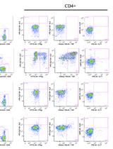
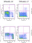
Production of cytokines plays an important role in the immune response. Cytokines are involved in many different pathways including the induction of many anti-viral proteins by IFN gamma, the induction of T cell proliferation by IL-2 and the inhibition of viral gene expression and replication by TNF alpha. Cytokines are not preformed factors but are rapidly produced and secreted in response to cellular activation. Intracellular cytokine detection by flow cytometry has emerged as the premier technique for studying cytokine production at the single-cell level. It detects the production and accumulation of cytokines within the endoplasmic reticulum after cell stimulation, allowing direct TH1 versus TH2 determination. It can also be used in combination with other flow cytometry protocols for immunophenotyping using cell surface markers or with MHC multimers to detect an antigen specific response, making it an extremely flexible and versatile method. This capability, combined with the high throughput nature of the instrumentation, gives intracellular cytokine staining an enormous advantage over existing single-cell techniques such as ELISPOT, limiting dilution, and T cell cloning. The principle steps of intracellular cytokine staining is as follows:1. Cells are activated for a few hours using either a specific peptide or a non-specific activation cocktail;2. An inhibitor of protein transport (e.g. Brefeldin A) is added to retain the cytokines within the cell;3. Next, EDTA is added to remove adherent cells from the activation vessel;4. After washing, antibodies to cell surface markers can be added to the cells;5. The cells are then fixed in paraformaldehyde and permeabilized;6. The anti-cytokine antibody is added and the cells can be analyzed by flow cytometer.

Phenotyping of Live Human PBMC using CyTOF<sup>TM</sup> Mass Cytometry

Phenotyping of Live Human PBMC using CyTOFTM Mass Cytometry

4 Favorites
19702 Views
Jan 20, 2015

Single-cell analysis has become an method of importance in immunology. Fluorescence flow cytometry has been a major player. However, due to issues such as autofluorescence and emission spillover between different fluorophores, alternative techniques are being developed. In recent years, mass cytometry has emerged, wherein antibodies labeled with metal ions are detected by ICP-MS. In order for a cell to be seen, a metal in the mass window must be present; there is no analogous parameter to forward or side scatter. The current mass window selected is approximately AW 103-196, which includes the lanthanides used for most antibody labeling, as well as iridium and rhodium for DNA intercalators.In this protocol, we use a cocktail of antibodies labeled with MAXPAR metal-chelating polymers to surface-stain live PBMC that have been previously cryopreserved. Many of these markers were taken from a standard fluorescence phenotyping panel (Maecker et al., 2012). No intracellular antibodies are used. We use a CyTOFTM (Cytometry by Time-Of-Flight) mass cytometer to acquire the ICP-MS data. Subsequent analysis of the dual count signal data using FlowJo software allows for cell types to be analyzed based on the dual count signal in each mass channel. The percentage of each cell type is determined and reported as a percent of the parent cell type.

Intracellular Cytokine Staining on PBMCs Using CyTOF<sup>TM</sup> Mass Cytometry

Intracellular Cytokine Staining on PBMCs Using CyTOFTM Mass Cytometry

6 Favorites
23374 Views
Jan 5, 2015

In this protocol, we use a CyTOFTM mass cytometry to collect single-cell data on a large number of cytokines/chemokines as well as cell-surface proteins that characterize T cells and other immune cells. The current selected mass window in AW 103-203 includes the lanthanides used for most antibody labeling, along with iridium and rhodium for DNA intercalators. The output data are in the format as .txt and .fcs files, which is compatible with many analysis programs. This protocol could be adapted to include tetramers into the staining panel, but we have not optimized for that purpose. The principal steps of intracellular cytokine staining are as follows: First, cells are activated for a few hours using either a specific peptide or a non-specific activation cocktail. An inhibitor of protein transport (e.g. Brefeldin A) is added to retain the cytokines within the cell. Next, EDTA is added to remove adherent cells from the activation vessel. After washing, antibodies to cell surface markers are added to the cells. The cells are then fixed in paraformaldehyde and permeabilized. We use a gentle detergent, saponin, as the permealization buffer because it is less destructive to surface and intracellular epitopes compared to harsh detergents or methanol. After permeabilization, the metal-conjugated anti-cytokine antibodies are added into the cell suspension. The stained cells are then sequentially introduced into the mass cytometry for signal intensity analysis.

Assessment of Human Dendritic Cell Antigen Uptake by Flow Cytometry

Assessment of Human Dendritic Cell Antigen Uptake by Flow Cytometry

6 Favorites
18037 Views
Nov 20, 2013

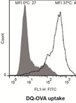
Antigen uptake by dendritic cells is the first key step towards induction of antigen-specific T-cell responses. This flow cytometry-based protocol describes the analysis of dendritic cell uptake of soluble antigens through two different mechanisms: non-specific macropinocytosis (using Lucifer Yelloy CH), and receptor-mediated endocytosis (using DQTM Ovalbumin). The protocol is generated based on data presented in Olivar et al. (2013).

Isolation of Human PBMCs

Isolation of Human PBMCs

18 Favorites
97392 Views
Feb 5, 2013

Peripheral blood mononuclear cells (PBMCs) are chiefly lymphocytes and monocytes. PBMCs are separated from the whole blood by a density gradient centrifugation method using Ficoll Histopaque.

<em>In vitro</em> Culture of Human PBMCs

In vitro Culture of Human PBMCs

29 Favorites
113225 Views
Feb 5, 2013

Peripheral blood mononuclear cells (PBMCs) consist of chiefly of lymphocytes and monocytes. Purified PBMCs are used in vitro to evaluate a variety of functions of lymphocytes viz; a) proliferation to mitogenic (PHA, Con-A) stimulation, b) monitoring of prior sensitisation in antigen recall assays by scoring lymphocyte proliferation, c) immunophenotyping for surface markers as well as intracellular molecules in monocytes and lymphocytes etc. Activation of monocytes/macrophages by small molecules, cytokines and pathogen components can also be monitored. PBMCs can also be used for a variety of structural and functional studies for addressing issues in human immunology such as scoring for apoptosis and production of cytokines as well as other mediators in vitro.

<em>Ex vivo</em> Human Antigen-specific T Cell Proliferation and Degranulation

Ex vivo Human Antigen-specific T Cell Proliferation and Degranulation

5 Favorites
15373 Views
Dec 5, 2012

Proliferative capacity and degranulation are important features of antigen-specific CD8+ T cells. By combining tetramer staining with a CFSE staining, we were able to enumerate the total number of antigen-specific T cells, as well as their number of divisions upon antigen-specific stimulation during a week. In addition, we performed restimulation of these cells, to analyze their ability to secrete cytolytic granules, visualized by CD107a staining.

Whole Blood Staining of Human Monocyte Subsets for Flow Cytometry

Whole Blood Staining of Human Monocyte Subsets for Flow Cytometry

3 Favorites
41585 Views
May 20, 2011

This is a general protocol to stain whole human blood for flow analysis with minimal spontaneous activation of monocytes. This protocol was developed or modified in Dr. Anne Davidson’s lab at Feinstein Institute for Medical Research.

Other special issues