Protocols for Coronavirus/COVID-19 Research

Protocols for Coronavirus/COVID-19 Research

Protocols for Coronavirus/COVID-19 Research

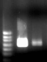
With the spread of the COVID-19 (SARS-CoV-2) pandemic across the world, sharing accurate and reproducible methods that can be readily available to the biomedical community became crucial. Therefore, Bio-protocol decided to dedicate a special issue to protocols used in Coronavirus/COVID-19 research in May 2020. By November 2021, we have published 19 high quality protocols used in basic science and clinical research, including diagnostic methods for COVID-19. Additionally, 9 protocols published before May 2020, used in tangential research on other coronaviruses, are also included in this special issue.

More new protocols will be welcomed to be included in this special issue, and we hope it will become a comprehensive resource for detailed protocols relevant to Coronavirus/COVID-19 research. Typically, we only accept protocols previously used in published articles reporting original research. However, due to the rapid rate of preprint publications during the pandemic, we also include protocols used in preprint articles and preprint versions of the protocols.


Editorial Committee Protocol List

Managing Editor

Gal Haimovich

Gal Haimovich

Weizmann Institute of Science, Israel

Associate Editor

Alba Blesa

Alba Blesa

Universidad Francisco de Vitoria

Alessandro Didonna

Alessandro Didonna

University of California

Alexandros Alexandratos

Alexandros Alexandratos

National and Kapodistrian University of Athens

Alka Mehra

Alka Mehra

Institute of Genomics and Integrative Biology

Antoine de Morree

Antoine de Morree

Stanford university

David Paul

David Paul

MRC Laboratory of Molecular Biology

Ivan Zanoni

Ivan Zanoni

Harvard Medical School

Khyati Hitesh Shah

Khyati Hitesh Shah

Sutro BioPharma

Longping Victor Tse

Longping Victor Tse

University of North Carolina at Chapel Hill

Luis Alberto Sánchez Vargas

Luis Alberto Sánchez Vargas

University of Rhode Island

Nicoletta Cordani

Nicoletta Cordani

University of Milano-Bicocca

Pilar Villacampa Alcubierre

Pilar Villacampa Alcubierre

Josep Carreras Leukaemia Research Institute

Reviewer

Angela Corona

Angela Corona

University of Cagliari

Anna Sloutskin

Anna Sloutskin

Bar-Ilan University

Balaji Olety Amaranath

Balaji Olety Amaranath

University of Massachusetts medical School

Chhuttan L Meena

Chhuttan L Meena

National Chemical laboratory NCL Pune

David Paul

David Paul

Cambridge

Day-Yu Chao

Day-Yu Chao

National chung-Hsing University

Emily Cope

Emily Cope

Northern Arizona University

George William Carnell

George William Carnell

University of Cambridge

Joyita Mukherjee

Joyita Mukherjee

The Francis Crick Institute

Kenji Sugiyama

Kenji Sugiyama

University of Tsukuba

Kirsten A. Copren

Kirsten A. Copren

University of Pennsylvania

Masfique Mehedi

Masfique Mehedi

University of North Dakota

Meenal Sinha

Meenal Sinha

University of California San Francisco

Mirko Cortese

Mirko Cortese

Telethon Institute of Genetics and Medicine

Molly M. Leung

Molly M. Leung

University of California Los Angeles

Nathan Rhys James

Nathan Rhys James

MRC Laboratory of Molecular Biology

Prasgant Sharma

Prasgant Sharma

University of Wisconsin School of Medicine and Public Health

Ran Chen

Ran Chen

Zhejiang JFK Biological Technology Co. Ltd.

Sarajo Mohanta

Sarajo Mohanta

Ludwig Maximilians University

Sesha Lakshmi Arathi Paluri

Sesha Lakshmi Arathi Paluri

Michigan State University

Shanie Saghafian-Hedengren

Shanie Saghafian-Hedengren

Karolinska Institutet

Shyam Solanki

Shyam Solanki

Washington State University

Smita Nair

Smita Nair

University of Würzburg, Germany

Sonali Chaturvedi

Sonali Chaturvedi

Gladstone Institute of Virology and Immunology

Suresh Panthee

Suresh Panthee

Teikyo University Institute of Medical Mycology

Tharmarajan Ramprasath

Tharmarajan Ramprasath

Georgia State University

Thibaud T. Renault

Thibaud T. Renault

CNRS / University of Bordeaux

Timo A Lehti

Timo A Lehti

R&D Specialist

Vaibhav B. Shah

Vaibhav B. Shah

The University of New South Wales

Vasudevan Achuthan

Vasudevan Achuthan

Harvard Medical School

Vikas Duhan

Vikas Duhan

QIMR Berghofer Medical Research Institute

Xiaoyi Zheng

Xiaoyi Zheng

Thomas Jefferson University

Zhenying Liu

Zhenying Liu

University of Pennsylvania

Production of Recombinant Replication-defective Lentiviruses Bearing the SARS-CoV or SARS-CoV-2 Attachment Spike Glycoprotein and Their Application in Receptor Tropism and Neutralisation Assays

Production of Recombinant Replication-defective Lentiviruses Bearing the SARS-CoV or SARS-CoV-2 Attachment Spike Glycoprotein and Their Application in Receptor Tropism and Neutralisation Assays

1 Favorites
3703 Views
Nov 5, 2021

For enveloped viruses, such as SARS-CoV-2, transmission relies on the binding of viral glycoproteins to cellular receptors. Conventionally, this process is recapitulated in the lab by infection of cells with isolated live virus. However, such studies can be restricted due to the availability of high quantities of replication-competent virus, biosafety precautions and associated trained staff. Here, we present a protocol based on pseudotyping to produce recombinant replication-defective lentiviruses bearing the SARS-CoV or SARS-CoV-2 attachment Spike glycoprotein, allowing the investigation of viral entry in a lower-containment facility. Pseudoparticles are produced by cells transiently transfected with plasmids encoding retroviral RNA packaging signals and Gag-Pol proteins, for the reconstitution of lentiviral particles, and a plasmid coding for the viral attachment protein of interest. This approach allows the investigation of different aspects of viral entry, such as the identification of receptor tropism, the prediction of virus host range, and zoonotic transmission potential, as well as the characterisation of antibodies (sera or monoclonal antibodies) and pharmacological inhibitors that can block entry.Graphic abstract: SARS-CoV and SARS-CoV-2 pseudoparticle generation and applications.

A Nucleocapsid-based Transcomplementation Cell Culture System of SARS-CoV-2 to Recapitulate the Complete Viral Life Cycle

A Nucleocapsid-based Transcomplementation Cell Culture System of SARS-CoV-2 to Recapitulate the Complete Viral Life Cycle

3 Favorites
3297 Views
Nov 5, 2021

The ongoing COVID-19 pandemic is caused by severe acute respiratory syndrome coronavirus 2 (SARS-CoV-2). As this virus is classified as a biosafety level-3 (BSL-3) agent, the development of countermeasures and basic research methods is logistically difficult. Recently, using reverse genetics, we developed a BSL-2 cell culture system for production of transcription- and replication-component virus-like-particles (trVLPs) by genetic transcomplementation. The system consists of two parts: SARS-CoV-2 GFP/ΔN genomic RNA, in which the nucleocapsid (N) gene, a critical gene for virion packaging, is replaced by a GFP reporter gene; and a packaging cell line for ectopic expression of N (Caco-2-N). The complete viral life cycle can be recapitulated and confined to Caco-2-N cells, with GFP positivity serving as a surrogate readout for viral infection. In addition, we utilized an intein-mediated protein splicing technique to split the N gene into two independent vectors and generated the Caco-2-Nintein cells as a packaging cell line to further enhance the security of this cell culture model. Altogether, this system provides for a safe and convenient method to produce trVLPs in BSL-2 laboratories. These trVLPs can be modified to incorporate desired mutations, permitting high-throughput screening of antiviral compounds and evaluation of neutralizing antibodies. This protocol describes the details of the trVLP cell culture model to make SARS-CoV-2 research more readily accessible.

Production, Titration, Neutralisation, Storage and Lyophilisation of Severe Acute Respiratory Syndrome Coronavirus 2 (SARS-CoV-2) Lentiviral Pseudotypes

Production, Titration, Neutralisation, Storage and Lyophilisation of Severe Acute Respiratory Syndrome Coronavirus 2 (SARS-CoV-2) Lentiviral Pseudotypes

0 Favorites
4908 Views
Nov 5, 2021

This protocol details a rapid and reliable method for the production and titration of high-titre viral pseudotype particles with the SARS-CoV-2 spike protein (and D614G or other variants of concern, VOC) on a lentiviral vector core, and use for neutralisation assays in target cells expressing angiotensin-converting enzyme 2 (ACE2) and transmembrane serine protease 2 (TMPRSS2). It additionally provides detailed instructions on substituting in new spike variants via gene cloning, lyophilisation and storage/shipping considerations for wide deployment potential. Results obtained with this protocol show that SARS-CoV-2 pseudotypes can be produced at equivalent titres to SARS-CoV and Middle East respiratory syndrome coronavirus (MERS-CoV) pseudotypes, neutralised by human convalescent plasma and monoclonal antibodies, and stored at a range of laboratory temperatures and lyophilised for distribution and subsequent application.

Identification and Quantitation of Neutrophil Extracellular Traps in Human Tissue Sections

Identification and Quantitation of Neutrophil Extracellular Traps in Human Tissue Sections

4 Favorites
4367 Views
Sep 20, 2021

Neutrophils are one of the first innate immune cells recruited to tissues during inflammation. An important function of neutrophils relies on their ability to release extracellular structures, known as Neutrophil Extracellular Traps or NETs, into their environment. Detecting such NETs in humans has often proven challenging for both biological fluids and tissues; however, this can be achieved by quantitating NET components (e.g., DNA or granule/histone proteins) or by directly visualizing them by microscopy, respectively. Direct visualization by confocal microscopy is preferably performed on formalin-fixed paraffin-embedded (FFPE) tissue sections stained with a fluorescent DNA dye and antibodies directed against myeloperoxidase (MPO) and citrullinated histone 3 (Cit-H3), two components of NETs, following paraffin removal, antigen retrieval, and permeabilization. NETs are defined as extracellular structures that stain double-positive for MPO and Cit-H3. Here, we propose a novel software-based objective method for NET volume quantitation in tissue sections based on the measurement of the volume of structures exhibiting co-localization of Cit-H3 and MPO outside the cell. Such a technique not only allows the unambiguous identification of NETs in tissue sections but also their quantitation and relationship with surrounding tissues.Graphic abstract:Graphical representation of the methodology used to stain and quantitate NETs in human lung tissue.

Optimised Method for the Production and Titration of Lentiviral Vectors Pseudotyped with the SARS-CoV-2 Spike

Optimised Method for the Production and Titration of Lentiviral Vectors Pseudotyped with the SARS-CoV-2 Spike

1 Favorites
3693 Views
Aug 20, 2021

The use of recombinant lentivirus pseudotyped with the coronavirus Spike protein of SARS-CoV-2 would circumvent the requirement of biosafety-level 3 (BSL-3) containment facilities for the handling of SARS-CoV-2 viruses. Herein, we describe a fast and reliable protocol for the transient production of lentiviruses pseudotyped with SARS-CoV-2 Spike (CoV-2 S) proteins and green fluorescent protein (GFP) reporters. The virus titer is determined by the GFP reporter (fluorescent) expression with a flow cytometer. High titers (>1.00 E+06 infectious units/ml) are produced using codon-optimized CoV-2 S, harbouring the prevalent D614G mutation and lacking its ER retention signal. Enhanced and consistent cell entry is achieved by using permissive HEK293T/17 cells that were genetically engineered to stably express the SARS-CoV-2 human receptor ACE2 along with the cell surface protease TMPRSS2 required for efficient fusion. For the widespread use of this protocol, its reagents have been made publicly available.Graphic abstract:Production and quantification of lentiviral vectors pseudotyped with the SARS-CoV-2 Spike glycoprotein

Production of the Receptor-binding Domain of the Viral Spike Proteins from  2003 and 2019 SARS CoVs and the Four Common Human Coronaviruses for Serologic Assays and Inhibitor Screening

Production of the Receptor-binding Domain of the Viral Spike Proteins from 2003 and 2019 SARS CoVs and the Four Common Human Coronaviruses for Serologic Assays and Inhibitor Screening

2 Favorites
5772 Views
May 20, 2021

The recombinant receptor-binding domain (RBD) of the viral spike protein from SARS-CoV-1 and 2 are reliable antigens for detecting viral-specific antibodies in humans. We and others have shown that the levels of RBD-binding antibodies and SARS-CoV-2 neutralizing antibodies in patients are correlated. Here, we report the expression and purification of properly folded RBD proteins from SARS and common-cold HCoVs in mammalian cells. RBD proteins were produced with cleavable tags for affinity purification from the cell culture medium and to support multiple immunoassay platforms and drug discovery efforts.Graphic abstract:High-Yield Production of Viral Spike RBDs for Diagnostics and Drug Discovery

COVID-19 Sample Pooling: From RNA Extraction to Quantitative Real-time RT-PCR

COVID-19 Sample Pooling: From RNA Extraction to Quantitative Real-time RT-PCR

4 Favorites
8134 Views
May 5, 2021

The COVID-19 pandemic requires mass screening to identify those infected for isolation and quarantine. Individually screening large populations for the novel pathogen, Severe Acute Respiratory Syndrome Coronavirus 2 (SARS-CoV-2), is costly and requires a lot of resources. Sample pooling methods improve the efficiency of mass screening and consume less reagents by increasing the capacity of testing and reducing the number of experiments performed, and are therefore especially suitable for under-developed countries with limited resources. Here, we propose a simple, reliable pooling strategy for COVID-19 testing using clinical nasopharyngeal (NP) and/or oropharyngeal (OP) swabs. The strategy includes the pooling of 10 NP/OP swabs for extraction and subsequent testing via quantitative real-time reverse transcription polymerase chain reaction (RT-qPCR), and may also be applied to the screening of other pathogens.

Computational Analysis and Phylogenetic Clustering of SARS-CoV-2 Genomes

Computational Analysis and Phylogenetic Clustering of SARS-CoV-2 Genomes

3 Favorites
5949 Views
Apr 20, 2021

COVID-19, the disease caused by the novel SARS-CoV-2 coronavirus, originated as an isolated outbreak in the Hubei province of China but soon created a global pandemic and is now a major threat to healthcare systems worldwide. Following the rapid human-to-human transmission of the infection, institutes around the world have made efforts to generate genome sequence data for the virus. With thousands of genome sequences for SARS-CoV-2 now available in the public domain, it is possible to analyze the sequences and gain a deeper understanding of the disease, its origin, and its epidemiology. Phylogenetic analysis is a potentially powerful tool for tracking the transmission pattern of the virus with a view to aiding identification of potential interventions. Toward this goal, we have created a comprehensive protocol for the analysis and phylogenetic clustering of SARS-CoV-2 genomes using Nextstrain, a powerful open-source tool for the real-time interactive visualization of genome sequencing data. Approaches to focus the phylogenetic clustering analysis on a particular region of interest are detailed in this protocol.

Colorimetric RT-LAMP and LAMP-sequencing for Detecting SARS-CoV-2 RNA in Clinical Samples

Colorimetric RT-LAMP and LAMP-sequencing for Detecting SARS-CoV-2 RNA in Clinical Samples

0 Favorites
5677 Views
Mar 20, 2021

During pandemics, such as the one caused by SARS-CoV-2 coronavirus, simple methods to rapidly test large numbers of people are needed. As a faster and less resource-demanding alternative to detect viral RNA by conventional qPCR, we used reverse transcription loop-mediated isothermal amplification (RT-LAMP). We previously established colorimetric RT-LAMP assays on both purified and unpurified SARS-CoV-2 clinical specimens and further developed a multiplexed sequencing protocol (LAMP-sequencing) to analyze the outcome of many RT-LAMP reactions at the same time (Dao Thi et al., 2020). Extending on this work, we hereby provide step-by-step protocols for both RT-LAMP assays and read-outs.

Primer ID Next-Generation Sequencing for the Analysis of a Broad Spectrum Antiviral Induced Transition Mutations and Errors Rates in a Coronavirus Genome

Primer ID Next-Generation Sequencing for the Analysis of a Broad Spectrum Antiviral Induced Transition Mutations and Errors Rates in a Coronavirus Genome

0 Favorites
6394 Views
Mar 5, 2021

Next generations sequencing (NGS) has become an important tool in biomedical research. The Primer ID approach combined with the MiSeq platform overcomes the limitation of PCR errors and reveals the true sampling depth of population sequencing, making it an ideal tool to study mutagenic effects of potential broad-spectrum antivirals on RNA viruses. In this report we describe a protocol using Primer ID sequencing to study the mutations induced by antivirals in a coronavirus genome from an in vitro cell culture model and an in vivo mouse model. Viral RNA or total lung tissue RNA is tagged with Primer ID-containing cDNA primers during the initial reverse transcription step, followed by two rounds of PCR to amplify viral sequences and incorporate sequencing adaptors. Purified and pooled libraries are sequenced using the MiSeq platform. Sequencing data are processed using the template consensus sequence (TCS) web-app. The Primer ID approach provides an accurate sequencing protocol to measure mutation error rates in viral RNA genomes and host mRNA. Sequencing results suggested that β-D-N4-hydroxycytidine (NHC) greatly increased the transition substitution rate but not the transversion substitution rate in the viral RNA genomes, and cytosine (C) to uridine (U) was found as the most frequently seen mutation.

A One-enzyme RT-qPCR Assay for SARS-CoV-2, and Procedures for Reagent Production

A One-enzyme RT-qPCR Assay for SARS-CoV-2, and Procedures for Reagent Production

0 Favorites
5202 Views
Jan 20, 2021

Given the scale of the ongoing COVID-19 pandemic, the need for reliable, scalable testing, and the likelihood of reagent shortages, especially in resource-poor settings, we have developed an RT-qPCR assay that relies on an alternative to conventional viral reverse transcriptases, a thermostable reverse transcriptase/DNA polymerase (RTX) (Ellefson et al., 2016). Here we show that RTX performs comparably to the other assays sanctioned by the CDC and validated in kit format. We demonstrate two modes of RTX use – (i) dye-based RT-qPCR assays that require only RTX polymerase, and (ii) TaqMan RT-qPCR assays that use a combination of RTX and Taq DNA polymerases (as the RTX exonuclease does not degrade a TaqMan probe). We also provide straightforward recipes for the purification of this alternative reagent RTX. We anticipate that in low resource or point-of-need settings researchers could obtain the available constructs and begin to develop their own assays, within whatever regulatory framework exists for them.

Evaluation of the Sequence Variability within the PCR Primer/Probe Target Regions of the SARS-CoV-2 Genome

Evaluation of the Sequence Variability within the PCR Primer/Probe Target Regions of the SARS-CoV-2 Genome

0 Favorites
4877 Views
Dec 20, 2020

Severe acute respiratory syndrome coronavirus 2 (SARS-CoV-2; initially named 2019-nCoV) is responsible for the recent coronavirus disease (COVID-19) pandemic, and polymerase chain reaction (PCR) is the current standard method for diagnosis from patient samples. As PCR assays are prone to sequence mismatches due to mutations in the viral genome, it is important to verify the genomic variability at primer/probe binding regions periodically. This step-by-step protocol describes a bioinformatics approach for an extensive evaluation of the sequence variability within the primer/probe target regions of the SARS-CoV-2 genome. The protocol can be applied to any molecular diagnostic assay of choice using freely available software programs and the ready-to-use multiple sequence alignment (MSA) file provided.Graphic abstract:Overview of the sequence tracing protocol. The figure was created using the Library of Science and Medical Illustrations from somersault18:24 licensed under a CC BY-NC-SA 4.0 license (https://creativecommons.org/licenses/by-nc-sa/4.0/). Video abstract: https://youtu.be/M1lV1liWE9k

Colorimetric RT-LAMP Methods to Detect Severe Acute Respiratory Syndrome Coronavirus 2 (SARS-CoV-2)

Colorimetric RT-LAMP Methods to Detect Severe Acute Respiratory Syndrome Coronavirus 2 (SARS-CoV-2)

1 Favorites
5116 Views
Nov 5, 2020

Standard diagnostic methods of Coronavirus Disease 2019 (COVID-19) rely on RT-qPCR technique which have limited point-of-care test (POCT) potential due to necessity of dedicated equipment and specialized personnel. LAMP, an isothermal nucleic acid amplification test (NAAT), is a promising technique that may substitute RT-qPCR for POCT of genomic materials. Here, we provide a protocol to perform reverse transcription LAMP targeting SARS-CoV-2. We adopted both real-time fluorescence detection and end-point colorimetric detection approaches. Our protocol would be useful for screening diagnosis of COVID-19 and be a baseline for development of improved POCT NAAT.

A Protocol for Simple, Rapid, and Direct Detection of SARS-CoV-2 from clinical samples, using Reverse Transcribed Loop-Mediated Isothermal Amplification (RT-LAMP)

A Protocol for Simple, Rapid, and Direct Detection of SARS-CoV-2 from clinical samples, using Reverse Transcribed Loop-Mediated Isothermal Amplification (RT-LAMP)

1 Favorites
4450 Views
Oct 20, 2020

SARS-CoV-2 has quickly spread all around the globe causing illness and wide damages. Most countries were unprepared for such a rapid spread and crisis. This led to various strategies for effective control of the new pandemic. A key aspect in all countries was to effectively test the population for the virus. Most countries chose a lockdown strategy in which many workplaces and activities are completely closed, leading to substantial economy costs. Here, we present a protocol we recently developed that allows rapid and simple detection of SARS-CoV-2 for the large population, eliminating costs and involvement of professional teams and laboratories. This protocol is based on Reverse Transcribed Loop-Mediated Isothermal Amplification (RT-LAMP). We tested this protocol directly on patient samples, both nasal and throat clinical swabs as well as saliva. Notably, this protocol is simple, cheap and can be easily applied to other pathogens as well.

A Standard Operative Procedure for Safe-handling of Remains and Wastes of COVID-19 Patients

A Standard Operative Procedure for Safe-handling of Remains and Wastes of COVID-19 Patients

0 Favorites
2183 Views
Oct 20, 2020

The ongoing coronavirus disease-2019 (COVID-19) pandemic has raised significant public health issues which need to be attended. To enable efficient handling of the situation and prevent the spread of the epidemic, healthcare professionals require various standard operating procedures (SOPs). Emerging evidence suggests high infectivity of the novel coronavirus strain–severe acute respiratory syndrome virus-2 (SARS-CoV-2)–causing COVID-19. The remains and wastes of COVID-19 patients can be a potential source of the virus exposure to the healthcare professionals if not handled with adequate precaution. Various institutions have issued SOPs in this regard, but a comprehensive approach is missing, which creates difficulty in interpretation and application of these SOPs. We have developed a comprehensive protocol for disposal of remains and wastes of the COVID-19 patients.We are following this protocol at our institution without any untoward event until date.

Extraction, Purification and Detection of SARS-CoV-2 Nucleic Acid

Extraction, Purification and Detection of SARS-CoV-2 Nucleic Acid

Vendor protocol
2 Favorites
2522 Views
Sep 5, 2020

Nucleic acid extraction and purification kit is used for isolating high-quality pathogen nucleic acids (DNA/RNA) from a variety of specimens. Nucleic acid can be directly extracted from liquid samples such as blood, serum, plasma, urine, nasopharyngeal swabs, and cell culture. And soil samples such as stool, tissue samples need to be pre- treated, obtaining a supernatant and carrying out extraction and purification to obtain the nucleic acids. The obtained nucleic acids can be directly used in related experiments such as Fluorescence quantitative PCR, RT-PCR, biochip analysis, second-generation sequencing.The SARS-CoV-2 Nucleic Acid Detection Kit is a molecular in vitro diagnostic test that aids in the detection and diagnosis of 2019-nCoV and is based on widely used nucleic acid amplification technology. The product contains oligonucleotide primers and dual-labeled hydrolysis probes and control material used in RT-PCR for the in vitro qualitative detection of SARS-CoV-2 RNA in nasopharyngeal swab, oropharyngeal swab and sputum specimens.The oligonucleotide primers and probes for detection of SARS-CoV-2 were selected from regions of the virus ORF1ab gene and nucleocapsid (N) gene. The panel is designed for specific detection of the SARS-CoV-2 (two primer/probe sets). An additional primer/probe set to detect the human RNase P gene (RNP) in control samples and clinical specimens is also included in the panel.

Immunophenotyping and Intracellular Staining of Fixed Whole Blood  for Mass Cytometry (CyTOF)

Immunophenotyping and Intracellular Staining of Fixed Whole Blood for Mass Cytometry (CyTOF)

2 Favorites
5269 Views
Sep 5, 2020

In this report, we present the implementation of mass cytometry for intracellular staining using fixed whole blood. In our assay described here, 250 µl of whole blood, is stimulated in vitro with PMA/ionomycin (or left unstimulated), in the presence of secretion inhibitors (brefeldin A and monensin), lysed-fixed using SMART TUBE buffers, barcoded (optional), surface stained, fixed, stained for intracellular markers, fixed and DNA stained. Using 250 µl of whole blood from a healthy donor, we show that the expression of major lineage populations such as T cells, B cells, NK cells and monocytes, as well as cytokines such as CD4+ and CD8+ IFNγ and TNFα across multiple batches (n = 27) is consistent, with the co-efficient of variation (CVs) ≤ 21%, implying minimum inter-variability. For each major cell type, the percentage is reported as a percent of singlets. The percentage of cytokine expression in response to stimulation is reported as a percent of the immediate parent cell type. This protocol has a number of benefits: from a biological perspective, it can be applied to clinical studies especially where blood draw volumes are limiting. Technically, the protocol can be adapted for barcoding, which adds the benefits of more uniform sample staining as well as antibody conservation especially for large study cohorts. Finally, for studies involving infectious diseases including the current global COVID-19 pandemic, this protocol permits infectious samples to be fixed prior to processing and staining, thereby reducing biosafety risks.

Heterologous Expression and Purification of SARS-CoV2 Nucleocapsid Protein

Heterologous Expression and Purification of SARS-CoV2 Nucleocapsid Protein

1 Favorites
4496 Views
Aug 5, 2020

This protocol describes a step by step method for heterologous expression of SARS-CoV2 Nucleocapsid (N) protein in Escherichia coli. Moreover, this protocol includes steps to purify the N protein to high purity and homogeneity. Thus, purified protein can be used for ligand binding assays and other biochemical experiments.

Protein Structure Analysis Method of SARS-COV-2 M Protein for Possible Clues Regarding Virion Stability, Longevity and Transmission

Protein Structure Analysis Method of SARS-COV-2 M Protein for Possible Clues Regarding Virion Stability, Longevity and Transmission

1 Favorites
2756 Views
May 19, 2020

The Severe Acute Respiratory Syndrome Coronavirus 2 or SARS-COV-2 has been the cause of a global pandemic in 2020. With the numbers infected rising well above a 1.9 million and confirmed deaths above 122,000 as of 15th April 2020, it has become the paramount health concern for the global community at present. The SARS-COV-2 genome has since been sequenced and its predicted proteome identified. In this study, we looked at the expected SARS-COV-2 proteins and compare them to its close relative, the Severe Acute Respiratory Syndrome-Related Coronavirus. In particular we focussed on the M protein which is known to play a significant role in the virion structure of Coronaviruses. The rationale here was that since the major risk factor associated with SARS-COV-2 was its ease of spread, we wished to focus on the viral structure and architecture to look for clues that may indicate structural stability, thus prolonging the time span for which it can survive free of a host. As a result of the study, we found some rather interesting differences between the M protein for SARS-COV-2 and the SARS-CoV virus M protein. This included amino acid changes from non-polar to polar residues in regions important for anchoring the protein in the envelope membrane.

SARS-CoV-2 (2019-nCoV) Nucleoprotein/NP ELISA Kit

SARS-CoV-2 (2019-nCoV) Nucleoprotein/NP ELISA Kit

Vendor protocol
1 Favorites
1243 Views
Apr 5, 2020
Detection of the SARS-CoV-2 Nucleocaspid Protein (NP) Using Immunohistochemistry

Detection of the SARS-CoV-2 Nucleocaspid Protein (NP) Using Immunohistochemistry

1 Favorites
3108 Views
Apr 5, 2020

While lymphocytopenia is a common characteristic of patients infected by the novel severe acute respiratory syndrome coronavirus 2 (SARS-CoV-2), the mechanisms responsible for this depletion are unclear. With the tissue samples of the spleens and lymph nodes (LNs) from six cases, immunohistochemistry demonstrated ACE2 (angiotensin-converting enzyme 2), the potential receptor of SARS-CoV-2, expresses on tissue-resident CD169+ macrophages in spleens and LNs.

An Optimized Method for the Production Using PEI, Titration and Neutralization of SARS-CoV Spike Luciferase Pseudotypes

An Optimized Method for the Production Using PEI, Titration and Neutralization of SARS-CoV Spike Luciferase Pseudotypes

4 Favorites
15779 Views
Aug 20, 2017

The protocol outlined represents a cost-effective, rapid and reliable method for the generation of high-titre viral pseudotype particles with the wild-type SARS-CoV spike protein on a lentiviral vector core using the widely available transfection reagent PEI. This protocol is optimized for transfection in 6-well plates; however it can be readily scaled to different production volumes according to application. This protocol has multiple benefits including the use of readily available reagents, consistent, high pseudotype virus production Relative Luminescence Units (RLU) titres and rapid generation of novel coronavirus pseudotypes for research into strain variation, tropism and immunogenicity/sero-prevalence.

Simultaneous Intranasal/Intravascular Antibody Labeling of CD4<sup>+</sup> T Cells in Mouse Lungs

Simultaneous Intranasal/Intravascular Antibody Labeling of CD4+ T Cells in Mouse Lungs

1 Favorites
11960 Views
Jan 5, 2017

CD4+ T cell responses have been shown to be protective in many respiratory virus infections. In the respiratory tract, CD4+ T cells include cells in the airway and parenchyma and cells adhering to the pulmonary vasculature. Here we discuss in detail the methods that are useful for characterizing CD4+ T cells in different anatomic locations in mouse lungs.

Affinity Pulldown of Biotinylated RNA for Detection of Protein-RNA Complexes

Affinity Pulldown of Biotinylated RNA for Detection of Protein-RNA Complexes

9 Favorites
25266 Views
Dec 20, 2016

RNA-binding proteins (RBPs) have recently emerged as crucial players in the regulation of gene expression. The interactions of RBPs with target mRNAs control the levels of gene products by altering different regulatory steps, including pre-mRNA splicing and maturation, nuclear mRNA export, and mRNA stability and translation (Glisovic et al., 2008). There are several methodologies available today to identify RNAs bound to specific RBPs; some detect only recombinant molecules in vitro, others detect recombinant and endogenous molecules, while others detect only endogenous molecules. Examples include systematic evolution of ligands by exponential enrichment (SELEX), biotinylated RNA pulldown assay, RNA immunoprecipitation (RIP) assay, electrophoretic mobility shift assay (EMSA), RNA footprinting analysis, and various UV crosslinking and immunoprecipitation (CLIP) methods such as CLIP, PAR-CLIP, and iCLIP (Popova et al., 2015). Here, we describe a simple and informative method to study and identify the RNA region of interaction between an RBP and its target transcript (Panda et al., 2014 and 2016). Its reproducibility and ease of use make this protocol a fast and useful method to identify interactions between RBPs and specific RNAs.

Murine Leukemia Virus (MLV)-based Coronavirus Spike-pseudotyped  Particle Production and Infection

Murine Leukemia Virus (MLV)-based Coronavirus Spike-pseudotyped Particle Production and Infection

1 Favorites
14157 Views
Dec 5, 2016

Viral pseudotyped particles (pp) are enveloped virus particles, typically derived from retroviruses or rhabdoviruses, that harbor heterologous envelope glycoproteins on their surface and a genome lacking essential genes. These synthetic viral particles are safer surrogates of native viruses and acquire the tropism and host entry pathway characteristics governed by the heterologous envelope glycoprotein used. They have proven to be very useful tools used in research with many applications, such as enabling the study of entry pathways of enveloped viruses and to generate effective gene-delivery vectors. The basis for their generation lies in the capacity of some viruses, such as murine leukemia virus (MLV), to incorporate envelope glycoproteins of other viruses into a pseudotyped virus particle. These can be engineered to contain reporter genes such as luciferase, enabling quantification of virus entry events upon pseudotyped particle infection with susceptible cells. Here, we detail a protocol enabling generation of MLV-based pseudotyped particles, using the Middle East respiratory syndrome coronavirus (MERS-CoV) spike (S) as an example of a heterologous envelope glycoprotein to be incorporated. We also describe how these particles are used to infect susceptible cells and to perform a quantitative infectivity readout by a luciferase assay.

Virus Infection and Titration of SARS-CoV in Mouse Lung

Virus Infection and Titration of SARS-CoV in Mouse Lung

2 Favorites
13618 Views
Mar 20, 2014

Two critical steps when investigating an animal model of a virus infection are consistently successfully infecting animals and accurately determining viral titers in tissue throughout the course of infection. Here we discuss in detail how to infect mice with SARS-CoV and then quantify the titer of virus in the lung.

Measurement of CD8 and CD4 T Cell Responses in Mouse Lungs

Measurement of CD8 and CD4 T Cell Responses in Mouse Lungs

3 Favorites
17024 Views
Mar 20, 2014

Study of the adaptive immune response to a viral challenge in an animal model often includes analysis of the T cell response. Here we discuss in detail the methods that are used to characterize the CD8 and CD4 T cell response following viral challenge in the lung.

RNA Isolation and Northern Blot Analysis

RNA Isolation and Northern Blot Analysis

7 Favorites
32943 Views
Mar 20, 2014

The northern blot is a technique used in molecular biology research to study gene expression by detection of RNA in a sample. With northern blotting it is possible to observe particular gene expression levels during differentiation, morphogenesis, as well as abnormal or diseased conditions. Here, we examine ATF3, ATF4, and GADD153 gene expression profiles by northern blot in Vero cells and H1299 cells after IBV infection. RNA was extracted in IBV (infectious bronchitis virus) infected cells and electrophoresis was used to separate the RNA sample. RNA was transferred from the electrophoresis gel to the blotting membrane by capillary transfer. Specific mRNA was detected with hybridization probes complementary to part of target sequence. The probes were prepared by RT-PCR and labeled by digoxigenin (DIG) using DIG labeling kit.

Biochemical Assays for MTase Activity

Biochemical Assays for MTase Activity

0 Favorites
10191 Views
Jan 20, 2014

Methyltransferase (MTase) transfers a methyl group (-CH3) from the donor S-adenosyl-L-methionine (AdoMet or SAM) to biologically active molecules such as hormones, neurotransmitters, lipids, proteins and nucleic acids. The addition of a methyl group causes a change in the physicochemical properties of the molecules. The mRNA cap structure is essential for cell and virus. Guanine-N7-methyltransferase (N7-MTase) methylates the GpppN cap at the N7 position of guanine, resulting in cap-0 structure (m7GpppN), and Ribose 2'-O-MTase further methylates the first nucleotide of higher eukaryotic cellular and viral mRNAs at the ribose 2'-OH position to form cap-1 (m7GpppNm) structures. Here, we describe a biochemical assay to detect the activities of mRNA capping MTases.

RNA-Affinity Chromatography

RNA-Affinity Chromatography

2 Favorites
15097 Views
Jul 5, 2013

RNA-affinity chromatography assays are used to identify proteins binding specific RNA sequences. These proteins represent potential factors contributing to the function of RNA molecules. In our lab, we have used this protocol to identify proteins binding sequence motifs involved in replication and transcription of positive strand RNA viruses. The assay described in this protocol consists on the immobilization of 5’-biotinylated RNA oligonucleotides (30-40 nt) on a streptavidin-conjugated, paramagnetic solid matrix. Then, cytoplasmic protein extracts pre-cleared on the solid matrix to decrease nonspecific binding, were incubated with the immobilized RNA molecules in the presence of a nonspecific competitor. RNA-protein complexes immobilized on the paramagnetic solid matrix were isolated using a magnet and the bound proteins were separated by polyacrylamide gel electrophoresis for proteomic analysis.

Updated in 2021 更新于2021年
Other special issues