Volume:16 Issue: 19 | Bio-protocol

Current Issue in 2026

Volume: 16, Issue: 19

left

Oct

5

Sep

20

Sep

5

Aug

20

Aug

5

Jul

20

Jul

5

Jun

20

Jun

5

May

20

May

5

Apr

20

Apr

5

Mar

20

Mar

5

Feb

20

Feb

5

Jan

20

Jan

5

right

Bioinformatics and Computational Biology
Tapenade: Spatial Quantification of Mechanical and Genetic Fields in Dense 3D Organoids From Cell to Tissue Scale

Tapenade: Spatial Quantification of Mechanical and Genetic Fields in Dense 3D Organoids From Cell to Tissue Scale

AG Alice Gros
JV Jules Vanaret
ST Sham Tlili
LG Léo Guignard
241 Views
Oct 5, 2026
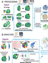
Whole-mount 3D imaging of multilayered biological tissues enables quantitative analysis of cell states and organization in their spatial context. However, extracting unbiased and meaningful quantitative information from dense, multilayered samples remains challenging due to imaging artifacts, increased density, and limited signal-to-noise ratio. Open source bioimage analysis workflows tailored to this type of analysis are scarce, and analysis bottlenecks like image curation or cell segmentation are seldom available without coding expertise. Here, we present a step-by-step computational protocol for the analysis of dense 3D organoid datasets using the Tapenade (Thorough Analysis PipEliNe for Advanced DEep imaging) workflow. Starting from multichannel image stacks, the protocol guides users through software installation, registration and fusion of multi-view datasets, preprocessing, and nuclei segmentation. It further details the generation of quantitative outputs, including morphometric measurements, deformation fields, and spatial correlation analyses. The workflow can be executed through open-source Python scripts or user-friendly Napari interfaces, allowing interactive parameter tuning and 3D visualization at each stage. This pipeline provides an accessible and modular framework for nonspecialist users to perform reproducible, multiscale quantitative analysis of 3D organoid images, while retaining flexibility for advanced users to customize individual steps.

Cancer Biology

Immune-Complex-Based In Vitro Deubiquitination Assay

Immune-Complex-Based In Vitro Deubiquitination Assay

FD Florence Dô
SM Sylvain Meloche
MS Marc J. Servant
196 Views
Oct 5, 2026
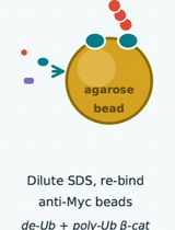
Deubiquitinases (DUBs) are attractive therapeutic targets within the ubiquitin-proteasome system, in part because four of the five DUB subfamilies are cysteine proteases amenable to the development of potent, selective inhibitors, as recently demonstrated for USP7. Identifying DUBs that deubiquitylate and stabilize specific human oncogenic proteins is therefore a promising approach to discovering new mechanism-based targets for cancer therapy. Several complementary experimental strategies are typically required to identify bona fide DUB–substrate pairs. Here, we present an efficient, straightforward in vitro immune-complex protocol to validate USP17-mediated deubiquitylation of the transcriptional co-activator β-catenin. In this assay, both β-catenin and USP17 are immunopurified from transiently transfected 293T cells and then combined to assess USP17 enzymatic activity. The protocol describes the in vitro enzymatic assay performed on immunopurified complexes and the immunoblot-based readout. It can be readily adapted to other DUBs and substrates for mechanistic studies.

Cell Biology

Applications of OptoProfilin in Living Cells for the Imaging of Focal Adhesions and Stress-Associated Phenotypes

Applications of OptoProfilin in Living Cells for the Imaging of Focal Adhesions and Stress-Associated Phenotypes

Clayton J. Brown Clayton J. Brown
RH Robert M. Hughes
171 Views
Oct 5, 2026
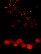
Cellular stress induces profound changes in cytoskeletal organization and biomolecular condensate formation. Traditional approaches for monitoring cellular stress often require multi-component biosensors, endpoint staining procedures, or indirect biochemical measurements. Here, we describe a protocol for the use of OptoProfilin, a genetically encoded single-component optogenetic biosensor derived from Profilin-1 fused to Cryptochrome 2 (Cry2) and mCherry. Following transient expression in mammalian cells, OptoProfilin exhibits light-dependent localization to focal adhesions under non-stressed conditions and transitions to punctate condensates under energetic, oxidative, osmotic, and senescence-associated stress conditions. The protocol includes transient transfection, induction of cellular stress, live-cell imaging, immunofluorescence validation, and quantitative image analysis. While this protocol describes imaging on a Leica widefield fluorescence microscope, it can readily be extended to other microscopy platforms. As a stand-alone biosensor that produces visually distinct responses in stressed versus non-stressed cells, OptoProfilin provides a convenient platform for investigating stress-associated cytoskeletal remodeling and biomolecular condensate formation.
Step-by-Step Protocol for Fluorescence-Based Analysis of Uptake in Transporter-Expressing Xenopus laevis Oocytes
Step-by-Step Protocol for Fluorescence-Based Analysis of Uptake in Transporter-Expressing Xenopus laevis Oocytes

Vd Víctor de Prado Parralejo
HN Hussam H. Nour-Eldin
CK Christa Kanstrup
110 Views
Oct 5, 2026
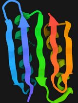
Xenopus laevis oocytes are widely used as a heterologous expression system for investigating the function of membrane proteins due to robust expression of heterologous protein and a low endogenous transport background. Traditionally, transporter activity in oocytes has been assessed using electrophysiology or radiolabeled uptake assays, approaches that are constrained by the requirement for electrogenicity of the transport process, availability of radiolabeled compounds, and instrumentation. Here, we describe a fluorescence-based uptake assay that enables direct and rapid quantification of transporter activity using a fluorescence plate reader. The protocol uses the Arabidopsis thaliana sucrose transporter 1 (SUC1) and its fluorescent substrate esculin as a case for how to set up the assay. The workflow includes optimizing assay conditions, sample preparation, fluorescent measurements, and downstream data analysis using R. This method can readily be adapted to other transporter-substrate pairs, and it supports applications such as transporter inhibitor screening, mutational analysis, characterization of kinetic properties, or indirect substrate specificity testing through competition assays. Overall, this protocol provides a simple and scalable alternative to traditional techniques, eliminating the need for radiolabeled compounds or electrophysiology while enabling easy quantitative assessment of transporter activity.
Identifying Differentially Expressed Proteins via Plasma Exosomal Proteomics

Identifying Differentially Expressed Proteins via Plasma Exosomal Proteomics

LZ Li-Li Zhang
GH Guo-Wei He
82 Views
Oct 5, 2026
Extracellular vesicle (EV) proteomics can be used to study intercellular communication and find biomarkers of non-invasive diseases. Traditional separation methods (ultracentrifugation, size exclusion chromatography) and data-dependent acquisition (DDA) mass spectrometry usually have the drawbacks of copurification of pollutants, poor reproducibility, and insufficient sampling of low-abundance peptides. This protocol describes a workflow for label-free extracellular vesicle proteomics, which combines size exclusion chromatography for vesicle separation, data-independent acquisition (DIA) for deep discovery, and parallel reaction monitoring (PRM) for targeted verification. Plasma-derived extracellular vesicles are treated by standardized lysis, digestion, and LC-MS/MS procedures, so that the protein group of extracellular vesicles can be identified and quantified robustly. The main advantages of this scheme are that it can achieve high quantitative reproducibility, unbiased detection of low-intensity peptides, and seamless transition from discovery to targeted verification, while also being suitable for liquid biopsy samples and various cardiovascular diseases.

Developmental Biology

3D Iterative Immunofluorescence Imaging on Whole-Mount Samples
3D Iterative Immunofluorescence Imaging on Whole-Mount Samples

MW Marvin F. Wyss
MH Max Hess
SS Shayan Shamipour
LP Lucas Pelkmans
247 Views
Oct 5, 2026
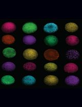
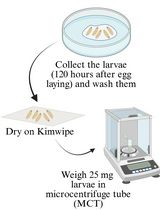
Multicellular organization relies on reciprocal interactions between molecular events, such as gene expression and protein state, and higher-scale properties, such as spatial patterning and tissue architecture. Understanding these processes requires methods that enable quantitative measurements at subcellular resolution, while maintaining the three-dimensional tissue organization. Conventional immunofluorescence imaging captures spatial information but is limited to the number of fluorescence markers that can be imaged simultaneously, whereas dissociation-based single-cell approaches can profile multimodal cellular states but lack positional information. Here, we describe 3D in toto iterative immunofluorescence imaging, termed 3D-4i, which enables up to ten-plex protein and protein state measurements in early zebrafish embryos. Leveraging sample immobilization on 96-well plates together with a gentle liquid handling system and high-content spinning disc confocal microscopy, this method comprises repeated rounds of antibody staining, optical clearing, confocal imaging, and antibody elution. Subsequent image analysis allows segmentation of nuclei and cells, extraction of quantitative single-cell features, and integration of molecular measurements with spatial context. Altogether, 3D-4i provides a scalable platform for investigating diverse biological processes in intact embryos, while maintaining both subcellular resolution and three-dimensional context.
Quantification of 20-Hydroxyecdysone From Drosophila Larvae at the Onset of Metamorphosis

Quantification of 20-Hydroxyecdysone From Drosophila Larvae at the Onset of Metamorphosis

JK Jyotsna Kawadkar
LB Lisupriya Baral
RM Ram Kumar Mishra
85 Views
Oct 5, 2026
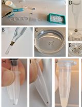
Metamorphosis in class Insecta is defined as the drastic transition of an organism from a larval stage to a morphologically very different adult form. Insects such as Drosophila melanogaster undergo metamorphosis, which requires precise temporal control, exerted by ecdysteroids, the primary regulators of these developmental transitions. The active form of ecdysone is 20-hydroxyecdysone (20E), which acts in pulses that trigger the onset of different developmental stages. A massive pulse of 20E occurs between the prepupal and pupal stages, triggering pupariation. Precise quantification of 20E levels is critical for understanding how this hormonal signaling governs the developmental timing of different stages. This protocol is sensitive for detecting low levels of 20E and describes a methanol-based extraction method coupled with a competitive ELISA to quantify whole-body 20E levels in third-instar Drosophila larvae. Further, this protocol enables detection of differences in ecdysone titers among genotypes affecting ecdysone biosynthesis, particularly at the late third-instar larval stage. This protocol can also be utilized for disease-causing insects (Aedes aegypti), as well as economically relevant beneficial insects (Apis mellifera), to study the effects of ecdysteroid analogs during development.

Mechanobiology

Live-Cell Detection of Relative Intracellular Tension Dynamics Using Non-FRET α-Actinin and α-Catenin Tension Indicators

Live-Cell Detection of Relative Intracellular Tension Dynamics Using Non-FRET α-Actinin and α-Catenin Tension Indicators

MH Maretoshi Hirai
KF Keita Fujiwara
191 Views
Oct 5, 2026
Molecular tension sensors enable the visualization of forces acting on specific intracellular proteins in living cells. Most established genetically encoded sensors rely on Förster resonance energy transfer (FRET), requiring donor–acceptor imaging and correction for spectral bleed-through, which can complicate their application in heterogeneous tissues. We developed non-FRET α-actinin and α-catenin tension indicators containing a force-responsive tension sensor (TS) module composed of an optimized circularly permuted enhanced green fluorescent protein (cpEGFP) scaffold and an elastic (GPGGA)8 linker. The TS module is incorporated into α-actinin or α-catenin, while a C-terminal mCherry serves as a force-insensitive reference for indicator abundance. This protocol describes indicator expression in cultured cells, validation using the myosin II inhibitor blebbistatin, two-color time-lapse imaging and live-cell super-resolution imaging using SRRF-Stream reconstruction based on super-resolution radial fluctuations (SRRF), and calculation of the green/red fluorescence ratio and normalized relaxation ratio. The protocol also specifies essential quality-control procedures, including imaging under nonsaturating conditions with fixed channel-specific acquisition settings across comparisons, registration of the green and red channels, and the use of appropriate vehicle and negative controls. For SRRF-Stream analysis, the green and red channels are reconstructed separately using identical reconstruction settings before ratio calculation. Representative reconstructed images should be compared with the corresponding conventional images to check for reconstruction artifacts. Optional procedures describe imaging of isolated cardiomyocytes and freshly isolated organs from tension-indicator mice. Because molecular loading is inferred from changes in TS fluorescence normalized to mCherry rather than from energy transfer between two fluorophores, the method avoids FRET-specific bleed-through correction; nevertheless, controlled acquisition and channel registration remain necessary. The indicators report relative changes in molecular loading and are particularly useful for resolving protein-specific and subcellular heterogeneity in tension dynamics.

Plant Science

Radial Water Loss Assay in Rice Roots Grown in Different Media
Radial Water Loss Assay in Rice Roots Grown in Different Media

LP Lucas L. Peralta Ogorek
SJ Samuel Jamieson
BP Bipin K. Pandey
125 Views
Oct 5, 2026
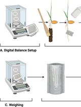
Plants growing in soil are exposed to several environmental stresses, such as soil compaction, flooding, or drought, and need to quickly acclimate to these conditions to survive. Such acclimations include suberization and lignification of the outer and inner parts of the root. Visualization of suberin and lignin depositions in roots can be done with several well-established staining techniques followed by microscopy, but these methods are mostly qualitative. Radial water loss assays provide a quick, robust, and quantitative method to evaluate the formation of suberized and/or lignified outer apoplastic barriers and how tightly they regulate water loss. Here, we provide a detailed step-by-step protocol, from harvesting the roots from soil conditions and cleaning them, to preparing them for water loss measurements, while using basic lab equipment. Since the data obtained is water loss as mass, quantitative and statistical comparisons can be performed. Finally, while in this protocol roots were grown in soil, the method can be applied to roots growing in hydroponics, agar plates, or any other growth conditions that allow non-destructive harvesting of roots. In summary, the protocol provides a fast and reliable method to assess differences in outer apoplastic barrier development of root tissues, allowing for fast root tissue screenings while obtaining quantitative data without expensive or highly specialized equipment.
A Simplified Density Gradient–Free Method for Isolating Functional Symbiosomes From Legume Nodules

A Simplified Density Gradient–Free Method for Isolating Functional Symbiosomes From Legume Nodules

FL Fuyu Li
YH Yining Hou
JM Jeremy D. Murray
125 Views
Oct 5, 2026
Symbiosomes are plant membrane–bound, organelle-like structures that enclose single or multiple bacteroids (differentiated rhizobia) within legume cells, serving specifically as the site for N2 fixation. Although isolated symbiosomes have been used to study nitrogen fixation, ion transport, and metabolite translocation, conventional density gradient–based isolation methods for their isolation are labor-intensive. Here, we describe a simplified, density gradient–free protocol for rapid enrichment of symbiosome-containing fractions from mature legume nodules using the Medicago truncatula–Sinorhizobium meliloti symbiosis as an example. The method combines razor blade homogenization, filtration through a 40 μm cell strainer, and sequential low-speed centrifugation to remove debris and enrich symbiosome-associated fractions. The resulting material is compatible with downstream mass spectrometry–based proteomics and mutant phenotypic analyses. Compared with conventional approaches, this protocol is rapid, simple, and suitable for higher-throughput biochemical and proteomic studies of symbiosomes.

Stem Cell

Efficient Generation of Fetal Hepatic Stellate Cells From hiPSC

Efficient Generation of Fetal Hepatic Stellate Cells From hiPSC

XY Xia Yang
HT Hideki Taniguchi
YN Yun-Zhong Nie
156 Views
Oct 5, 2026
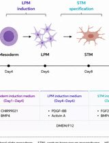
Human induced pluripotent stem cell (hiPSC)-derived liver organoids have emerged as valuable models for studying human liver development. However, existing organoid systems often lack developmentally matched cell populations, particularly fetal hepatic stellate cells (HSCs), limiting their ability to recapitulate key developmental processes. Current approaches for generating HSCs rely on primary cells, immortalized cell lines, or hiPSC differentiation methods that frequently produce activated HSC-like cells and often require cell sorting. Here, we describe an efficient protocol for generating expandable fetal-like HSCs from hiPSCs through a stepwise differentiation strategy that mimics embryonic HSC development. The resulting cells can be robustly expanded while maintaining characteristic molecular and functional features of fetal HSCs. This protocol provides a reproducible and scalable source of fetal-like HSCs without cell sorting and supports the generation of multicellular liver organoids containing developmentally relevant stromal components. Beyond the validation of the protocol in studies of liver maturation and vascularization, it can be applied to investigations of HSC biology and congenital liver diseases.