- Protocols

- Articles and Issues
- For Authors
- About
- Become a Reviewer

Past Issue in 2013
Volume: 3, Issue: 21
Biochemistry
Quantification of Bacterial Fatty Acids by Extraction and Methylation
β1 Integrin Cell-surface Immunoprecipitation (Selective Immunoprecipitation)

Cell Biology
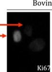
Ki67 Immunofluorescence on Bovine Cell Lines

Immunology
Bacterial Counts in Spleen

Microbiology
Immunoplaque Assay (Influenza Virus)

H2O2 Kill Assays of Biofilm Bacteria

H2O2 Kill Assays of Planktonic Stationary Phase Bacteria
Adhesion of Moraxella catarrhalis to Respiratory Tract Epithelial Cells


Analysis of Moraxella catarrhalis Outer Membrane Protein Profiles
Neuroscience
Dissection of Different Areas from Mouse Hippocampus
Implantation of Dkk-1-soaked Beads into the Neural Tube of Chicken Embryos
Mitochondrial Isolation and Purification from Mouse Spinal Cord
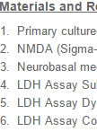
NMDA-induced Excitotoxicity and Lactate Dehydrogenase Assay in Primary Cultured Neurons
Plant Science
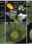
Ovule Clearing Method for Solanaceous Species
Visualization and Quantification of Actin Dynamics in Rice Protoplasts


Stem Cell
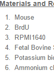
In vivo BrdU Incorporation Assay for Murine Hematopioetic Stem Cells
