


- Protocols

- Articles and Issues
- For Authors
- About
- Become a Reviewer


PrimeFlowTM Assay for Cell Type–Specific Co-detection of Transgene RNA and Protein in Mouse Spleens From Preclinical Studies

Published: Vol 16, Iss 12, Jun 20, 2026 DOI: 10.21769/BioProtoc.5748 Views: 390
Reviewed by: Kif Liakath-AliSalma MerchantAnonymous reviewer(s)
 PDF
PDF  Ask a question
Advertisement


Protocol Collections
Comprehensive collections of detailed, peer-reviewed protocols focusing on specific topics




Abstract
The PrimeFlowTM assay is a flow cytometry–based method for the co-detection of RNAs and proteins in cells. When combined with cell characterization by immunophenotyping, PrimeFlowTM can be used to simultaneously detect RNA and proteins in a cell type–specific manner in complex heterogeneous samples, offering an advantage over bulk tissue analysis methods. Here, we describe the implementation of the PrimeFlowTM assay protocol for the detection of transgene mRNA and protein expression in spleen samples from mice treated in vivo with luciferase mRNA-lipid nanoparticles (LNPs). This protocol involves spleen tissue dissociation for cell isolation, followed by cell fixation and permeabilization to allow immunolabeling of intracellular luciferase protein. The immunophenotyping strategy is based on immunolabeling with mouse CD marker antibodies for the identification of T cells, B cells, monocytes, granulocytes/macrophages, NK cells, and non-hematopoietic cells. The RNAs of luciferase and a housekeeping gene, β-actin, are detected with sequence-specific probe sets by employing sequential oligonucleotide annealing steps and fluorescent labeling using a branched DNA (bDNA) technology. Samples are analyzed by flow cytometry. Based on our analysis, we conclude it is feasible to apply the PrimeFlowTM approach for evaluating successful drug targeting to the cell types of interest and any potential differences in the kinetics of RNA delivery and protein expression in various tissue cells, supporting the discovery and development of RNA therapeutics.
Key features
• This protocol can be employed for cell-specific simultaneous detection of up to four different RNAs (using available commercial reagents) and multiple proteins by flow cytometry.
• This protocol requires application-specific optimization of the RNA-binding probe sets and antibody reagents for transgene detection and immunophenotyping panel.
Keywords: PrimeFlowTM assay Flow cytometry
RNA therapeutics
Transgene expression
Immunophenotyping
RNA analysis
Flow-based in situ hybridization
Branched DNA (bDNA)
Flow cytometry
RNA therapeutics
Transgene expression
Immunophenotyping
RNA analysis
Flow-based in situ hybridization
Branched DNA (bDNA)
Graphical overview
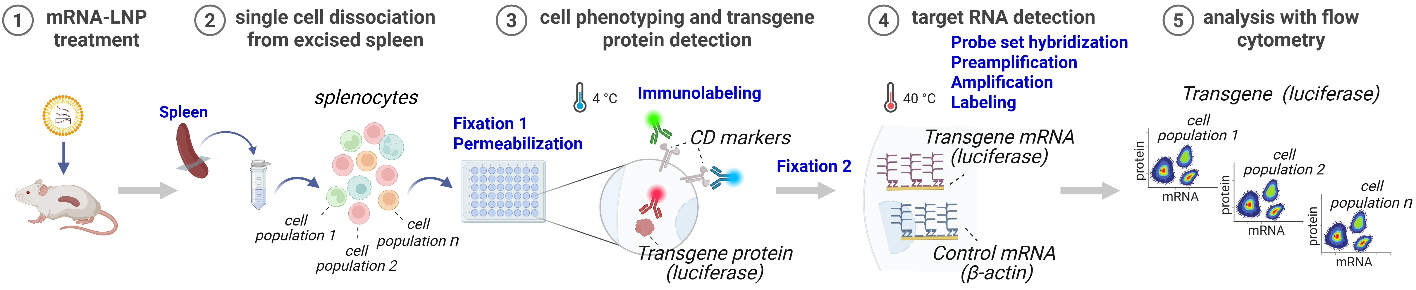
Overview of the PrimeFlowTM assay workflow for mouse spleen analysis. Cells are dissociated from the spleens of mRNA-lipid nanoparticle (LNP)-injected mice. Then, immunophenotyping with target protein labeling is conducted, followed by target-specific oligonucleotide probe hybridization and RNA signal amplification by bDNA reaction [1]. The cells are analyzed with flow cytometry. Created in Biorender.com.
Background
Development of gene therapies and vaccines relies on efficient transgene RNA delivery to target cell types, which necessitates a detailed understanding of RNA level and transgene protein expression kinetics in different cell populations. However, current bioanalytical methods, such as PCR for RNA and liquid chromatography–mass spectrometry (LC-MS) for protein measurements, usually do not allow simultaneous RNA and protein measurement in the same sample. More importantly, these methods rely on bulk tissue analysis and, therefore, do not provide cell type–specific resolution.
The PrimeFlowTM technique, commercially available as Thermo Fisher Scientific PrimeFlowTM RNA Assay, can potentially mitigate the above-mentioned issues. The PrimeFlowTM assay has been developed by combining flow cytometry with in-situ hybridization and the branched DNA (bDNA) amplification technique [1–5]. The detection of target proteins, either membrane or intracellular, is conducted with fluorescently labeled antibodies that are found compatible with the assay conditions. The RNA detection by the PrimeFlowTM approach requires in-situ hybridization with target-specific probe pairs, provided by Thermo Fisher, either off the shelf or custom-generated. Then, the bound probe signal is amplified (reportedly, ~8,000–16,000 fold [1–5]), through a series of steps: the bDNA reaction causes oligonucleotide pre-amplifiers to anneal the pre-bound target probe pairs, then multiple amplifiers bind each pre-amplifier, and, subsequently, multiple fluorescently tagged label probes bind to each amplifier, generating a tree-like structure on the target RNA molecule with a high number of associated fluorescent molecules. Studies have shown that the PrimeFlowTM assay has high sensitivity, with a detection limit of ~10 copies/cell for mRNA and ~20 copies/cell for miRNA [5], while also known to be specific [1]. Currently, the available commercial kit can be multiplexed to detect four RNAs and multiple target proteins [1,4].
Further, when combined with cell immunophenotyping, the PrimeFlowTM assay can provide valuable information on the presence of mRNA and protein in predefined cell populations in a tissue. Thus, we advanced the implementation of the PrimeFlowTM approach for the detection of luciferase transgene mRNA and protein in cells from the spleens of mice treated with luciferase mRNA-lipid nanoparticles (LNPs) in vivo. We found that, despite challenging execution (e.g., identifying and qualifying compatible reagents, temperature requirements, multistep labor-intensive protocol) and the need for further optimization (e.g., cell loss), the PrimeFlowTM assay can support simultaneous multiplex detection of transgene mRNA and protein in a cell type–specific manner in samples from preclinical studies with potential to provide otherwise unattainable information for development of RNA-based and other types of biotherapeutics.
Materials and reagents
Biological materials
1. Male BALB/c mice (8–10 weeks old, 25–30 g) (Charles River Labs)
Reagents
1. PrimeFlowTM RNA Assay Kit (Thermo Fisher Scientific, catalog number: 88-18005-210), containing:
a. PrimeFlowTM RNA Fixation Buffer 1A (catalog number: 00-18100-54)
b. PrimeFlowTM RNA II Fixation Buffer 1B (catalog number: 00-18200-54)
c. PrimeFlowTM RNA Permeabilization Buffer (10×) (catalog number: 00-18300-43)
d. PrimeFlowTM RNA Fixation Buffer 2 (8×) (catalog number: 00-18400-53)
e. PrimeFlowTM RNA Wash Buffer (catalog number: 00-19180-26)
f. PrimeFlowTM RNA Target Probe Diluent (catalog number: 00-19185-12)
g. PrimeFlowTM RNA PreAmp Mix (catalog number: 00-16000-53)
h. PrimeFlowTM RNA Amp Mix (catalog number: 00-16001-53)
i. PrimeFlowTM RNA Label Probe Diluent (catalog number: 00-19183-100)
j. PrimeFlowTM RNase Inhibitors (100×) (catalog number: 00-16002-19)
k. PrimeFlowTM Compensation Kit (catalog number: 88-17009-42)
l. IC Fixation Buffer (catalog number: 00-8222-758)
m. Label Probes, 100× (catalog number: 00-16003-32)
Note: Expiration dates and storage and handling instructions for all materials in the kit are provided by the manufacturer.
2. Antibodies
Note: All antibodies and antibody mixes were stored refrigerated and protected from light. Freezing and thawing were not examined and are not recommended.
a. Rat anti-mouse CD45-BUV496, clone 30-F11 [BD Biosciences (BD HorizonTM), catalog number: 569673]
b. Rat anti-mouse Ly-6C-BUV805, clone HK1.4 [BD Biosciences (BD OptiBuildTM), catalog number: 755202]
c. Mouse anti-mouse H2Kd/H2Dd (MHC-I)-BV421, clone 34-1-2S [BD Biosciences (BD HorizonTM), catalog number: 567294]
d. Rat anti-mouse CD19-BV605, clone 6D5 (BioLegend, catalog number: 115540)
e. Mouse anti-mouse NK1.1-BV711, clone PK136 [BD Biosciences (BD HorizonTM), catalog number: 569723]
Note: Immunolabeling of the NK1.1 marker cannot be employed with every mouse strain, as some strains are considered NK1.1-negative despite having an NK cell population. Strain-appropriate alternatives should be used.
f. Hamster anti-mouse CD3-BV750, clone 145-2C11 [BD Biosciences (BD OptiBuildTM), catalog number: 746988]
g. Rabbit anti-firefly luciferase-PE, clone EPR17789 (Abcam, catalog number: AB237253)
h. Rat anti-mouse CD11b-APC-eFluor780, clone M1/70 [Thermo Fisher Scientific (eBioscienceTM), catalog number: 47-0112-82]
3. Target probe sets, stored frozen (-20 °C), as recommended by the manufacturer:
a. PrimeFlowTM Type 4 Mouse β-Actin target probe set for Alexa Fluor 488 [Thermo Fisher Scientific (Invitrogen), catalog number: PF-210, VB4-10432]
b. PrimeFlowTM Type 1 luciferase target probe set for Alexa Fluor 647 (custom-designed VPWCWFC Synthetic G6 Luc co) [Thermo Fisher Scientific (Invitrogen), catalog number: PF-210]
4. BD OMICS-Guard sample preservation buffer (BD Biosciences, catalog number: 570911), stored refrigerated
5. BD PharmingenTM stain buffer (BSA) (BD Biosciences, catalog number: 554657), stored refrigerated
6. BD PharmingenTM stain buffer (FBS) (BD Biosciences, catalog number: 554656), stored refrigerated
7. Water (nuclease-free) (Fisher BioReagents, catalog number: BP2484-50), stored at ambient temperature
8. VitaStainTM AO/PI staining solution (Nexcelom Bioscience, catalog number: CS2-0106-5mL), stored refrigerated
Solutions
1. Permeabilization buffer (see Recipes)
2. Fixation buffer 2 (see Recipes)
3. RNA wash buffer with 1× RNase inhibitors (see Recipes)
Recipes
1. Permeabilization buffer
| Reagent | Dilution | Final concentration |
|---|---|---|
| PrimeFlowTM RNA permeabilization buffer (10×) | 1:10 | 1× |
| PrimeFlowTM RNase inhibitors (100×) | 1:100 | 1× |
Dilute in DNase/RNase-free water.
2. Fixation buffer 2
| Reagent | Dilution | Final concentration |
|---|---|---|
| PrimeFlowTM RNA fixation buffer 2 (8×) | 1:8 | 1× |
| PrimeFlowTM RNase inhibitors (100×) | 1:100 | 1× |
Dilute in PrimeFlowTM RNA wash buffer.
3. RNA wash buffer with 1× RNase inhibitors
| Reagent | Dilution | Final concentration in RNA wash buffer |
|---|---|---|
| PrimeFlowTM RNase inhibitors (100×) | 1:100 | 1× |
Laboratory supplies
Note: Based on the information provided by the manufacturers (website description, certificate of analysis), the supplies listed below are considered RNase-free.
1. 5 mL polystyrene round-bottom tube (12 × 75 mm style) (FACS tube) (Falcon, catalog number: 352054)
2. V-bottom 96-well plate (Costar, Corning, catalog number: 3896); supplemented with a lid from a flat-bottom 96-well plate (Costar, Corning, catalog number: 3596)
3. 50 mL polypropylene conical tube (30 × 115 mm style) (Falcon, catalog number: 352070)
4. 15 mL high-clarity polypropylene conical tube (17 × 120 mm style) (Falcon, catalog number: 352096)
5. 40 μm cell strainer (filter) (Fisher Scientific, catalog number: 07-201-430)
Equipment
1. BD FACSymphonyTM A5 Cell Analyzer (BD Biosciences, catalog number: 660964; any flow cytometer with appropriate optical setup can be used)
2. 40 °C incubator (Quincy Labs, model: 10E)
3. ViewRNATM Temperature Validation kit (Thermo Fisher Scientific, catalog number: QV0523)
4. Centrifuge (Thermo Fisher Scientific Sorvall X4R Pro-MD, catalog number: 75009521)
5. Cellometer Vision Cell Profiler (Nexcelom, model: Cellometer Vision)
Software and datasets
1. BD FACSDivaTM (BD Biosciences, version 9.1)
2. FlowJoTM (BD Biosciences, version 10)
3. Prism [GraphPad (Dotmatics), version 10]
4. Excel (Microsoft)
Procedure
A. Spleen dissociation
1. Store harvested spleen samples from LNP-treated mice in 50 mL tubes containing 20 mL of BD OMICS-Guard sample preservation buffer. Alternative sample preservation and storage buffers can be used.
Notes:
1. The spleens were harvested the day before and stored overnight refrigerated.
2. Each spleen sample corresponds to ~1/3 of the spleen of a mouse obtained by physical sectioning.
3. Red blood cell lysis is not required for this protocol.
2. Press the spleens through 40-μm filters kept on individual 50 mL tubes containing 5 mL of BD stain buffer (FBS) using the back of 5 mL syringe plungers. Keep the tubes on ice.
3. Wash filters with 5 mL of cold BD stain buffer (FBS).
4. Centrifuge samples at 400× g for 5 min and remove the supernatants.
5. Resuspend cells in 3 mL of BD stain buffer (FBS).
6. Count the cells with AO/PI (20 μL of cell suspension + 20 μL of AO/PI solution).
Note: Observed cell viability in such samples is ~70%.
7. Transfer the required number of viable cells from each spleen into 15 mL tubes and centrifuge the samples at 400× g for 5 min.
Note: 5 × 10⁶ cells per sample is recommended. An equal number of cells per sample should be used.
8. Aspirate the supernatant.
B. Immunophenotyping and target protein labeling
1. Prepare fixation buffer 1 on the day of the experiment by mixing equal parts of PrimeFlowTM RNA Fixation Buffer 1A and PrimeFlowTM RNA Fixation Buffer 1B. Mix gently by inverting several times.
Caution: See General note 8.
2. Resuspend each splenocyte sample in fixation buffer 1 to achieve 25 × 106 cells/mL. Dispense 200 μL/well of a 96-well V-bottom plate. The number of cells per well is 5 × 10⁶.
Notes:
1. The cellularity of a healthy mouse spleen depends on the strain and age of the animal. For an 8–10-week-old adult mouse, ~50 × 106 viable cells were obtained from the provided tissue sample.
2. Cells from each spleen sample are added in duplicate wells.
3. All subsequent incubation steps are conducted in plates covered with lids.
3. Incubate for 30 min at 4 °C.
4. Centrifuge the plate at 1,000× g for 4 min and aspirate supernatants.
5. Resuspend the cells in 200 μL/well of cold permeabilization buffer.
6. Centrifuge cells at 1,000× g for 4 min and aspirate supernatants.
7. Repeat washing and centrifugation with permeabilization buffer as described in steps B5–6.
8. Resuspend the splenocyte samples in 100 μL/well of permeabilization buffer with the indicated dilutions of all antibodies (See Reagents and Table 1).
Table 1. Immunolabeling antibodies description and flow cytometer configuration
| Target protein | Host | Fluorophore | Dilution | Laser | Filter |
|---|---|---|---|---|---|
| CD45 | Rat | BUV496 | 1:80 | UV | 515/30 |
| Ly-6C | Rat | BUV805 | 1:50 | UV | 820/60 |
| H2Kd/H2Dd (MHC-I) | Mouse | BV421 | 1:50 | Violet | 450/50 |
| CD19 | Rat | BV605 | 1:50 | Violet | 610/20 |
| NK1.1 | Mouse | BV711 | 1:50 | Violet | 710/50 |
| CD3 | Hamster | BV750 | 1:50 | Violet | 740/35 |
| Firefly luciferase | Rabbit | PE | 1:50 | Yellow/Green | 586/15 |
| CD11b | Rat | APC-eFluor780 | 1:160 | Red | 780/60 |
9. Incubate for 40 min at 4 °C.
10. Add 150 μL/well of permeabilization buffer to wells and centrifuge at 1,000× g for 4 min. Aspirate the supernatant.
11. Wash samples with 200 μL/well of permeabilization buffer. Centrifuge at 1,000× g for 4 min. Aspirate the supernatant.
12. Resuspend samples in 200 μL/well of fixation buffer 2.
13. Incubate in the dark at room temperature for 1 h.
14. Centrifuge cells at 1,000× g for 4 min. Aspirate the supernatant.
15. Wash samples with 200 μL/well of RNA wash buffer with 1× RNase inhibitors and centrifuge at 1,000× g for 4 min. Aspirate the supernatant.
16. Add 100 μL/well of RNA wash buffer with 1× RNase inhibitors and resuspend.
Pause point: (Optional) Before proceeding to target RNA detection in section C, samples can be stored overnight at 4 °C protected from light. Assay performance after longer storage was not assessed.
C. Target RNA signal amplification and detection
1. Warm PrimeFlowTM RNA target probe diluent to 40 °C for ~1 h.
Caution: See General note 8.
Note: Use the 40 °C incubator to warm the target probe diluent.
2. Thaw the target probes at room temperature for ~1 h.
3. Take the 96-well V-bottom plate out from 4 °C and keep it at room temperature in the dark until the plate is up to room temperature.
Notes:
1. This step can be omitted if sections B and C are performed on the same day.
2. All subsequent incubation steps are conducted in plates covered with lids.
4. Prepare the target probe master mix by diluting the following target probes at 1:20 v/v in PrimeFlowTM RNA target probe diluent:
a. Mouse β-Actin target probe set: Type 4.
b. Luciferase target probe set: Type 1.
Critical: Incorrect dilution of the target probes could cause weak/no signal or high background for target RNA.
5. Add 100 μL/well of the target probe master mix to wells containing 100 μL/well of RNA wash buffer with 1× RNase inhibitors. Resuspend samples by pipetting.
Optional: At this step, the cells can be transferred to a new V-bottom 96-well plate to combine samples from different treatments for ease of processing.
6. Incubate for 2 h at 40 °C.
Critical: See General note 6.
7. Centrifuge samples at 1,000× g for 4 min and aspirate the supernatant.
8. Wash samples with 200 μL/well of RNA wash buffer with 1× RNase inhibitors.
9. Centrifuge samples at 1,000× g for 4 min and aspirate the supernatant.
10. Resuspend samples in 100 μL/well of RNA wash buffer with 1× RNase inhibitors and store overnight at 4 °C.
Pause point: Samples can be stored overnight at 4 °C protected from light. Assay performance after longer storage was not assessed.
11. The following day, warm PrimeFlowTM RNA PreAmp mix, PrimeFlowTM RNA Amp mix, and PrimeFlowTM RNA label probe diluent to 40 °C.
Caution: See General note 8.
12. Warm the samples stored overnight at 4 °C (in the dark) to room temperature.
13. Add 100 μL/well of PrimeFlowTM RNA PreAmp mix to all samples and mix by pipetting.
14. Incubate for 90 min at 40 °C.
Critical: See General note 6.
15. Centrifuge samples at 1,000× g for 4 min and aspirate the supernatant.
16. Wash samples with 200 μL/well of RNA wash buffer with 1× RNase inhibitors, centrifuge samples at 1,000× g for 4 min, and aspirate the supernatant.
17. Resuspend samples in 100 μL/well of RNA wash buffer with 1× RNase inhibitors and add 100 μL/well of PrimeFlowTM RNA Amp mix. Mix by pipetting.
18. Incubate for 90 min at 40 °C.
Critical: See General note 6.
19. Centrifuge samples at 1,000× g for 4 min and aspirate the supernatant.
20. Wash samples with 200 μL/well of RNA wash buffer with 1× RNase inhibitors, centrifuge samples at 1,000× g for 4 min, and aspirate the supernatant.
21. Dilute RNA label probes at 1:100 in PrimeFlowTM RNA label probe diluent.
Note: This step can be performed during centrifugation steps C19 and/or C20.
22. Resuspend samples in 100 μL/well of RNA wash buffer with 1× RNase inhibitors. Add 100 μL/well of diluted label probes and mix by pipetting.
23. Incubate for 60 min at 40 °C.
Critical: See General note 6.
24. Centrifuge samples at 1,000× g for 4 min and aspirate the supernatant.
25. Wash samples with 200 μL/well of RNA wash buffer with 1× RNase inhibitors, centrifuge samples at 1,000× g for 4 min, and aspirate the supernatant.
26. Resuspend samples in 150 μL/well of BD stain buffer (BSA).
Pause point: Samples can be stored for a few hours at 4 °C protected from light until acquisition by flow cytometry. Longer sample stability should be assessed.
D. Cell analysis by flow cytometry
1. Transfer cells from the V-bottom 96-well plate to 12 × 75 mm FACS tubes.
Note: (Optional) Cells can be acquired directly from the 96-well plate if a flow cytometer with plate acquisition capability is available.
2. Start the BD FACSymphonyTM A5 flow cytometer and open BD FACSDiva acquisition software.
Note: Any multi-parameter flow cytometer with the appropriate optical setup and compatible acquisition software can be used for cell analysis.
3. Set up and/or verify detector voltages for fluorescent channels and forward scatter (FSC) and side scatter (SSC) parameters. Use compensation samples to verify voltages for fluorescent channels (see below for compensation sample preparation).
4. Acquire samples and record events (see Tables 1 and 2 for immunolabeling, RNA labeling, and cytometer configuration details).
Critical: Acquire all compensation controls with the same detector voltages as the splenocyte samples.
Notes:
1. Acquisition flow rate should not exceed ~5,000 events per second to reduce variability and acquisition errors.
2. The number of acquired events per sample should be adequate for the intended assay to obtain sufficient cell counts in the analyzed gates.
Table 2. RNA target probe set details and cytometer configuration
| Target RNA | Fluorophore | Laser | Filter |
|---|---|---|---|
| Mouse β-actin | AlexaFluor488 | Blue | 515/20 |
| Firefly luciferase | AlexaFluor647 | Red | 670/30 |
5. Compensation sample preparation
Prepare compensation samples using PrimeFlowTM Compensation Kit based on the manufacturer’s instructions.
a. Label a FACS tube for each fluorochrome used in the experiment, plus one additional tube for the unstained control.
b. Mix UltraComp eBeads by pulse-vortexing and add one drop to each tube.
c. Add 5 μL of the appropriate Alexa Fluor compensation controls compatible with target probe sets for RNA.
Notes:
1. Compensation control Alexa Fluor 488 (for mouse β-Actin Type 4 target probe set).
2. Compensation control Alexa Fluor 647 (for luciferase Type 1 target probe set).
d. For each antibody used for immunolabeling, add the volume that would be contained in 100 μL of staining buffer (see Table 1).
e. Mix tubes briefly by pulse-vortexing.
f. Incubate at 4 °C for 30 min in the dark.
g. Add 2 mL of BD staining buffer (BSA) to each tube and centrifuge at 600× g for 5 min.
h. Decant supernatant and vortex briefly to resuspend beads in residual volume.
i. Add 100 μL of IC fixation buffer to each tube and mix briefly by pulse-vortexing.
Caution: See General note 8.
j. Keep at 4 °C until ready to proceed to the next step (samples are kept for 1 h in this experiment).
k. Incubate at room temperature for 25 min in the dark.
l. Add 2 mL of BD staining buffer (BSA) to each tube and centrifuge at 600× g for 5 min.
m. Decant supernatant and add 0.5 mL of BD staining buffer (BSA) to each tube.
n. Mix briefly by pulse-vortexing and store at 4 °C until analysis.
o. Acquire samples on the flow cytometer.
Data analysis
1. Export the FCS files from the flow cytometry acquisition software (BD FACSDivaTM v9.1 in this study) and import them into a flow cytometry data analysis software (FlowJoTM v10 in this study).
2. Generate the spillover matrix in FlowJoTM v10. Use the untreated compensation control beads as the negative control.
3. Apply the compensation to samples in FlowJoTM v10.
4. Gate the cells to individual populations (see Figures 1 and S1 for the gating strategy and Table 3 for a list of gated cell populations) and quantify in FlowJoTM v10.
The PrimeFlowTM assay requires the sequence-specific annealing of target probe sets to RNA and sequential bDNA reaction for RNA signal amplification. Therefore, it is important to select the cells that have successfully undergone the PrimeFlowTM reaction (PrimeFlow+ cells) while eliminating the other cells for accurate data analysis. The PrimeFlowTM assay uses RNA from housekeeping genes for this purpose, as positive controls for assay performance. Here, the mouse β-actin was used as the positive control gene to select the PrimeFlow+ cells (an FMO without β-actin probe can be used to define the positive gate) (Figures 1 and S1). The gates for luciferase mRNA and protein in each cell population were created based on the background signal from vehicle-treated cells, which are negative for luciferase presence (Figures 1 and S1). Additionally, the monocytes, which are Ly6C+, exist as two separate populations in in vivo–treated mice based on their CD11b expression. Both these populations were analyzed as a single monocyte population for the purpose of this analysis (Figure 1).
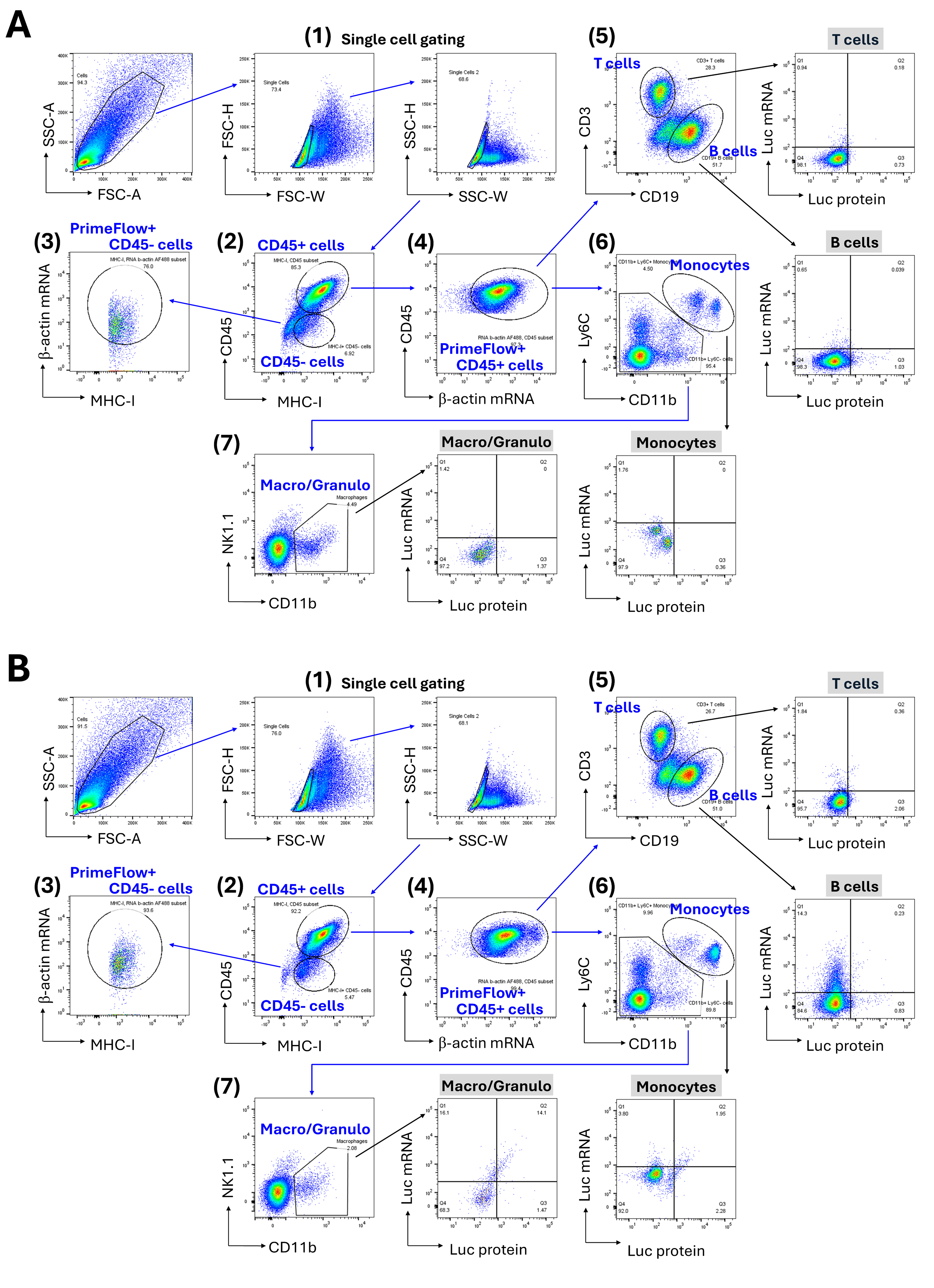
Figure 1. Gating strategy for mouse splenocyte immunophenotyping and luciferase RNA and protein co-detection. Representative pseudocolor scatterplots are shown as examples for samples from vehicle-treated (A) and LNP-treated (B) mice, illustrating a hierarchical gating strategy, as pointed by blue arrows (1–7). The primary gate was set on “single cells” by gating on the main population, followed by two consecutive single-cell gates (1). Then, using MHC-I vs. CD45 labeling, CD45- (non-hematopoietic) and CD45+ (hematopoietic) cell gates were created (2). For the subsequent analysis, β-actin mRNA vs. CD45 labeling signals were used to define “PrimeFlow+ CD45-” or “PrimeFlow+ CD45+” events (3 and 4, respectively). This “PrimeFlow+ CD45+” population gate was implemented to further gate “T cells” and “B cells” based on mutually exclusive CD3 and CD19 signals (5), and “Monocytes” were gated based on CD11b and Ly6C staining signals (6). The CD11b-/low and Ly6C- population was used to then gate on NK1.1- macrophages and granulocytes (“Macro/Granulo”) (7). See also Figure S1 for an example of gating on NK1.1+ NK cells in splenocytes from a C57Bl/6 mouse. For each of the identified splenocyte cell populations, the median fluorescence intensity of luciferase mRNA and protein is assessed. Furthermore, as pointed out by black arrows, bivariate plots of luciferase protein (Luc protein) vs. luciferase mRNA (Luc mRNA) signal were created to report % positive cells in each subpopulation identified with the employed immunophenotyping strategy. For most of the samples, ≥100,000 events were acquired. SSC: side scatter, FSC: forward scatter, A: area, H: height, W: width.
Table 3. Cell types gated in the data analysis. The following cell types were identified and analyzed using the immunophenotyping markers as indicated.
| Cell type | Immunophenotyping |
|---|---|
| Non-hematopoietic cells | MHC-I+ β-actin mRNA+ CD45- |
| T cells | MHC-I+ β-actin mRNA+ CD45+ CD3+ CD19- |
| B cells | MHC-I+ β-actin mRNA+ CD45+ CD3- CD19+ |
| Monocytes | MHC-I+ β-actin mRNA+ CD45+ CD11b+ Ly6C+ |
| Natural killer cells | MHC-I+ β-actin mRNA+ CD45+ CD11b- Ly6C- NK1.1+ |
| Macrophages and granulocytes | MHC-I+ β-actin mRNA+ CD45+ CD11blow Ly6C- NK1.1- |
5. Export the quantified data for cell types to Microsoft Excel.
a. Calculate the % total luciferase protein positive by adding the % of luciferase protein and mRNA positive cells to the % of cells only positive for luciferase protein.
b. Calculate the % total luciferase mRNA detection by adding the % of luciferase protein and mRNA positive cells to the % of cells only positive for luciferase mRNA.
6. Export the calculated % values and median fluorescent intensity (MFI) values for luciferase protein and mRNA detection in individual cell types (values of corresponding cell population in vehicle-treated samples were subtracted) to GraphPad Prism 10.
7. In Prism, generate data tables for each data analysis parameter for individual cell types in a grouped format containing duplicate values. Graph the results with the Column family.
Validation of protocol
1. Two technical replicates from each spleen were prepared and analyzed, from 38 total mice.
2. Vehicle (LNP dilution buffer)-treated samples were used as negative controls to determine the gating strategy for luciferase mRNA+ and protein+ cell populations and for background subtraction.
3. The specificity of the probe set for luciferase mRNA was characterized using positive and negative control samples during method development.
4. Mouse β-actin mRNA was used as an internal RNA control to evaluate sample processing quality.
5. This protocol has been used to analyze samples from three preclinical studies, and a manuscript is currently in preparation describing the protocol implementation for the study sample analysis.
General notes and troubleshooting
General notes
1. This protocol can also be used for the analysis of mouse splenocytes transfected with mRNA-LNP ex vivo (see Figure S1).
2. The protocol for sections B and C generally follows the manufacturer’s recommended protocol for the PrimeFlowTM assay. Refer to “PrimeFlowTM RNA Assay User Manual and Protocol, Appendix 7: Protocol using 96-well plates” by Invitrogen (Thermo Fisher Scientific) [4] for more information.
3. For the flow cytometry immunophenotyping panel design, make sure that the antibodies (if post-fixation immunolabeling is used) and the associated fluorophores are compatible with the PrimeFlowTM assay conditions of strong fixation and high temperature in situ hybridization (refer to the manufacturer’s instructions for more information). Some fluorophores, such as PerCP, PerCP-Cyanine5.5, and PerCP-eFluorTM 710, are already identified by the vendor as not compatible with the PrimeFlowTM assay [4]. Test key reagents experimentally to ensure compatibility.
4. The PrimeFlowTM assay involves a higher number of washing steps compared to more common flow cytometry sample preparation approaches, as well as high temperature and strong fixation incubation steps that impact cell integrity, causing substantial cell loss during sample preparation. Therefore, be cautious when using this protocol with samples with low cell numbers. In the protocol steps that require removing supernatants after sample centrifugation and mixing cells with reagents, consider gentle pipetting and several pulse-vortexing before reagent addition. Additionally, conducting surface CD marker immunolabeling together with intracellular transgene immunolabeling after cell permeabilization reduces the number of sample washing steps and may help in ameliorating cell loss.
5. The PrimeFlowTM probe sets are available in four different types. It is important to note that Type 1 (Alexa Fluor 647) and Type 10 (Alexa Fluor 568) are highly sensitive and should be used for targets with low/unknown expression. Type 4 (Alexa Fluor 488) and Type 6 (Alexa Fluor 750) have intermediate to low sensitivity, making them optimal to detect medium to high expressing targets [4]. Lower sensitivity probes can also be used for the RNA not used for quantification, such as that of housekeeping genes. If custom-made probe sets are needed, discuss probe set design and requirements with the manufacturer.
6. It is important to ensure, prior to the assay execution, that the temperature in the incubator for in situ hybridization is at 40 ± 1 °C and is maintained constant during the hybridization steps, since improper temperature could cause a weak signal and/or high background. To this end, the ViewRNATM Temperature Validation kit is recommended. Refer to “PrimeFlowTM RNA Assay User Manual and Protocol, Appendix 6: Temperature validation procedure for incubator” by Invitrogen (Thermo Fisher Scientific) [4] for more information. Additionally, do not stack plates in the incubator during the incubation steps at 40 °C.
7. It is highly recommended to include an internal RNA control (such as a housekeeping gene RNA) in the experimental design to evaluate the efficiency of in situ hybridization and bDNA amplification reactions and analyze only the cells that underwent this process successfully.
8. PrimeFlowTM RNA fixation buffer 1A contains formaldehyde. PrimeFlowTM RNA target probe diluent, PrimeFlowTM RNA PreAmp mix, and IC fixation buffer contain formamide. Therefore, these reagents should be used with caution due to health/safety concerns.
9. Prepare the permeabilization buffer, fixation buffer 2, and RNA wash buffer with 1× RNase inhibitors fresh on the day of use; these are not intended for storage.
10. For critical assay reagents, evaluate lot-to-lot variability by setting performance criteria and confirming that each new lot meets these standards through comparability testing against a reference lot to ensure consistent assay results.
11. All centrifugation steps are performed while refrigerated, with maximum acceleration and brake settings. Washing/resuspension following centrifugation and aspirating supernatants are performed by pulse-vortexing followed by reagent addition and mixing by pipetting several times.
12. All incubations are conducted with the samples being protected from light, with no shaking, at the temperatures required for the respective steps.
Troubleshooting
Problem 1: Weak/no signal for immunolabeled proteins.
Possible causes: Fluorophores conjugated to antibodies are not compatible with the PrimeFlowTM assay, and/or antibody clones are not compatible with staining post-fixation.
Solution: Identify and use fluorophores compatible with the PrimeFlowTM assay and antibody clones compatible with post-fixation staining.
Problem 2: Weak/no signal or high background for luciferase RNA.
Possible causes:
i. Incorrect incubation temperature during target probe hybridization and/or bDNA reaction.
ii. Probe sets do not bind or are not target-specific.
iii. Sub-optimal probe type sensitivity for low-expressing or highly expressed targets.
Solutions:
i. Ensure that the incubator temperature stays at 40 ± 1 °C during the target probe hybridization and bDNA reaction steps and that samples are uniformly heated.
ii. Confirm probe set performance with positive and negative controls. Design new probes using bioinformatics tools.
iii. Change the probe type to optimize sensitivity: Type 1/Type 10 (high sensitivity) for low expressing/unknown targets, and Type 4/Type 6 (intermediate to low sensitivity) for medium to high expressing targets (see General note 5).
iv. Examine different probe set dilutions to improve signal-to-noise.
Refer to “PrimeFlowTM RNA Assay User Manual and Protocol, Appendix 1: Troubleshooting” by Invitrogen (Thermo Fisher Scientific) [4] for more information.
Supplementary information
The following supporting information can be downloaded here:
1. Figure S1. Example of gating strategy for immunophenotyping and luciferase RNA and protein co-detection in isolated mouse splenocytes transfected with mRNA-LNP ex vivo.
Acknowledgments
Conceptualization, P.G.; Investigation, S.G.K., P.G.; Study coordination, X.F.; mRNA-LNP design, S.K., O.V., J.S.R.; Writing—Original Draft, S.G.K.; Writing— Review & Editing, S.G.K., P.G.; Supervision, P.G., J.C., H.N. Funding for the study was from Pfizer, Inc. The authors would like to thank Margaret Taylor in the Pharmacokinetics, Dynamics and Metabolism Department, Pfizer Inc., for assay development efforts, and colleagues in the Comparative Medicine Department, Pfizer Inc., for performing animal-related work.
Competing interests
All authors were employees of Pfizer, Inc. at the time this work was conducted, and may hold equity in Pfizer, Inc.
Ethical considerations
All procedures performed on animals were in accordance with regulations and established guidelines and were reviewed and approved by an Institutional Animal Care and Use Committee or through an ethical review process.
References
- PrimeFlowTM RNA Assay Technology Validation Paper. (n.d.). https://documents.thermofisher.com/TFS-Assets/LSG/brochures/FC04271-1_Prime-FlowRNA-Assay-TechValPaper_v13_102914_web.pdf
- Alter, H. J., Sanchez-Pescador, R., Urdea, M. S., Wilber, J. C., Lagier, R. J., Bisceglie, A. M. D., Shih, J. W. and Neuwald, P. D. (1995). Evaluation of branched DNA signal amplification for the detection of hepatitis C virus RNA. J Viral Hepat. 2(3): 121–132. https://doi.org/10.1111/j.1365-2893.1995.tb00017.x
- Tsongalis, G. J. (2006). Branched DNA technology in molecular diagnostics. Am J Clin Pathol. 126(3): 448–453. https://doi.org/10.1309/90bu6kdxanfln4rj
- PrimeFlow® RNA Assay. (n.d.). https://documents.thermofisher.com/TFS-Assets/LSG/manuals/88-18005.pdf
- Lai, C., Stepniak, D., Sias, L. and Funatake, C. (2018). A sensitive flow cytometric method for multi-parametric analysis of microRNA, messenger RNA and protein in single cells. Methods. 134–135: 136–148. https://doi.org/10.1016/j.ymeth.2017.12.016
Article Information
Publication history
Received: Mar 17, 2026
Accepted: May 31, 2026
Available online: Jun 15, 2026
Published: Jun 20, 2026
Copyright
© 2026 The Author(s); This is an open access article under the CC BY-NC license (https://creativecommons.org/licenses/by-nc/4.0/).
How to cite
Gallolu Kankanamalage, S., Fan, X., Kundu, S., Villamizar, O., Roh, J. S., Cohen, J., Neubert, H. and Goihberg, P. (2026). PrimeFlowTM Assay for Cell Type–Specific Co-detection of Transgene RNA and Protein in Mouse Spleens From Preclinical Studies. Bio-protocol 16(12): e5748. DOI: 10.21769/BioProtoc.5748.

 Download Citation in RIS Format
Category
Immunology > Immune cell staining > Flow cytometry
Molecular Biology > RNA > RNA detection
Do you have any questions about this protocol?
Post your question to gather feedback from the community. We will also invite the authors of this article to respond.
 Post a Question


