


- Protocols

- Articles and Issues
- For Authors
- About
- Become a Reviewer


Enriching Bacteria-Specific RNA From Host Samples Before NGS With Transcript-Capture

(§Technical contact: elamont@uw.edu) Published: Vol 16, Iss 12, Jun 20, 2026 DOI: 10.21769/BioProtoc.5712 Views: 278
Reviewed by: Alba BlesaJiadong ZhaoKyle Stewart Skalenko
 PDF
PDF  Ask a question
Original research article
The authors used this protocol in:
Dec 2025
Advertisement


Protocol Collections
Comprehensive collections of detailed, peer-reviewed protocols focusing on specific topics




Related protocols
Abstract
Pathogen gene expression from host samples is often challenging to study due to low signal and high host RNA background. PCR probes have been recently used to hybridize and extract bacterial sequences from next-generation sequencing (NGS) libraries generated from in vitro and animal models of infection; however, these strategies require purchasing commercially synthesized probes that often do not capture the entire transcriptome. Transcript-capture sequencing is a novel capture approach for extracting RNA of a target bacterial species from samples in which there is substantial contamination by the host or other microbes. Biotinylated 150-base-pair DNA probes are generated in-house from bacterial DNA spanning the entire bacterial genome. Probes are hybridized to the cDNA of NGS sequencing libraries prepared from host samples to capture and enrich for bacterial-specific RNA reads before sequencing. This method results in a >200-fold increase in bacterial RNA reads from infected host samples (including in vitro, animal, and human samples) and generates complete bacterial transcriptomes with high gene coverage (>80%). Use of this protocol on infected host samples reveals a snapshot of bacterial activity during disease that may improve understanding of the physiological state of pathogens within their hosts.
Key features
• Generates single-stranded biotinylated DNA probes in-house from bacterial gDNA.
• Uses DNA probes in a hybridization capture approach to enrich for cDNA of a target species >200-fold in standard next-generation RNA sequencing libraries.
• Allows generation of complete bacterial transcriptomes (>80% gene coverage) from samples in which transcriptomic signal-to-noise is limiting, such as from animal models and clinical samples.
Keywords: DNA probes Bacterial transcriptomics
NGS
Infected host samples
Dot blot
Bacterial transcriptomics
NGS
Infected host samples
Dot blot
Graphical overview
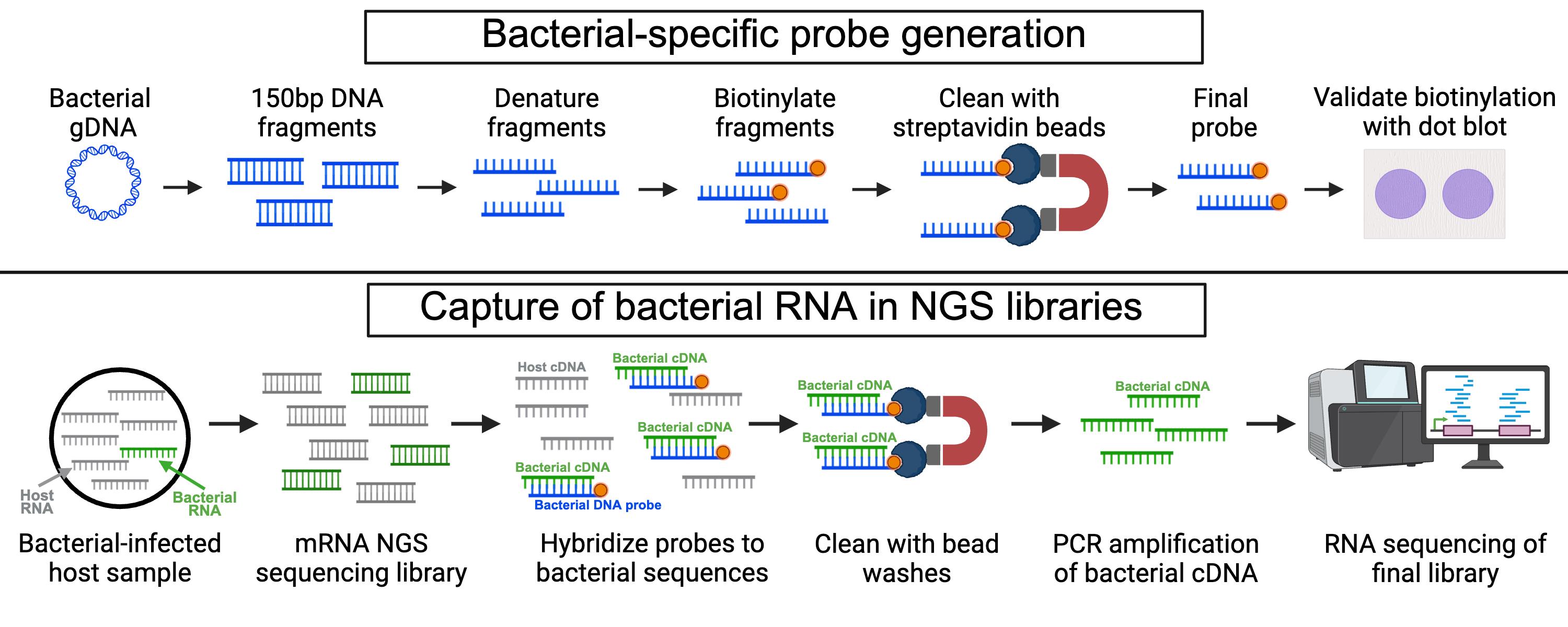
Background
Measuring messenger RNA (mRNA) from living bacteria is a promising method to understand the physiological state of pathogens within host environments. However, bacterial activity in vivo is not well described as it is technically difficult to process the minimal amount of bacterial mRNA present in most infected host material [1–4]. For example, previous work using dual-RNA next-generation sequencing (NGS) on clinical tuberculosis sputum samples has mapped only 0.85% (<300,000) bacterial reads to the infecting pathogen Mycobacterium tuberculosis (Mtb) due to an abundance of host RNA [5]. Several methods have previously been used to specifically enrich for bacterial RNA out of mixed samples for NGS applications, including using commercially synthesized biotinylated oligonucleotides that hybridize to matching sequences in an NGS sequencing library [6–10]. This strategy has limitations: custom probe design and synthesis is expensive, pre-designed probe libraries often fail to capture all genes present, and these methods have not been well-validated in clinical samples.
Here, we describe the transcript-capture sequencing protocol to enrich for bacterial RNA out of mixed samples, including animal models and clinical samples. Like the recently described DNA-capture technique genome capture sequencing (GenCap-Seq) [11,12], we produce biotinylated probes generated in-house from bacterial gDNA in a cost-effective and unbiased approach to sequence complete bacterial transcriptomes from infected host material. Using this protocol, we have generated complete bacterial transcriptomes (>1 million bacterial reads and >60% of bacterial genes having ≥10 reads aligning) from samples with as few as 1 × 10⁴ bacterial cells, with partial transcriptomes recovered from samples with as few as 1 × 10² cells [13]. We used probes generated from Mtb DNA for validation of transcript-capture; however, probe generation is customizable and can be used to determine the transcriptomes of other target species, with the potential for wide applications across different sample types and microbial species. For example, this protocol could be adapted to generate DNA probes of any microbial species or strain (including bacteria, viruses, parasites, or fungi) to enrich specific RNA from complex mixed samples such as infected host tissue, microbiome samples, or environmental sources like soil and seawater.
Materials and reagents
Biological materials
1. Bacterial DNA (see General notes 1 and 2)
2. NGS library for RNA sequencing prepared from bacterial-infected host sample (see General notes 3–5)
Reagents
1. DEPC H2O (Sigma-Aldrich, catalog number: 693520)
2. 1 M Tris-HCl pH 8 (Fisher Scientific, catalog number: 15-568-025)
3. 5 M sodium chloride (NaCl) (Invitrogen, catalog number: AM9759)
4. 0.5 M EDTA (Invitrogen, catalog number: AM9261)
5. Saline sodium citrate (SSC) buffer, 20× concentrated (Sigma-Aldrich, catalog number: SRE0068)
6. Formamide (Thermo Scientific, catalog number: 17899)
7. Tween-20 (Sigma-Aldrich, catalog number: P1379)
8. Ampure XP beads (Beckman Coulter, catalog number: A63882)
9. Shrimp alkaline phosphatase (rSAP) and rCutSmart buffer (NEB, catalog number: M0371S)
10. Terminal transferase and cobalt (II) chloride (CoCl2) (NEB, catalog number: M0315S)
11. Biotin-11-ddATP (Revvity, catalog number: NEL548001EA)
12. Qubit ssDNA Assay kit (ThermoFisher, catalog number: Q10212)
13. Qubit dsDNA HS Assay kit (ThermoFisher, catalog number: Q32851)
14. Monarch PCR & DNA Cleanup kit (NEB, catalog number: T1030)
15. Dynabeads MyOne Streptavidin C1 (Invitrogen, catalog number: 65002)
16. Bovine serum albumin (BSA) (Sigma-Aldrich, catalog number: A5611)
17. Streptavidin-AP conjugate (Roche, catalog number: 11089161001)
18. BCIP/NBT liquid substrate system (Sigma, catalog number: B1911-100ML)
19. xGen Human Cot DNA (IDT, catalog number: 1080768)
20. xGen Universal blockers (IDT, catalog number: 1075475)
21. xGen Hybridization and Wash v2 kit (IDT, catalog number: 1080584)
22. Dynabeads M-270 Streptavidin (Fisher, catalog number: 65-305)
23. 200 proof ethanol (EtOH) (Sigma-Aldrich, catalog number: E7023)
24. Nuclease-free H2O (Invitrogen, catalog number: 10977015)
25. 2× KAPA HiFi HotStart ReadyMix (KAPA Biosystems, catalog number: KK2602)
26. P5 primer 5′ AATGATACGGCGACCACCGA 3′ ordered from IDT at 25 nmol concentration and purified with standard desalting
27. P7 primer 5′ CAAGCAGAAGACGGCATACGA 3′ ordered from IDT at 25 nmol concentration and purified with standard desalting
Solutions
1. 10 μM Biotin-11-ddATP (see Recipes)
2. 2× binding and wash buffer (see Recipes)
3. 1× binding and wash buffer (see Recipes)
4. 0.1× SSC (see Recipes)
5. Formamide solution (see Recipes)
6. 10× Tris-buffered saline (TBS) (see Recipes)
7. 1× Tris-buffered saline with Tween-20 (TBST) (see Recipes)
8. Blocking buffer (see Recipes)
9. 1:2,000 Streptavidin-AP conjugate (see Recipes)
10. 80% EtOH (see Recipes)
11. 95% EtOH (see Recipes)
12. 10 mM Tris-HCl, pH 8.5 (see Recipes)
Recipes
1. 10 μM Biotin-11-ddATP
| Reagent | Final concentration | Quantity or volume |
|---|---|---|
| Biotin-11-ddATP | 10 μM | 1 μL |
| DEPC H2O | 99 μL |
Store at 4 °C for up to 1 year.
2. 2× binding and wash buffer
| Reagent | Final concentration | Quantity or volume |
|---|---|---|
| 1 M Tris-HCl | 10 mM | 500 μL |
| 5 M NaCl | 300 mM | 3 mL |
| 0.5 M EDTA | 1 mM | 100 μL |
| DEPC H2O | 46.4 mL |
Sterilize by filtering through a 0.22 μm filter. Store at room temperature.
3. 1× binding and wash buffer
| Reagent | Final concentration | Quantity or volume |
|---|---|---|
| 2× binding and wash buffer | 1× | 10 mL |
| DEPC H2O | 10 mL |
Store at room temperature.
4. 0.1× SSC
| Reagent | Final concentration | Quantity or volume |
|---|---|---|
| 20× SSC | 0.1× | 250 μL |
| DEPC H2O | 49.75 mL |
Sterilize by filtering through a 0.22 μm filter. Store at room temperature.
5. Formamide solution
| Reagent | Final concentration | Quantity or volume |
|---|---|---|
| Formamide | 95% | 950 μL |
| 0.5 M EDTA | 10 mM | 20 μL |
| DEPC H2O | 30 μL |
Aliquot 100 μL into 1.5 mL tubes. Store at -20 °C. Avoid freeze-thawing.
6. 10× TBS
| Reagent | Final concentration | Quantity or volume |
|---|---|---|
| 1 M Tris-HCl pH 8 | 200 mM | 100 mL |
| 5 M NaCl | 1.5 M | 150 mL |
| DEPC H2O | 250 mL |
Filter through a 0.22 μm filter to sterilize. Store at room temperature.
7. 1× TBST
| Reagent | Final concentration | Quantity or volume |
|---|---|---|
| 10× TBS | 1× | 50 mL |
| Tween 20 | 0.1% | 500 μL |
| DEPC H2O | 450 mL |
Filter through a 0.22 μm filter to sterilize. Store at room temperature.
8. Blocking buffer
| Reagent | Final concentration | Quantity or volume |
|---|---|---|
| BSA | 2% | 10 g |
| 1× TBST | 500 mL |
Filter through a 0.22 μm filter to sterilize. Store at room temperature.
9. 1:2,000 Streptavidin-AP conjugate
| Reagent | Final concentration | Quantity or volume |
|---|---|---|
| Streptavidin-AP conjugate | 1:2,000 | 7.5 μL |
| Blocking buffer | 15 mL |
Prepare immediately before use. Do not store.
10. 80% EtOH
| Reagent | Final concentration | Quantity or volume |
|---|---|---|
| 200 proof EtOH | 80% | 40 mL |
| DEPC H2O | 10 mL |
Filter through a 0.22 μm filter to sterilize. Store at room temperature and use within 1 week.
11. 95% EtOH
| Reagent | Final concentration | Quantity or volume |
|---|---|---|
| 200 proof EtOH | 95% | 47.5 mL |
| DEPC H2O | 2.5 mL |
Filter through a 0.22 μm filter to sterilize. Store at room temperature and use within 1 week.
12. 10 mM Tris-HCl, pH 8.5
| Reagent | Final concentration | Quantity or volume |
|---|---|---|
| 1 M Tris-HCl | 10 mM | 100 μL |
| DEPC H2O | 9.9 mL |
Check pH with a pH reader and adjust to pH 8.5. Filter through a 0.22 μm filter to sterilize. Store at room temperature.
Laboratory supplies
1. Covaris microTUBE AFA fiber pre-slit snap-cap 6 × 16 mm (Fisher Scientific, catalog number: NC9871616)
2. FlashGel system (Lonza, catalog number: 57067)
3. 1.5 mL DNA LoBind tubes (Eppendorf, catalog number: 0030108051)
4. 5 mL DNA LoBind tubes (Eppendorf, catalog number: 0030108310)
5. 96-well LoBind PCR plates (Eppendorf, catalog number: 0030129512)
6. 0.2 mL PCR 8-strip tubes (VWR, catalog number: 490003-692)
7. Magnetic Stand-96 (Invitrogen, catalog number: AM10027)
8. DynaMag-2 magnet (Invitrogen, catalog number: 12321D)
9. 0.22 μm Stericup vacuum filtration system (Fisher Scientific, catalog number: S2GPU05RE)
10. 0.2 μm nitrocellulose membrane (Thermo Scientific, catalog number: 88024)
11. Microseal B PCR plate sealing film (Bio-Rad, catalog number: 17010701)
12. 20 μL BIOTIX XTIP Rainin LTS compatible filter low-retention sterile pipette tips (Fisher Scientific, catalog number: 12-111-400)
13. 200 μL BIOTIX XTIP Rainin LTS compatible filter low-retention sterile pipette tips (Fisher Scientific, catalog number: 12-111-362)
14. 1,000 μL BIOTIX XTIP Rainin LTS compatible filter low-retention sterile pipette tips (Fisher Scientific, catalog number: 12-111-364)
Equipment
1. Pipet-Lite LTS Pipette L-2×LS+ manual single-channel pipette, 0.1–2 μL (Mettler Toledo, catalog number: 17014393)
2. Pipet-Lite LTS Pipette L-20×LS+ manual single-channel pipette, 2–20 μL (Mettler Toledo, catalog number: 17014392)
3. Pipet-Lite LTS Pipette L-20×LS+ manual single-channel pipette, 20–200 μL (Mettler Toledo, catalog number: 17014391)
4. Pipet-Lite LTS Pipette L-20×LS+ manual single-channel pipette, 100–1,000 μL (Mettler Toledo, catalog number: 17014382)
5. Pipet-Lite XLS+ manual 12-channel pipette 2–20 μL (Mettler Toledo, catalog number: 17013808)
6. Pipet-Lite XLS+ manual 12-channel pipette 20–200 μL (Mettler Toledo, catalog number: 17013805)
7. M220 Focused-ultrasonicator (Covaris, catalog number: 500295)
8. Microcentrifuge (Eppendorf, catalog number: 13-864-454)
9. Dry block heater (VWR, catalog number: 75838-318)
10. Qubit 4 Fluorometer (ThermoFisher, catalog number: Q33238)
11. Two 96-well thermal cyclers (Bio-Rad, catalog number: 1861096)
12. Vortex mixer (VWR, catalog number: 97043-562)
13. Benchtop laboratory pH/mV meter (Fisher Scientific, catalog number: 13636AB315A)
14. UV crosslinker (Fisher Scientific, catalog number: 13-245-221)
15. Low-speed orbital shaker (Corning, catalog number: CLS6782FP)
16. SpeedVac integrated vacuum concentrator system (Thermo Scientific, catalog number: 1156U31)
17. Freezer (-20 °C)
18. Refrigerator (4 °C)
Procedure
A. Generate capture probes from bacterial gDNA
1. Start with gDNA from a single bacterial culture that has been extracted in a way to prevent shearing (no bead beating). See General notes 1 and 2.
2. Using a Covaris, shear DNA to an average of 150 base pair (bp) fragments.
a. Dilute DNA in DEPC H2O to 5 μg in 130 μL.
b. Add 130 μL of DNA (total 5 μg) to the Covaris microTUBE.
c. Shear according to conditions in Table 1. Some optimization of shearing treatment conditions may be required to generate 150 bp fragments for specific instruments.
Table 1. Shearing treatment conditions. Shearing conditions to generate 150 bp fragments of gDNA. Some optimization may be required.
| Treatment time | Setpoint temperature (°C) | Minimum temperature (°C) | Maximum temperature (°C) | Peak power | Cycles/burst | Duty factor |
|---|---|---|---|---|---|---|
| 585 s | 20.0 | 18.0 | 75.0 | 50 | 200 | 15% |
d. Repeat until all DNA has been sheared.
Note: We recommend starting with 10 × 5 μg of gDNA (1,300 μL), which will generate roughly 2–3 μg of final probe.
e. (Optional) Confirm shearing on a FlashGel (Figure 1).
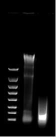
Figure 1. FlashGel image of gDNA before and after shearing. Lane 1: FlashGel DNA marker (100–4,000 bp); Lane 2: unsheared gDNA; Lane 3: gDNA sheared to 150 bp.
3. Store all sheared gDNA in a single LoBind tube (1.5 or 5 mL, depending on the amount sheared). Only use LoBind reagents throughout section A.
4. Clean sheared gDNA with Ampure XP beads.
a. Bring Ampure XP beads to room temperature for >30 min and vortex thoroughly to resuspend.
b. Aliquot 50 μL sheared DNA per well into a LoBind plate.
c. Add 150 μL (3×) of resuspended Ampure beads to each well. Pipette to mix thoroughly.
d. Incubate at room temperature for 5 min.
e. Place on a magnet to clear beads for ~5 min.
f. Discard supernatant without disturbing bead pellet.
g. Add 200 μL of 80% EtOH and incubate at room temperature for 30 s.
h. Discard supernatant without disturbing the bead pellet.
i. Repeat ethanol wash once.
j. Discard supernatant without disturbing the bead pellet and remove residual ethanol. Allow to air-dry for 5 min on a magnet and remove excess ethanol with a smaller tip.
k. Remove from the magnet and resuspend beads in 40 μL of DEPC H2O. Mix well.
l. Incubate for 5 min at room temperature.
m. Clear beads on a magnet for ~2 min.
n. While the plate is on the magnet, transfer the supernatant to the same 5 mL LoBind tube. Avoid bead carryover.
5. Quantitate using the Qubit dsDNA HS Assay kit.
Note: Expected dsDNA concentration: 10–30 ng/μL.
6. Aliquot 500 ng of DNA into individual wells of a LoBind PCR plate
Pause point: Samples can be stored overnight at 4 °C.
7. Dephosphorylate the ends of DNA.
a. Prepare the dephosphorylation reaction master mix in a 1.5 mL tube (Table 2).
Note: The H2O volume depends on the volume of 500 ng DNA. For example, if 500 ng of DNA is 16.6 μL, 25.9 μL of H2O should be added to the dephosphorylation reaction master mix.
Table 2. Master mix for the dephosphorylation reaction
| Reagent | Volume per sample |
|---|---|
| Cutsmart buffer (10×) | 5 μL |
| rSAP | 2.5 μL |
| Sheared + cleaned DNA (500 ng) | variable |
| H2O | to 50 μL |
| Total volume | 50 μL |
b. Add the dephosphorylation reaction master mix (Table 2) to each well of 500 ng DNA.
c. With the lid set to 105 °C, incubate in a thermocycler at 37 °C for 2 h, followed by 65 °C for 15 min.
8. Denature DNA.
a. Heat samples in a thermocycler at 100 °C for 10 min with the lid set to 105 °C.
b. Immediately flash freeze samples in dry ice + EtOH bath to prevent renaturation (alternatively, you can immediately place on ice for >5 min). Proceed immediately to biotinylation.
Critical: Make sure samples are cooled immediately after heat denaturation, or the biotinylation step will not work.
9. Biotinylate DNA.
a. Prepare the biotinylation master mix in a 1.5 mL tube (Table 3).
Table 3. Master mix for the biotinylation reaction
| Reagent | Volume per sample |
|---|---|
| 2.4 mM CoCl2 | 5 μL |
| 10 μM Biotin-11-ddATP | 2.5 μL |
| Terminal transferase | 1 μL |
| Total volume | 8.5 μL |
b. To each 50 μL sample from step A8, add 8.5 μL of biotinylation master mix (Table 3) for a final volume of 85.5 μL.
c. With the lid set to 105 °C, incubate in a thermocycler at 37 °C for 1.5 h, followed by heat inactivation at 70 °C for 10 min.
Pause point: Samples can be stored overnight at 4 °C.
d. Pool all samples into one 5 mL LoBind tube.
Note: Expected volume: 3–4 mL.
e. Quantify using Qubit ssDNA Assay kit.
Note: Expected ssDNA concentration: 10–30 ng/μL.
10. Remove unbound biotin with the Monarch PCR & DNA Cleanup kit following the ssDNA protocol.
a. Turn on a heat block to 50 °C and pre-heat DEPC H2O (100 μL per sample).
b. Aliquot DNA into 4 μg samples in separate 5 mL LoBind tubes.
Note: Samples should be 300–500 μL. Expect 8–10 samples if starting with 10 × 5 μg of sheared gDNA.
c. Add 100 μL of DNA cleanup binding buffer (from the kit) for every 50 μL of sample. Mix by pipetting.
Critical: Do not vortex!
Note: For example, for a 400 μL sample, add 800 μL of binding buffer.
d. Add 300 μL of 95% EtOH for every 50 μL sample. Mix by pipetting, do not vortex!
Note: For example, for a 400 μL sample, add 2,400 μL of 95% EtOH.
e. Insert the column into the collection tube and load 800 μL of sample onto the column. Centrifuge at 16,000× g for 1 min. Then, discard the flowthrough.
f. Repeat step A10e for each sample by adding more sample to the column without exceeding 800 μL. Repeat until all sample has gone through the column.
g. Re-insert the column into the collection tube. Add 500 μL of DNA wash buffer (from the kit) and centrifuge for 1 min. Discard the flowthrough.
h. Repeat step A10g two more times, for a total of three washes.
i. After discarding the flowthrough from the final wash, reinsert the column into the collection tube and centrifuge again to make sure the membrane is dry.
j. Transfer column to a new 1.5 mL LoBind tube.
k. Add 50 μL of warm DEPC H2O to the center of the matrix. Incubate at room temperature for 1 min, then centrifuge at 16,000× g for 1 min to elute DNA.
l. Add another 50 μL of H2O to the center of the matrix. Incubate at room temperature for 1 min, then centrifuge at 16,000× g for 1 min to elute DNA.
Note: The final volume of each sample is 100 μL.
Pause point: Samples can be stored overnight at 4 °C.
11. Purify DNA with MyOne Streptavidin C1 beads to eliminate non-biotinylated strands.
a. Turn on a heat block to 65 °C.
b. Wash MyOne Streptavidin C1 beads:
i. Aliquot 50 μL of Dynabeads per sample into a new 1.5 mL LoBind tube, to a maximum of 6 samples (300 μL Dynabeads) per tube.
ii. Place the tube containing the beads on a magnet until clear (~2 min).
iii. Discard the supernatant without disturbing the bead pellet.
iv. Remove from the magnet.
v. Add 1 mL of 1× binding and wash buffer and resuspend.
vi. Place the sample on a magnet until the solution is clear (~2 min).
vii. Discard the supernatant without disturbing the bead pellet.
viii. Wash three times as follows:
(1) Remove the tube from the magnet and resuspend the beads in a volume of 1× binding and wash buffer equal to the initial volume of beads taken (maximum 300 μL).
(2) Pipette to resuspend the beads.
(3) Clear beads on the magnet (~1 min).
(4) Discard the supernatant without disturbing the bead pellet.
ix. After three washes, resuspend beads in 2× binding and wash buffer at twice the original volume per reaction.
Note: For example, if 300 μL of Dynabeads were initially put in the LoBind tube, add 600 μL of 2× binding and wash buffer.
x. Aliquot 100 μL of resuspended beads into separate 1.5 mL LoBind tubes, one tube for each sample.
c. Add the 100 μL DNA samples from step A10l to the 100 μL washed beads.
i. Gently pipette to resuspend the beads.
ii. Incubate at room temperature for 15 min. Gently vortex tubes for 1–2 s to mix beads 3–4 times during incubation.
d. Place the tube on the magnet until clear (~2 min).
e. Discard the supernatant without disturbing the bead pellet.
f. Resuspend the sample in 200 μL of 1× binding and wash buffer.
g. Place the tube on a magnet until clear (~2 min).
h. Discard the supernatant without disturbing the bead pellet.
i. Remove the tube from magnet.
j. Resuspend beads in 100 μL of 0.1× SSC.
k. Place the tube on a magnet until clear (~2 min).
l. Discard the supernatant without disturbing the bead pellet.
m. Remove the tube from the magnet.
n. Resuspend beads in 50 μL of formamide mix.
12. Dissociate biotinylated DNA from beads using formamide.
a. Incubate the sample from step A11l in a 65 °C heat block for 5 min.
b. Place the sample on a magnet until the solution is clear (~2 min).
c. Transfer supernatant (50 μL) to a new 1.5 mL LoBind tube, avoiding bead carryover.
13. Remove unbound biotin with the Monarch PCR & DNA Cleanup kit following the ssDNA protocol.
a. Turn on the heat block to 50 °C and pre-heat the elution buffer bottle (from the kit).
b. Add 100 μL of DNA cleanup binding buffer (from the kit) for every 50 μL sample. Mix by pipetting, do not vortex!
c. Add 300 μL of 95% EtOH for every 50 μL sample. Mix by pipetting, do not vortex!
d. Insert the column into the collection tube and load 800 μL of sample onto the column. Centrifuge at 16,000× g for 1 min; then, discard the flowthrough.
e. Repeat step A13d for each sample by adding more sample to the column without exceeding 800 μL. Repeat until all sample has gone through the column.
f. Reinsert the column into the collection tube. Add 500 μL of DNA wash buffer (from the kit) and centrifuge for 1 min. Discard the flowthrough.
g. Repeat step A13f.
h. Transfer the column to a new 1.5 mL LoBind tube.
i. Add 11 μL of warm elution buffer (from the kit) to the center of the matrix. Wait for 1 min, then centrifuge at 16,000× g for 1 min to elute DNA.
j. Add another 11 μL of warm elution buffer to the center of the matrix. Wait for 1 min, then centrifuge at 16,000× g for 1 min to elute DNA.
k. Discard the column and store the final probe product at -20 °C.
Note: The final volume is 22 μL.
14. Quantitate the final probe product with the Qubit ssDNA Assay kit.
Note: Expected concentration: 15–50 ng/μL. Expected total volume: 170–220 μL.
B. Qualitatively confirm biotinylation of probes with dot blot
1. (Optional) Prepare dilutions of biotinylated ssDNA with known molarity as a positive control.
2. Apply 1 μL dots of the final probe from step A14 and the optional positive control to a 0.2 μm nitrocellulose membrane.
3. Immediately dry the blot with a UV crosslinker.
Note: If no UV crosslinker is available, wait for dots to dry on their own at room temperature.
4. Immerse the dried membrane in blocking buffer in a small dish (for example, the lid of a tip box) and incubate for 1 h at room temperature with gentle shaking.
5. Wash the membrane three times with 1× TBST. For each wash, immerse the membrane in 1× TBST and incubate for 5–10 min with gentle shaking.
6. Immerse the membrane in 1:2,000 Streptavidin-AP conjugate and incubate for 1 h at room temperature with gentle shaking.
7. Wash the membrane three times with 1× TBST. For each wash, immerse the membrane in 1× TBST and incubate for 10 min with gentle shaking.
8. Immerse the membrane in BCIP/NBT substrate solution for 10–30 min until color develops.
9. Stop the reaction by rinsing the membrane in H2O.
10. Air-dry the membrane and store in the dark at room temperature (Figure 2).
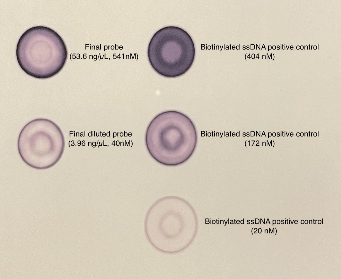
Figure 2. Example dot blot of final probe sample. Two dots of final probe (left column) and three dots of positive control biotinylated ssDNA (right column).
C. Capture bacterial RNA from NGS libraries with probes
Note: This protocol is based on the xGen Hybridization and Wash v2 kit (IDT) protocol.
1. Set WASH (Table 4) and HYB (Table 5) programs on two thermocyclers.
Table 4. WASH program conditions. Set thermocycler lid to 70 °C.
| Temperature | Duration |
|---|---|
| 65 °C | Infinity |
Table 5. HYB program conditions. Set thermocycler lid to 100 °C.
| Temperature | Duration |
|---|---|
| 95 °C | 10 min |
| 65 °C | 16 h |
| 65 °C | Infinity |
2. Start with NGS libraries prepared from RNA samples; see General notes 3–5.
3. Aliquot 500 ng of each sequencing library into individual 1.5 mL LoBind tubes. 500 ng libraries can be pooled into the same tube to minimize the use of expensive reagents. Pooling up to 5 libraries (5 × 500 ng total) in one tube has worked well. See General note 6.
Notes:
1. Make sure there is ≥500 ng in each LoBind tube. If using libraries with <500 ng total, pool low-concentration libraries together to maintain 500 ng in each tube.
2. Do not capture more than 32 samples of pooled libraries at a time to prevent extended time between washes.
4. Add 5 μL of Human Cot DNA to each tube.
5. Add 2 μL of xGen Universal blockers to each tube.
6. Vortex to mix well and briefly centrifuge (any speed, <5 s) to ensure contents are at bottom of tube.
7. Dry down the mixture in a SpeedVac set to autorun. Continue until all contents are dry (20–60 min).
Pause point: Dried samples can be stored at room temperature overnight, or at -20 °C for longer.
8. Perform hybridization:
a. Bring contents of the xGen Hybridization and Wash kit to room temperature. Inspect the tube of 2× hybridization buffer for any precipitate. If crystals are present, heat the tube at 65 °C, shaking intermittently by hand.
b. Create the hybridization master mix (Table 6) with custom probe concentration (Table 7).
Table 6. Hybridization master mix
| Reagent | Volume per sample |
|---|---|
| xGen 2× hybridization buffer | 8.5 μL |
| xGen hybridization buffer enhancer | 2.7 μL |
| Custom probes* + H2O | 5.8 μL* |
| Total volume | 17 μL |
*Custom probe concentration: Variable; capture of 500 ng libraries with as little as a 10 ng probe has been successful. Capture with <10 ng probe has not been tested.
Table 7. Custom probe concentration. Combine the desired concentration of probe and nuclease-free H2O to make 5.8 μL total.
| Number of pooled libraries in sample (ng of library) | Concentration of probe per sample |
|---|---|
| 1 (500 ng) | 20–25 ng |
| 2–3 (1,000–1,500 ng) | 40–50 ng |
| 4–5 (2,000–2,500 ng) | 50 ng |
c. Vortex to mix the hybridization master mix and add 17 μL to each of the tubes containing dried DNA.
d. Briefly centrifuge tubes and transfer contents to individual wells of a 96-well LoBind plate. Avoid using outside wells to limit evaporation (Figure 3). This is the sample plate.
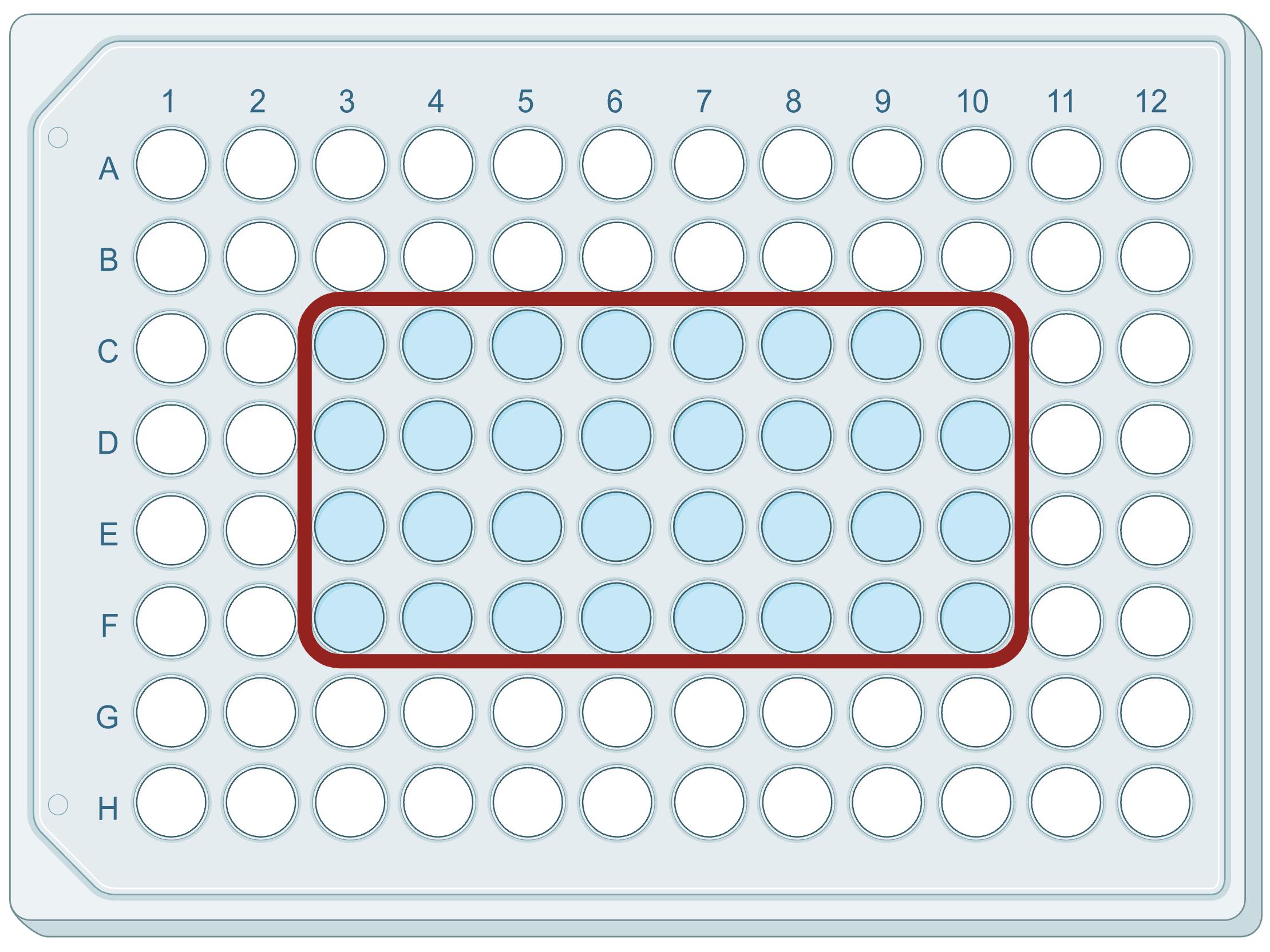
Figure 3. Plate showing recommended wells to use during capture reaction
e. Securely seal the plate with a Microseal B seal.
f. Incubate for 5 min at room temperature.
g. Vortex the samples, making sure that they are completely mixed.
h. Briefly centrifuge the samples.
i. Place the plate on the thermal cycler and start the HYB program.
Note: The HYB program will take 16 h to complete. We recommend running overnight.
9. Prepare buffers:
a. Dilute xGen buffers to create 1× working solutions (Table 8), multiplying by the required number of samples and adding 10% extra. Swirl to mix; do not vortex!
Notes:
1. If wash buffer 1 is cloudy, heat the bottle in a 65 °C water bath to allow resuspension.
2. The 1× working solutions are stable at room temperature (15–25 °C) for up to 4 weeks.
Table 8. xGen buffer dilutions
| Reagent | Nuclease-free H2O | Buffer | Total |
|---|---|---|---|
| xGen 2× bead wash buffer | 150 μL | 150 μL | 300 μL |
| xGen 10× wash buffer 1 | 225 μL | 25 μL | 250 μL |
| xGen 10× wash buffer 2 | 135 μL | 15 μL | 150 μL |
| xGen 10× wash buffer 3 | 135 μL | 15 μL | 150 μL |
| xGen 10× stringent wash buffer | 270 μL | 30 μL | 300 μL |
b. In PCR 8-strip tubes, aliquot buffers for each sample tube. Prepare one strip tube of 110 μL of wash buffer 1 per sample tube and two strip tubes of 160 μL of stringent wash buffer per sample tube. Seal strip tubes and set aside.
Notes:
1. For example, for 32 samples, there will be 32 strip tubes (4 rows of strips) of wash buffer 1 and 64 strip tubes of stringent wash buffer (8 rows of strips).
2. Do not discard the remaining wash buffer 1. The remaining buffer is needed to perform the room-temperature washes later in the protocol.
c. Prepare the bead resuspension mix in a LoBind tube (Table 9), multiplying by the required number of samples and adding 10% extra.
Table 9. Bead resuspension mix dilutions
| Reagent | Volume per sample |
|---|---|
| xGen 2× hybridization buffer | 8.5 μL |
| xGen hybridization buffer enhancer | 2.7 μL |
| Nuclease-free water | 5.8 μL |
10. Wash streptavidin beads immediately before the end of the HYB program:
a. Remove Dynabeads M-270 streptavidin beads from 4 °C and equilibrate the beads at room temperature for at least 30 min before washing.
b. Mix the beads thoroughly by vortexing for 15 s.
c. Add 50 μL of streptavidin beads to a new LoBind PCR plate, filling a well for every sample being captured.
d. Add 100 μL of 1× bead wash buffer to each well, then gently pipette to mix 10 times.
e. Place the plate containing beads on a magnet and allow the beads to fully separate from the supernatant (~1 min).
f. Discard supernatant without disturbing bead pellet.
g. Remove the plate containing beads from the magnet.
h. Perform the following wash:
i. Add 100 μL of 1× bead wash buffer to each well containing beads; then, gently pipette the mix 10 times.
ii. Place the plate on the magnet for approximately 1 min, allowing the beads to fully separate from the supernatant.
iii. Carefully remove and discard the clear supernatant without disturbing the bead pellet.
i. Perform an additional wash by repeating step C10h for a total of two washes.
j. Resuspend the beads in 17 μL of bead resuspension mix.
k. Mix thoroughly to ensure the beads are not left to dry in the well. If needed, briefly centrifuge the plate containing beads.
l. Seal plate with beads with a Microseal B seal and set aside.
11. Perform bead capture.
a. Start the WASH program in the second thermal cycler and add buffer strip tubes prepared in step C9b. Make sure the lid temperature is set to 70 °C for the WASH program. The buffer strip tubes need to warm up for at least 15 min before use.
b. After the 16 h HYB program is complete, remove the sample plate from the thermal cycler.
c. Once the sample plate has been removed from the instrument, stop the HYB program.
d. Immediately after the HYB program is complete, start the WASH program. At this point, both thermal cyclers should be running the WASH program.
e. Using a multichannel pipette and fresh filter tips, transfer the fully homogenized streptavidin beads from step C10l to the samples and gently pipette until fully mixed.
f. Securely seal the sample plate with a Microseal B.
g. Place the sample plate in the thermal cycler, running the WASH program for 45 min. During incubation, remove the plate every 10–12 min to quickly and gently vortex.
Note: It is safe to place the sample plate in the thermal cycler before the lid temperature has fully cooled to 70 °C when starting the incubation.
12. Perform heated washes.
Note: Always keep the buffer strip tubes on the thermal cycler during washes, to maintain their set temperature, and close the thermocycler lid between uses.
a. After the 45 min incubation with the WASH program, remove the sample plate from the thermal cycler.
b. With the buffer tubes remaining in the second thermal cycler, transfer 100 μL of heated wash buffer 1 to each sample and pipette to mix 10 times, being careful to minimize bubble formation.
c. Place the sample plate on the magnet for 1 min.
d. Discard supernatant without disturbing bead pellet.
e. Remove the sample plate from the magnet, then add 150 μL of heated stringent wash buffer to each well containing a sample.
f. Pipette to mix 10 times, being careful to minimize bubble formation.
g. Securely seal the sample plate with a Microseal B. Then, incubate for 5 min in the thermal cycler (WASH program).
h. Place the sample plate on the magnet for 1 min.
i. Discard the supernatant without disturbing the bead pellet.
j. Remove the sample plate from the magnet, then add 150 μL of heated stringent wash buffer to each well containing a sample.
k. Pipette to mix 10 times, being careful to minimize bubble formation.
l. Securely seal the sample plate with a Microseal B; then, incubate for 5 min on the thermal cycler (WASH program).
m. Place the sample plate on the magnet for 1 min.
13. Perform room-temperature washes.
Note: To ensure that the beads remain fully resuspended, vigorously vortex the sample plate during the room-temperature washes.
a. While the sample plate is on the magnet, discard the supernatant.
b. Add 150 μL of wash buffer 1 (kept at room temperature) to each well of the sample plate.
c. Securely seal the sample plate with a Microseal B, then vortex at full speed thoroughly, until fully resuspended.
Note: Liquid will touch the top of the seal; make sure the seal is secured.
d. Incubate for 2 min while alternating between vortexing for 30 s and resting for 30 s, to ensure the mixture remains homogenous.
e. Centrifuge the sample plate at 25× g for 5 s to avoid well-to-well contamination.
f. Place the sample plate on the magnet for 1 min, then remove and discard the seal.
g. Discard the supernatant without disturbing the bead pellet. Then, remove the sample plate from the magnet.
h. Add 150 μL of wash buffer 2; then, securely seal the sample plate with a fresh seal and vortex thoroughly until fully resuspended.
Note: Beads tend to stick to the side of the wells after using wash buffer 2.
i. Incubate for 2 min while alternating between vortexing for 30 s and resting for 30 s, to ensure the mixture remains homogenous.
j. After the incubation, briefly centrifuge the sample plate (at 25× g for 5 s).
k. After centrifuging, place the sample plate on the magnet for 1 min; then, remove and discard the seal.
l. Discard the supernatant without disturbing the bead pellet; then, remove the sample plate from the magnet.
m. Add 150 μL of wash buffer 3. Then, securely seal the sample plate with a fresh seal and vortex thoroughly until fully resuspended.
n. Incubate for 2 min while alternating between vortexing for 30 s and resting for 30 s, to ensure the mixture remains homogenous.
o. After the incubation, briefly centrifuge the sample plate (at 25×g for 5 s).
p. After centrifuging, place the sample plate on the magnet for 1 min, then remove and discard the seal.
q. Discard the supernatant without disturbing the bead pellet.
r. With the sample plate still on the magnet, use fresh pipette tips to ensure that all residual wash buffer 3 has been removed, then remove the plate from the magnet.
s. Add 20 μL of nuclease-free water to each capture.
t. Pipette to mix thoroughly to resuspend any beads stuck to the side of the well.
u. Do not discard the beads. Use the entire 20 μL of resuspended beads containing captured DNA in post-capture PCR.
14. Perform post-capture PCR.
a. In a tube, prepare the amplification reaction mix (Table 10), multiplied by the number of samples on the plate and adding 10% overfill.
Table 10. Amplification reaction mix
| Reagent | Volume per sample |
|---|---|
| 2× KAPA HiFi HotStart ReadyMix | 25 μL |
| 10 μM P5 primer | 2.5 μL |
| 10 μM P7 primer | 2.5 μL |
b. Add 30 μL of amplification reaction mix to each well of the sample plate containing 20 μL, for a final volume of 50 μL.
c. Pipette to mix thoroughly.
d. Seal the sample plate with Microseal B and gently vortex to help resuspend beads from the side of the wells.
e. Briefly centrifuge the sample plate.
f. Place the sample plate in a thermocycler and run the post-capture PCR (Table 11).
Table 11. Post-capture PCR cycling conditions. Set the thermocycler lid to 105 °C.
| Step | Temperature | Duration | Number of cycles |
| Polymerase activation | 98 °C | 45 s | 1 |
| Denaturation | 98 °C | 15 s | Variable (15–20) |
| Annealing | 60 °C | 30 s | |
| Extension | 72 °C | 30 s | |
| Final extension | 72 °C | 1 min | 1 |
| Hold | 4 °C | Infinity | NA |
Pause point: Amplified captured libraries can be stored at 4 °C overnight.
15. Purify post-capture PCR fragments with Ampure XP beads.
a. Bring Ampure XP beads to room temperature for >30 min and vortex thoroughly to resuspend.
b. Add 75 μL (1.5× volume) of Ampure XP beads to each amplified capture sample.
c. After adding the beads, pipette thoroughly to mix and incubate for 5–10 min.
d. Place the plate on the magnet until the supernatant is clear (2–5 min).
e. Discard the supernatant without disturbing the bead pellet.
f. While keeping the plate on the magnet, add 125 μL of 80% EtOH, then incubate for 1 min.
g. Remove the EtOH without disturbing the bead pellet, then repeat another wash with 125 μL of 80% EtOH.
h. Allow the beads to air dry for 1–3 min. Do not overdry the beads.
i. Remove the sample plate from the magnet and elute in 22 μL of 10 mM Tris-HCl, pH 8.5. Mix thoroughly.
j. Incubate for 5 min at room temperature.
k. Place the plate on a magnet until the supernatant is clear (1–2 min).
l. Transfer 20 μL of supernatant to a new LoBind plate, making sure that no beads are carried over.
Pause point: Purified PCR fragments may be stored at -20 °C for up to 1 week before sequencing.
16. Libraries are now ready to be sequenced. See General notes 7 and 8. (Optional) Check a subset of samples on a FlashGel for the presence of a PCR bubble (see Troubleshooting, Problem 2).
Validation of protocol
This protocol has been used and validated in the following research article:
Lamont et al. [13] Transcript-Capture sequencing enriches mRNA of Mycobacterium tuberculosis from host samples. NAR Molecular Medicine (Figures 2 and 3).
General notes and troubleshooting
General notes
1. We used a lab strain of Mycobacterium tuberculosis to validate this protocol; however, DNA from other bacterial species or strains can be used to generate capture probes.
2. Bacterial DNA for probes should be extracted using a method that does not shear the DNA. DNA can be eluted in any standard elution buffer (e.g., H2O, TE buffer, Tris-HCl).
3. Total RNA should be extracted from host samples containing bacterial RNA following standard extraction procedures for the bacterial species of interest. RNA extraction should be performed immediately after sample collection or from samples that have been immediately preserved (for example, in TRizol) to limit RNA degradation.
4. rRNA should be removed from total RNA samples following standard removal procedures for the bacterial species of interest. General rRNA removal protocols are available (e.g., Illumina Ribo-Zero Plus rRNA Depletion kit), as well as probe-based methods that can be tailored to a specific species (for example, [14]).
5. NGS libraries should be prepared from rRNA-depleted RNA samples using a standard Illumina library prep protocol. For example, the NEBNext Ultra II RNA Library Prep kit for Illumina (NEB, catalog number: E7770L) with NEBNext Multiplex Oligos for Illumina (NEB, catalog number: E7600S). Libraries should be quantified using Qubit or qPCR (e.g., with a KAPA Library Quantification kit) and not yet pooled.
6. If pooling libraries for capture, only combine libraries that have similar estimated bacterial loads. For example, do not pool a library containing ~100 bacterial cells with a library containing ~1,000,000 bacterial cells.
7. Captured libraries can be multiplexed and sequenced on a short-read sequencing platform following standard procedures. We have successfully sequenced captured libraries on Illumina NextSeq and Element AVITI platforms with 75 and 150 cycle conditions.
8. Sequencing efficiency can vary due to the number of bacterial cells in the host samples. For samples with 1 × 10⁴ bacterial cells, expect ~50% of total reads to be bacterial. If >1 million bacterial reads are desired, make sure to sequence with enough depth for each sample to have >2 million total reads. For more examples of sequency efficiency variability due to bacterial cell numbers, see Figure 2E in [13].
9. Always use sterile pipette tips and replace tips at every step.
Troubleshooting
Problem 1: When generating capture probes (section A), no biotinylated ssDNA is seen on the dot blot or a low concentration of final probe (<5 ng/μL).
Possible causes: Insufficient denaturation before biotinylation or excessive loss of sample during Monarch PCR & DNA cleanup.
Solutions: Make sure samples are heated to 100 °C for at least 10 min and are cooled (or frozen) immediately afterward. Perform a dot blot on samples after step A10 to confirm biotinylation. Perform Monarch PCR & DNA Cleanup with freshly prepared 95% EtOH.
Problem 2: PCR bubble in post-capture PCR fragments, usually observed with a Bioanalyzer or Tapestation trace when preparing libraries for sequencing.
Possible cause: Too many cycles during post-capture PCR.
Solutions: Perform a reconditioning PCR. Add 30 μL of amplification reaction mix (Table 10) and 12 μL of nuclease-free H2O to 8 μL of each post-capture library. Run a PCR with post-capture PCR cycling conditions (Table 11) but perform only two cycles. If samples have already been pooled for sequencing, the same reconditioning PCR can be run on the pooled library. After amplification, follow step C15 to purify the samples with Ampure XP beads.
Acknowledgments
Conceptualization, E.I.L., R.M.J., S.M., D.R.S.; Investigation, E.I.L., R.M.J., J.A.; Writing—original draft, E.I.L.; Writing—review & editing, E.I.L., R.M.J., J.A., S.M., D.R.S.; Funding acquisition, D.R.S.
Original research paper supported by National Institutes of Health (U19 AI162598 and P30AI168034); the Gates Foundation (INV-056401); and the Institute of Translational Science Translational Research Training program (TL1TR002318).
The initial protocol was derived from [11], and the current protocol was validated and used in [13]. The graphical abstract was created in BioRender: Lamont, E. (2026) https://BioRender.com/aomc86f. Figure 3 was created in BioRender: Lamont, E. (2026) https://BioRender.com/0xm6zfb.
Competing interests
The authors declare no conflicts of interest.
References
- Westermann, A. J., Gorski, S. A. and Vogel, J. (2012). Dual RNA-seq of pathogen and host. Nat Rev Microbiol. 10(9): 618–630. https://doi.org/10.1038/nrmicro2852
- Penaranda, C. and Hung, D. T. (2019). Single-Cell RNA Sequencing to Understand Host–Pathogen Interactions. ACS Infect Dis. 5(3): 336–344. https://doi.org/10.1021/acsinfecdis.8b00369
- Dartois, V. A. and Rubin, E. J. (2022). Anti-tuberculosis treatment strategies and drug development: challenges and priorities. Nat Rev Microbiol. 20(11): 685–701. https://doi.org/10.1038/s41579-022-00731-y
- Pisu, D., Huang, L., Grenier, J. K. and Russell, D. G. (2020). Dual RNA-Seq of Mtb-Infected Macrophages In Vivo Reveals Ontologically Distinct Host-Pathogen Interactions. Cell Rep. 30(2): 335–350.e4. https://doi.org/10.1016/j.celrep.2019.12.033
- Lai, R. P. J., Cortes, T., Marais, S., Rockwood, N., Burke, M. L., Garza-Garcia, A., Horswell, S., Sesay, A. K., O’Garra, A., Young, D. B., et al. (2021). Transcriptomic Characterization of Tuberculous Sputum Reveals a Host Warburg Effect and Microbial Cholesterol Catabolism. mBio. 12(6): e01766–21. https://doi.org/10.1128/mbio.01766-21
- Betin, V., Penaranda, C., Bandyopadhyay, N., Yang, R., Abitua, A., Bhattacharyya, R. P., Fan, A., Avraham, R., Livny, J., Shoresh, N., et al. (2019). Hybridization-based capture of pathogen mRNA enables paired host-pathogen transcriptional analysis. Sci Rep. 9(1): e1038/s41598–019–55633–6. https://doi.org/10.1038/s41598-019-55633-6
- Wynn, E. A., Dide-Agossou, C., Reichlen, M., Rossmassler, K., Al Mubarak, R., Reid, J. J., Tabor, S. T., Born, S. E. M., Ransom, M. R., Davidson, R. M., et al. (2023). Transcriptional adaptation of Mycobacterium tuberculosis that survives prolonged multi-drug treatment in mice mBio. 14(6): e02363–23. https://doi.org/10.1128/mbio.02363-23
- Peterson, E. J., Bailo, R., Rothchild, A. C., Arrieta‐Ortiz, M. L., Kaur, A., Pan, M., Mai, D., Abidi, A. A., Cooper, C., Aderem, A., et al. (2019). Path‐seq identifies an essential mycolate remodeling program for mycobacterial host adaptation. Mol Syst Biol. 15(3): e20188584. https://doi.org/10.15252/msb.20188584
- Chung, M., Teigen, L., Liu, H., Libro, S., Shetty, A., Kumar, N., Zhao, X., Bromley, R. E., Tallon, L. J., Sadzewicz, L., et al. (2018). Targeted enrichment outperforms other enrichment techniques and enables more multi-species RNA-Seq analyses. Sci Rep. 8(1): e1038/s41598–018–31420–7. https://doi.org/10.1038/s41598-018-31420-7
- Donaldson, G. P., Chou, W. C., Manson, A. L., Rogov, P., Abeel, T., Bochicchio, J., Ciulla, D., Melnikov, A., Ernst, P. B., Chu, H., et al. (2020). Spatially distinct physiology of Bacteroides fragilis within the proximal colon of gnotobiotic mice. Nat Microbiol. 5(5): 746–756. https://doi.org/10.1038/s41564-020-0683-3
- Hayden, H. S., Joshi, S., Radey, M. C., Vo, A. T., Forsberg, C., Morgan, S. J., Waalkes, A., Holmes, E. A., Klee, S. M., Emond, M. J., et al. (2022). Genome Capture Sequencing Selectively Enriches Bacterial DNA and Enables Genome-Wide Measurement of Intrastrain Genetic Diversity in Human Infections. mBio. 13(5): e01424–22. https://doi.org/10.1128/mbio.01424-22
- Long, D. R., Bryson-Cahn, C., Waalkes, A., Holmes, E. A., Penewit, K., Tavolaro, C., Bellabarba, C., Zhang, F., Chan, J. D., Fang, F. C., et al. (2024). Contribution of the patient microbiome to surgical site infection and antibiotic prophylaxis failure in spine surgery. Sci Transl Med. 16(742): eadk8222. https://doi.org/10.1126/scitranslmed.adk8222
- Lamont, E. I., Jones, R. M., Assadi, J., Morrison, R., Song, T., Yu, X., Weiner, D. M., Via, L. E., Winter, J., Ma, S., et al. (2025). Transcript-Capture sequencing enriches mRNA of Mycobacterium tuberculosis from host samples. NAR Mol Med. 3(1): e1093/narmme/ugaf045. https://doi.org/10.1093/narmme/ugaf045
- Culviner, P. H., Guegler, C. K. and Laub, M. T. (2020). A Simple, Cost-Effective, and Robust Method for rRNA Depletion in RNA-Sequencing Studies. mBio. 11(2): e00010–20. https://doi.org/10.1128/mbio.00010-20
Article Information
Publication history
Received: Feb 27, 2026
Accepted: Apr 27, 2026
Available online: May 19, 2026
Published: Jun 20, 2026
Copyright
© 2026 The Author(s); This is an open access article under the CC BY-NC license (https://creativecommons.org/licenses/by-nc/4.0/).
How to cite
Lamont, E. I., Jones, R. M., Assadi, J., Ma, S. and Sherman, D. R. (2026). Enriching Bacteria-Specific RNA From Host Samples Before NGS With Transcript-Capture. Bio-protocol 16(12): e5712. DOI: 10.21769/BioProtoc.5712.

 Download Citation in RIS Format
Category
Microbiology > Microbe-host interactions > Bacterium
Systems Biology > Transcriptomics > RNA-seq
Molecular Biology > RNA > RNA sequencing
Do you have any questions about this protocol?
Post your question to gather feedback from the community. We will also invite the authors of this article to respond.
 Post a Question


