


- Protocols

- Articles and Issues
- For Authors
- About
- Become a Reviewer


Parallelised Cloning, Mammalian Cell Expression, and Purification of Nanobodies Identified by Phage Display

Published: Vol 16, Iss 9, May 5, 2026 DOI: 10.21769/BioProtoc.5682 Views: 726
Reviewed by: Sébastien GillotinMithun SantraAnonymous reviewer(s)
 PDF
PDF  Ask a question
Advertisement



Protocol Collections
Comprehensive collections of detailed, peer-reviewed protocols focusing on specific topics




Related protocols
Abstract
Nanobodies are recombinant single-domain antibodies (VHHs) derived from the heavy chain–only subset of camelid immunoglobulins that can be reverse-engineered into bivalent antibodies by fusion to immunoglobulin Fc constant regions. Mammalian cells are the system of choice to produce VHH-Fcs to ensure authentic folding and post-translation glycosylation of the expressed VHH-Fcs. In a recent project to find neutralising VHH-Fc binders to the spike proteins of SARS-CoV-2 viruses, we identified a need for rapid expression and purification of multiple VHH-Fc fusions from nanobodies selected by phage display. Here, we present a protocol for the construction of expression vectors by parallel ligase-independent cloning, transient small-scale expression in mammalian cells (4 mL culture volume), screening antigen-binding activity, and midi-scale purification (30 mL culture volume) for downstream activity assays. The workflow is completely transferable between different vector formats, of which three are described herein: Fc fusion dimers, monomeric CD4 fusions, and His-tagged monomers.
Key features
• Miniaturised and parallelised methodology for the screening and production of large numbers of VHHs that bind antigens of interest.
• Streamlined and unified, high-throughput cloning strategy for use in multiple modular vectors for monomeric and dimeric VHH production in mammalian culture.
Keywords: Camelid heavy chain-only antibody Nanobody
VHH
Fc fusions
CD4 fusions
Modular vector system
Expi293
Mammalian expression system
Protein purification
High throughput
Nanobody
VHH
Fc fusions
CD4 fusions
Modular vector system
Expi293
Mammalian expression system
Protein purification
High throughput
Graphical overview
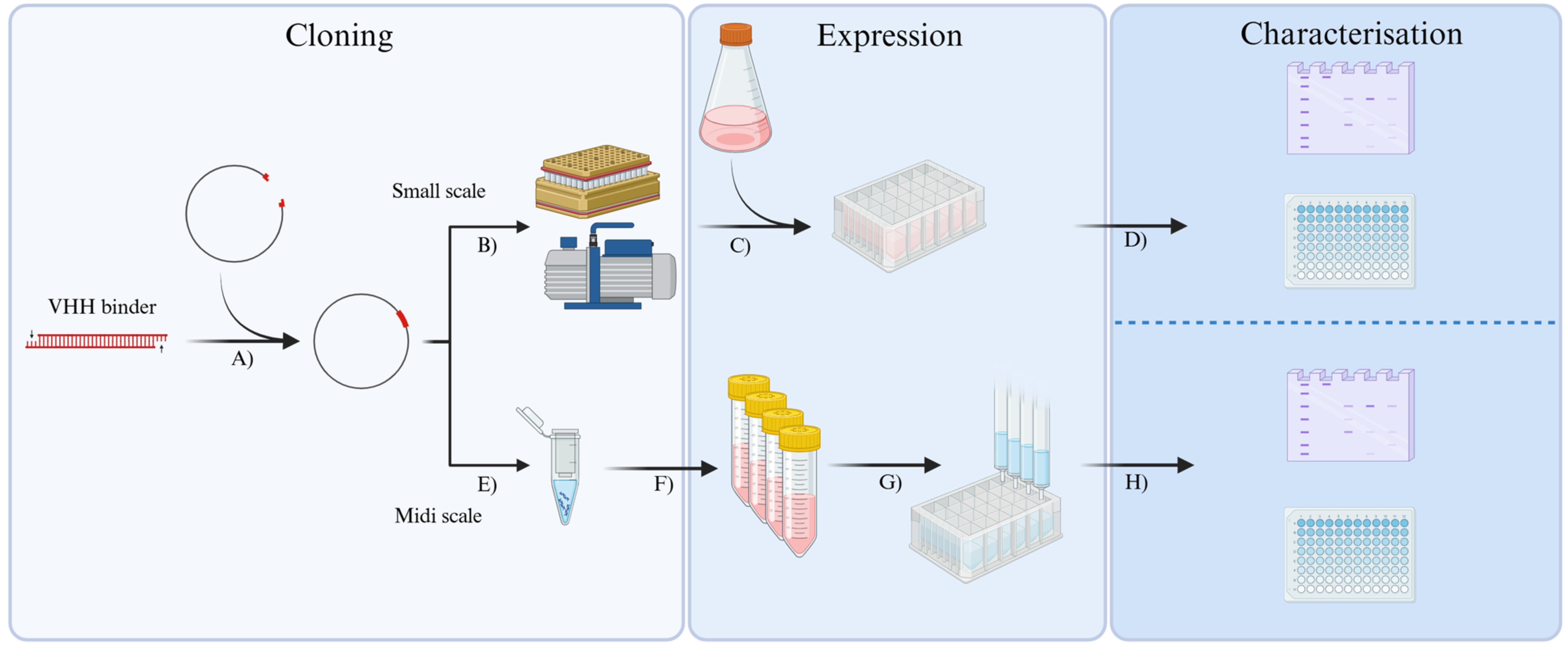
Schematic overview of the workflow outlined in this paper. Identified putative single-domain antibody (VHH) binders are (A) cloned into a selected vector from the pOPIN vector suite [1], (B) followed by plasmid DNA extraction and purification, and used for (C) small-scale (4 mL) mammalian transient transfection. (D) Secreted VHHs in the culture medium are quantified using densitometry on Coomassie-stained SDS-PAGE gels or observed by western blot, and binding to the target antigen is tested by titration enzyme-linked immunosorbent assay (ELISA). (E) Higher quantities of plasmid DNA from ELISA-positive clones are isolated by midi prep and used for (F) midi-scale (30 mL) mammalian transient transfection. (G) VHHs secreted into the culture medium are purified by affinity chromatography using a custom multi-column apparatus, which facilitates parallelised gravity purification. (H) The binding properties of the purified VHHs are observed and characterised by SDS-PAGE and titration ELISA before being used in subsequent pilot studies. Figure made using BioRender.
Background
Camelids (llamas, camels, and alpacas) produce unique heavy chain–only antibodies as part of their natural immune system, whose variable domains (VHHs or nanobodies) can be recombinantly expressed as monomeric antigen-binding proteins. VHHs have many applications in the biosciences, and their utilisation as anti-viral therapeutics has rapidly gathered momentum since the COVID-19 pandemic. Nanobodies are typically identified by screening phage display libraries of VHHs generated from either immunised camelids [2] or built by gene synthesis [3]. The small size of VHHs (14 kDa) means that they can easily be assembled into dimers and higher order multimers; a common format is genetic fusion to the Fc of immunoglobulin G (IgG). Bivalent nanobody-Fcs have increased binding activity compared to monomeric VHHs due to avidity and also gain the effector functions of IgGs. VHHs are generally produced in E. coli by secretion into the periplasm followed by purification using immobilised metal affinity chromatography [2,4,5]. However, transient expression in mammalian cells is the system of choice for producing VHH-Fcs as soluble authentic glycosylated products recoverable from the cell media by protein A affinity chromatography [6]. There is only one report of the expression of a soluble VHH-Fc in the cytoplasm of E. coli [7]. To accelerate the production of VHH-Fcs, we have developed a protocol for the streamlined construction and small-scale expression screening of VHH-Fcs using Expi293 cells. Given that not all VHHs are well expressed in E. coli in our experience, we have incorporated the production of monomeric nanobodies in mammalian cells into the workflow. These are expressed as either VHHs alone or fused to the extracellular domains of CD4, which can improve expression levels [8]. Using only common laboratory instrumentation and an easily made multi-column purification apparatus, this paper details an accessible, low-cost, and high-throughput method for the expression screening of VHHs in mammalian cells in three vector formats [1], midi scale up in 30 mL culture volume, and parallelised purification that generates sufficient material for characterising binding activity and testing in downstream functional assays. The identification of nanobodies that have been shown to bind to the spike protein of SARS-CoV-2 virus is presented to exemplify the methods.
Materials and reagents
Biological materials
1. StellarTM competent cells (TaKaRa, catalog number: 636766), store at -80 °C
2. Expi293FTM cells (Gibco, catalog number: A14527), store in liquid nitrogen vapour phase
Reagents
1. pOPINTTGneo_Fc (Addgene, catalog number: 252742), store at -20 °C
2. pOPINTTGneo_CD4 (Addgene, catalog number: 252740), store at -20 °C
3. pOPINTTGneo (Addgene, catalog number: 252739), store at -20 °C
4. Luria broth (LB) medium (FormediumTM, catalog number: LBL0103), store at room temperature (RT)
5. Bacto agar (FormediumTM, catalog number: AGR10), store at RT
6. Ampicillin (FormediumTM, catalog number: AMP100), store at 4 °C
7. QIAprep Spin Miniprep kit (QIAGEN, catalog number: 27104), store at RT
8. Nuclease-free water (not DEPC-treated) (InvitrogenTM, catalog number: AM9932), store at RT
9. rCutsmart® 10× buffer (New England Biolabs®, catalog number: B6004), store at -20 °C
10. KpnI-HFTM (New England Biolabs®, catalog number: R3142), store at -20 °C
11. PmeI (New England Biolabs®, catalog number: R0560L), store at -20 °C
12. Purelink PCR Purification kit (InvitrogenTM, catalog number: K310002), store at RT
13. TE buffer, Tris-EDTA, 1× solution, pH 8.0 (Fisher BioReagents, catalog number: BP2473), store at RT
14. Nb_Fc_Forward primer 5′ GCGTAGCTGAAACCGCCCAGGTGCAGCTGG 3′ (IDT), store at -20 °C
15. Nb_Fc_Reverse primer 5′ GTGATGGTGATGTTTCGAAGAGACGGTGACCTGG 3′ (IDT), store at -20 °C
16. Phusion flash high-fidelity PCR master mix (InvitrogenTM, catalog number: F548L), store at -20 °C
17. Highprep PCR magnetic beads (Magbio, catalog number: AC60050), store at 4 °C
18. Ethanol (EtOH) (Fisher BioReagentsTM, catalog number: E/0650DF/17), store at RT
19. ClonExpress II One-step Cloning kit (Vazymbiotech, catalog number: C112-02), store at -20 °C
20. Ultrapure IPTG (>99%) (NeoBiotech, catalog number: NB-45-00030-25G), store at -20 °C
21. X-gal ready to use (Thermo ScientificTM, catalog number: R0941), store at -20 °C
22. Terrific broth (TB), modified, granulated (Melford, catalog number: T15100), store at RT
23. Wizard® SV 96 plasmid DNA purification system (Promega, catalog number: A2255), store at RT
24. TTG_Forward primer 5′ CTGGCCATACACTTGAGTG 3′ (IDT), store at -20 °C
25. TTG_Reverse primer 5′ CCTTTATTAGCCAGAGGTCG 3′ (IDT), store at -20 °C
26. Taq polymerase with standard Taq reaction buffer (New England Biolabs, catalog number: M0273X), store at -20 °C
27. 25 mM dNTP (Thermo ScientificTM, catalog number: R1122), store at -20 °C
28. 5× GelPilot DNA loading dye (Qiagen, catalog number: 239901), store at 4 °C
29. Tris/borate/EDTA TBE buffer (Invitrogen, catalog number: 15581044), store at RT
30. SybrSafe DNA gel stain (Invitrogen, catalog number: S33102), store at 4 °C
31. HyperLadderTM 1 kb (Meridian Bioscience, catalog number: BIO-33053), store at 4 °C
32. Virkon® (Rely+onTM, catalog number: 12358667), store at RT
33. Expi293TM expression medium (GibcoTM, catalog number: A14351-01), store at 4 °C in the dark
34. Trypan Blue solution (Sigma, catalog number: T8154), store at RT
35. Opti-MEMTM I reduced serum medium (Gibco, catalog number: 31985062), store at RT in the dark
36. Polyethylenimine (PEI) MAX® 40K (Polysciences, catalog number: 24765-1), store at RT
37. HBSS, no calcium, no magnesium, no phenol red (GibcoTM, catalog number: 14175095), store at 4 °C
38. D (+)-glucose monohydrate (Thermo Fisher Scientific, catalog number: 450740050), store at RT
39. Valproic acid sodium salt (Sigma-Aldrich, catalog number: P4543), store at RT
40. Sodium propionate (Sigma-Aldrich, catalog number: P1880), store at RT
41. BSA (Sigma-Aldrich, catalog number: A2153), store at 4 °C
42. His-tagged VHH previously purified and quantified; store at -80 °C
43. 2× Laemmli buffer (Sigma-Aldrich, catalog number: S3401), store at 4 °C
44. Mark12TM unstained standard (InvitrogenTM, catalog number: LC5677), store at 4 °C
45. PageRulerTM Plus pre-stained protein ladder (ThermoScientific, catalog number: 26619), store at 4 °C
46. 20× NuPAGETM MES SDS running buffer (InvitrogenTM, catalog number: NP000202), store at RT
47. InstantBlue® Coomassie protein stain (Abcam, catalog number: ab119211), store at 4 °C
48. Ponceau S (Sigma, catalog number: P3504-10G), store at RT
49. Acetic acid (Thermo Scientific, catalog number: 11393358), store at RT
50. NaOH (Fluka, catalog number: 06203-5KG), store at RT
51. 10× PBS (Fisher BioReagentsTM, catalog number: BP399-20), store at RT
52. Skim milk powder (Oxoid, catalog number: LP0031), store at RT
53. Tween-20 (Sigma-Aldrich, catalog number: P1379), store at RT
54. Mouse monoclonal anti-polyhistidine-peroxidase antibody (Sigma, catalog number: A7058), store at -20 °C
55. Mouse monoclonal (JDC-10) anti-human IgG Fc (HRP) (Abcam, catalog number: ab99759), store at -20 °C
56. ECLTM western blotting detection reagent (Cytiva AmershamTM, catalog number: RPN2236), store at 4 °C
57. QuantaBluTM Fluorogenic Peroxidase Substrate kit (Thermo Scientific, catalog number: 15169), store at 4 °C
58. Plasmid Plus Midi kit (Qiagen, catalog number: 12945), store at RT
59. PierceTM Protein A plus agarose (Thermo ScientificTM, catalog number: 22811), store at 4 °C
60. TRIS base (Melford, catalog number: T60040), store at RT
61. Glycine (Melford, catalog number: G0709), store at RT
62. Hydrochloric acid (Sigma-Aldrich, catalog number: 30721-M), store at RT
63. Ni-NTA Superflow (Qiagen, catalog number: 1018142), store at 4 °C
64. Imidazole (Fluorochem, catalog number: F021690-1KG), store at RT
65. Sodium phosphate dibasic (Na2HPO4) (Sigma, catalog number: 71640-250G), store at RT
66. Sodium chloride (NaCl) (Fisher Chemical, catalog number: 10598630), store at RT
67. Ethylenediaminetetraacetic acid (EDTA) (Sigma, catalog number: 431788), store at RT
68. Nickel Sulphate (NiSO4) (Sigma, catalog number: N4882), store at RT
69. TRIS-HCl (Sigma, catalog number: 10812846001), store at RT
70. Sodium dodecyl sulfate (SDS) (Sigma, catalog number: L4509-10G), store at RT
Solutions
1. LB medium (see Recipes)
2. 100 mg/mL ampicillin (see Recipes)
3. 1% (w/v) agar LB plates containing 100 μg/mL ampicillin (see Recipes)
4. LB medium containing 100 μg/mL ampicillin (see Recipes)
5. 70% (v/v) EtOH (see Recipes)
6. 1 M IPTG (see Recipes)
7. 1% (w/v) agar LB containing 100 μg/mL ampicillin, 2 mM IPTG, and 40 μg/mL Xgal (see Recipes)
8. TB medium (see Recipes)
9. TB medium containing 100 μg/mL ampicillin (see Recipes)
10. 1× Tris/borate/EDTA (TBE) buffer (see Recipes)
11. 1% (w/v) agarose gel containing 1× SYBRTM Safe DNA gel stain (see Recipes)
12. 1 mg/mL PEI MAX 40K (see Recipes)
13. 45% w/v glucose (see Recipes)
14. 300 mM valproic acid (see Recipes)
15. 1 M sodium propionate (see Recipes)
16. 1× MES (see Recipes)
17. 0.1% Ponceau S (see Recipes)
18. 1 M NaOH (see Recipes)
19. 1× PBS (see Recipes)
20. 5% milk PBS (see Recipes)
21. 0.05% Tween-PBS (PBS-T) (see Recipes)
22. ECL western blotting substrate (see Recipes)
23. 2% milk PBS (see Recipes)
24. Antibody dilution buffer 0.1% (w/v) BSA-PBS (see Recipes)
25. QuantaBlu Fluorogenic Peroxidase Substrate kit (see Recipes)
26. Protein A neutralisation buffer (see Recipes)
27. Protein A elution buffer (see Recipes)
28. 0.5 M NaOH (see Recipes)
29. Ni-NTA wash buffer (see Recipes)
30. Ni-NTA elution buffer (see Recipes)
31. Stripping buffer (see Recipes)
32. Regeneration buffer (see Recipes)
Recipes
1. LB medium
| Reagent | Final concentration | Quantity or volume |
|---|---|---|
| LB medium mix | 25 g/L | 6.25 g |
| ddH2O | n/a | 250 mL |
| Total | n/a | 250 mL |
Autoclave and store at RT. Stable for at least one month.
2. 100 mg/mL ampicillin
| Reagent | Final concentration | Quantity or volume |
|---|---|---|
| Ampicillin | 100 mg/mL | 2 g |
| ddH2O | n/a | 20 mL |
| Total | n/a | 20 mL |
Filter-sterilise using a 0.22 μm filter. Prepare 500 μL aliquots and store at -20 °C. Stable for at least one year.
Caution: Ampicillin is a sensitiser.
3. 1% (w/v) agar LB plates containing 100 μg/mL ampicillin
| Reagent | Final concentration | Quantity or volume |
|---|---|---|
| LB medium mix | 25 g/L | 6.25 g |
| Bacto agar | 1% (w/v) | 2.5 g |
| ddH2O | n/a | 250 mL |
| Total | n/a | 250 mL |
| Ampicillin (Recipe 2) | 100 μg/mL | 250 μL |
Autoclave LB medium and Bacto agar mix and allow to cool to 55 °C before the addition of antibiotics. Invert to mix and pour into 8.5 cm Petri dishes in a safety cabinet. Allow to cool at RT. Prepare and use on the same day.
4. LB medium containing 100 μg/mL ampicillin
| Reagent | Final concentration | Quantity or volume |
|---|---|---|
| LB medium (Recipe 1) | n/a | 100 mL |
| Ampicillin (Recipe 2) | 100 μg/mL | 100 μL |
| Total | n/a | 100 mL |
Prepare just before use in a safety cabinet.
5. 70% (v/v) EtOH
| Reagent | Final concentration | Quantity or volume |
|---|---|---|
| Ethanol (absolute) | 70% (v/v) | 70 mL |
| H2O | n/a | 30 mL |
| Total | n/a | 100 mL |
Store at RT. Stable for at least 1 year.
6. 1 M IPTG
| Reagent | Final concentration | Quantity or volume |
|---|---|---|
| IPTG | 1 M | 4.766 g |
| ddH2O | n/a | 20 mL |
| Total | n/a | 20 mL |
Filter-sterilise using a 0.22 μm filter. Prepare 500 μL aliquots and store at -20 °C. Stable for at least one year.
7. 1% (w/v) agar LB containing 100 μg/mL ampicillin, 2 mM IPTG, and 40 μg/mL Xgal
| Reagent | Final concentration | Quantity or volume |
|---|---|---|
| LB medium mix | 25 g/L | 6.25 g |
| Bacto agar | 1% (w/v) | 2.5 g |
| ddH2O | n/a | Make up to 250 mL |
| Total | n/a | 250 mL |
| Ampicillin (Recipe 2) | 100 μg/mL | 250 μL |
| IPTG (Recipe 6) | 2 mM | 500 μL |
| X-gal (20 mg/mL) | 40 μg/mL | 500 μL |
Autoclave and allow to cool to 55 °C before the addition of additives. Invert to mix and pour into 8.5 cm Petri dishes in a safety cabinet. Allow to cool and set at RT. Store at 4 °C for one week.
8. TB medium
| Reagent | Final concentration | Quantity or volume |
|---|---|---|
| Bacto tryptone | 12 g/L | 3 g |
| Yeast extract | 24 g/L | 6 g |
| Glycerol | 0.4% (v/v) | 1 mL |
| KH2PO4 | 0.17 M | 0.5775 g |
| K2HPO4 | 0.72 M | 3.135 g |
| ddH2O | n/a | 250 mL |
| Total | n/a | 250 mL |
Autoclave and store at RT. Stable for at least one month.
9. TB medium containing 100 μg/mL ampicillin
| Reagent | Final concentration | Quantity or volume |
|---|---|---|
| TB medium (Recipe 8) | n/a | 100 mL |
| Ampicillin (Recipe 2) | 100 μg/mL | 100 μL |
| Total | n/a | 100 mL |
Prepare just before use in a safety cabinet.
10. 1× TBE buffer
| Reagent | Final concentration | Quantity or volume |
|---|---|---|
| TBE (10×) | 1× | 100 mL |
| H2O | n/a | 900 mL |
| Total | n/a | 1,000 mL |
Store at RT. Stable for at least one month.
11. 1% (w/v) agarose gel containing 1× SYBRTM Safe DNA gel stain
| Reagent | Final concentration | Quantity or volume |
|---|---|---|
| Agarose | 1% (w/v) | 1 g |
| TBE (Recipe 10) | 1× | 100 mL |
| Total | n/a | 100 mL |
| SYBRTM Safe DNA gel stain (1,000×) | 1× | 10 μL |
Heat in a microwave until the agarose has melted, and allow to cool to 55 °C before addition of the gel stain. Pour into the gel casting apparatus and allow to solidify. Prepare on the day of use.
12. 1 mg/mL PEI MAX 40K
| Reagent | Final concentration | Quantity or volume |
|---|---|---|
| PEI MAX 40K | 1 mg/mL | 100 mg |
| ddH2O | n/a | 100 mL |
| Adjust pH to 7 | ||
| Total | n/a | 100 mL |
Filter using a 0.22 μm membrane in a sterile environment. Store at 4 °C for one month or at -20 °C for up to 6 months.
13. 45% w/v glucose
| Reagent | Final concentration | Quantity or volume |
|---|---|---|
| Glucose | 45% (w/v) | 90 g |
| HBSS | n/a | 200 mL |
| Total | n/a | 200 mL |
Filter using a 0.22 μm membrane in a sterile environment. Store at 4 °C for one month or at -20 °C for up to 6 months.
Note: Enhancers can be prepared in sterile HBSS, cell media, or PBS. Preparation in HBSS may see precipitation of salts when storing, which can be resolved by warming in a water bath or preparing in an alternative solution.
14. 300 mM valproic acid
| Reagent | Final concentration | Quantity or volume |
|---|---|---|
| Valproic acid | 300 mM | 10 g |
| HBSS | n/a | 200 mL |
| Total | n/a | 200 mL |
Filter using a 0.22 μm membrane in a sterile environment. Store at 4 °C for one month or at -20 °C for up to 6 months.
Note: Enhancers can be prepared in sterile HBSS, cell media, or PBS. Preparation in HBSS may see precipitation of salts when storing, which can be resolved by warming in a water bath or preparing in an alternative solution.
15. 1 M sodium propionate
| Reagent | Final concentration | Quantity or volume |
|---|---|---|
| Sodium propionate | 1 M | 20 g |
| HBSS | n/a | 200 mL |
| Total | n/a | 200 mL |
Filter using a 0.22 μm membrane in a sterile environment. Store at 4 °C for one month or at -20 °C for up to 6 months.
Note: Enhancers can be prepared in sterile HBSS, cell media, or PBS. Preparation in HBSS may see precipitation of salts when storing, which can be resolved by warming in a water bath or preparing in an alternative solution.
16. 1× MES
| Reagent | Final concentration | Quantity or volume |
|---|---|---|
| MES (20×) | 1× | 100 mL |
| H2O | n/a | 1900 mL |
| Total | n/a | 2000 mL |
Store at RT. Stable for at least 1 year.
17. 0.1% Ponceau S
| Reagent | Final concentration | Quantity or volume |
|---|---|---|
| Ponceau S | 0.1% (w/v) | 0.1 g |
| Acetic acid | 1.5% (v/v) | 1.5 mL |
| ddH2O | n/a | 100 mL |
| Total | n/a | 100 mL |
Store at RT. Stable for at least 1 year.
18. 1 M NaOH
| Reagent | Final concentration | Quantity or volume |
|---|---|---|
| NaOH | 1 M | 40 g |
| ddH2O | n/a | 1,000 mL |
| Total | n/a | 1,000 mL |
Store at RT. Stable for at least 1 year.
19. 1× PBS
| Reagent | Final concentration | Quantity or volume |
|---|---|---|
| PBS (10×) | 1× | 100 mL |
| ddH2O | n/a | 900 mL |
| Total | n/a | 1,000 mL |
Store at RT. Stable for at least 1 month.
20. 5% milk PBS
| Reagent | Final concentration | Quantity or volume |
|---|---|---|
| Skim milk powder | 5% (w/v) | 2.5 g |
| PBS (Recipe 19) | 1× | 50 mL |
| Total | n/a | 50 mL |
Prepare on the day of use.
21. 0.05% Tween-PBS (PBS-T)
| Reagent | Final concentration | Quantity or volume |
|---|---|---|
| PBS (Recipe 19) | 1× | 1000 mL |
| Tween-20 | 0.05% (v/v) | 500 μL |
| Total | n/a | 1000 mL |
Store at RT. Stable for at least 1 month.
22. ECL western blotting substrate
| Reagent | Final concentration | Quantity or volume |
|---|---|---|
| Solution A | 50% (v/v) | 2.5 mL |
| Solution B | 50% (v/v) | 2.5 mL |
| Total | n/a | 5 mL |
Make and use immediately.
23. 2% milk PBS
| Reagent | Final concentration | Quantity or volume |
|---|---|---|
| Skim milk powder | 2% (w/v) | 1 g |
| PBS (Recipe 19) | 1× | 50 mL |
| Total | n/a | 50 mL |
Prepare on the day of use.
24. Antibody dilution buffer 0.1% (w/v) BSA-PBS
| Reagent | Final concentration | Quantity or volume |
|---|---|---|
| BSA | 0.1% (w/v) | 50 mg |
| PBS (Recipe 19) | 1× | 50 mL |
| Total | n/a | 50 mL |
Prepare on the day of use.
25. QuantaBlu Fluorogenic Peroxidase Substrate kit
| Reagent | Final concentration | Quantity or volume |
|---|---|---|
| QuantaBlu substrate solution | 90% (v/v) | 9 mL |
| QuantaBlu peroxide solution | 10% (v/v) | 1 mL |
| Total | n/a | 10 mL |
Prepare immediately before use.
26. Protein A neutralisation buffer
| Reagent | Final concentration | Quantity or volume |
|---|---|---|
| Tris base | 1 M | 12.11 g |
| ddH2O | n/a | 100 mL |
| Adjust pH to 9 | ||
| Total | n/a | 100 mL |
27. Protein A elution buffer
| Reagent | Final concentration | Quantity or volume |
|---|---|---|
| Glycine | 0.1 M | 751 mg |
| ddH2O | n/a | 100 mL |
| Adjust pH to 3.5 | ||
| Total | n/a | 100 mL |
Store at RT for up to one week.
28. 0.5 M NaOH
| Reagent | Final concentration | Quantity or volume |
|---|---|---|
| NaOH | 0.5 M | 20 g |
| ddH2O | n/a | 1000 mL |
| Total | n/a | 1000 mL |
Store at RT. Stable for at least 1 year.
29. Ni-NTA wash buffer
| Reagent | Final concentration | Quantity or volume |
|---|---|---|
| Imidazole | 30 mM | 1.02 g |
| PBS (Recipe 19) | 1× | 500 mL |
| Adjust pH to 7.4 | ||
| Total | n/a | 500 mL |
Store at RT. Stable for at least one month.
30. Ni-NTA elution buffer
| Reagent | Final concentration | Quantity or volume |
|---|---|---|
| Imidazole | 300 mM | 10.21 g |
| PBS (Recipe 19) | 1× | 500 mL |
| Adjust pH to 7.4 | ||
| Total | n/a | 500 mL |
Store at RT. Stable for at least one month.
31. Stripping buffer
| Reagent | Final concentration | Quantity or volume |
|---|---|---|
| Na2HPO4 | 20 mM | 2.84 g |
| NaCl | 500 mM | 29.22 g |
| EDTA | 50 mM | 14.61 g |
| ddH2O | n/a | 1,000 mL |
| Adjust pH to 7.5 | ||
| Total | n/a | 1,000 mL |
Store at RT. Stable for at least 6 months.
32. Regeneration buffer
| Reagent | Final concentration | Quantity or volume |
|---|---|---|
| NiSO4 | 0.1 M | 26.29 g |
| ddH2O | n/a | 1,000 mL |
| Total | n/a | 1,000 mL |
Store at RT. Stable for at least 6 months.
Laboratory supplies
1. 96-well PP 1.2 mL cluster tubes (Corning®, catalog number: CLS4411-960EA)
2. 8.5 cm Petri dishes (Greiner, Bio-One, catalog number: 633181)
3. 24-deep well plate (Sigma-Aldrich, Axygen, catalog number: AXYPDW10ML24C)
4. Cell culture adhesive seal (Azenta Life Sciences, catalog number: 4ti-0517)
5. PCR tubes and caps (VWR®, catalog number: 20170-010)
6. Microcentrifuge tube 1.5 mL (Greiner, Bio-One, catalog number: 616201)
7. 96-well PCR plate, non-skirted (StarLab, catalog number: E1403-1200-C)
8. 1.25 mL RitipsTM dispenser tips (Ritter PlasticTM, catalog number: 16610302)
9. Combitips® advanced 0.5, 2.5, 5, 10 mL (Eppendorf, catalog numbers: 0030089421, 0030089448, 0030089456, and 0030089464)
10. PCR foil seal (Azenta Life Sciences, catalog number: 4ti-0550)
11. 24-well tissue culture plate (Cell Scientific, catalog number: CC060024)
12. 96-well 2.2 mL deep-well block (Sigma Aldrich, Axygen, catalog number: AXYP2MLSQCS)
13. 1 L polycarbonate Erlenmeyer flask with vent cap (Corning®, catalog number: 431-147)
14. 15 mL conical centrifuge tube (Greiner, Bio-One, catalog number: 188261)
15. 24-well blocks RB (24) (Qiagen, catalog number: 19583)
16. NuPAGETM Bis-Tris Midi protein gels, 4%–12%, 1.0 mm (InvitrogenTM, catalog number: WG1403BOX)
17. iBlotTM 2 transfer stacks, nitrocellulose, regular size (Invitrogen, catalog number: IB23001)
18. Western blot box (Merck, catalog number: Z742095)
19. 384-well microplate without lid, black, high-binding, sterile (Greiner, Bio-One, catalog number: 781077)
20. Reagent reservoirs, non-sterile (VWR®, catalog number: 613-1176)
21. TPP® TubeSpin bioreactor tubes (Merck, catalog number: Z761028)
22. 50 mL centrifuge tube storage box (Merck, catalog number: EP0030140591)
23. 50 mL centrifuge tube, conical bottom (Greiner Bio-One, catalog number: 227261)
24. Poly-Prep® chromatography columns (Bio-Rad, catalog number: 7311550)
25. 96-deep-well 2 mL plate (FisherbrandTM, catalog number: 11391555)
26. NuPAGETM Bis-Tris Mini protein gels, 4%–12%, 1.0–1.5 mm (InvitrogenTM, catalog number: NP0329BOX)
27. PD Miditrap 10 (Sigma Aldrich, Cytiva, catalog number: GE28-9180-08)
Equipment
1. Class 2 microbiological safety cabinet (Contained Air solutions, catalog number: BioMAT 2)
2. Orbital shaking incubator (Shel Lab, catalog number: SI6/SI6R)
3. HeracellTM VIOS 160i CO2 Incubator (Thermo Scientific, catalog number: 51033559)
4. PCR thermal cycler (Applied Biosystems, catalog number: 4375305)
5. FrescoTM 21 microcentrifuge (Thermo Scientific, catalog number: 75002555)
6. NanoDropTM One/OneC microvolume UV-Vis spectrophotometer (Thermo Scientific, catalog number: ND-ONE-W)
7. Multi-sub electrophoresis system (Thistle Scientific, catalog number: 41105340)
8. HandyStepTM Touch Electronic Repeating Pipette (BRAND, catalog number: 16328007)
9. Picus® 2 Electronic 8-Channel Pipette 0.5–10 μL (Sartorius, catalog number: LH-747321)
10. Picus® 2 Electronic 8-Channel Pipette 10–300 μL (Sartorius, catalog number: LH-747361)
11. Picus® 2 Electronic 8-Channel Pipette 50–1,200 μL (Sartorius, catalog number: LH-747391)
12. DynaMagTM 96-side magnet (Invitrogen, catalog number: 12331D)
13. NanoPhotometer® N120 (Implen, catalog number: 16309021)
14. Multifuge X4 Pro-MD (Thermo Scientific, catalog number: 75009500)
15. Chemical duty pump (Millipore, catalog number: WP6122050)
16. Vac-Man® 96 vacuum manifold (Promega, catalog number: A2291)
17. Multipipette® M4 (Eppendorf, catalog number: 4982000012)
18. PowerPac basic power supply (Bio-Rad, catalog number: 1645050EDU)
19. XCell4 SureLockTM Midi-Cell (Invitrogen, catalog number: WR0100)
20. ChemiDocTM imaging system (Bio-Rad, catalog number: 12003154)
21. iBLOTTM 2 gel transfer device (Invitrogen, catalog number: IB21001)
22. Dry heating block (Grant, catalog number: QBD2)
23. WellwashTM microplate washer (ThermoFisher, catalog number: 5165000)
24. Digital microplate shaker (Corning, catalog number: S2020-P4-COR)
25. CLARIOstar Plus plate reader (BMG Labtech, catalog number: 430-501S-FL)
26. Custom multi-column purification apparatus
27. Roller mixer (Stuart, catalog number: SRT9)
Software and datasets
1. ImageLab Software (Bio-Rad, version 6.1.0 build7); requires a license but ImageJ can be used for free (detailed below)
2. ImageJ (NIH, version 1.54g); public domain
3. SnapGene (Dotmatics, version 8.2); requires a license
Procedure
The procedure is summarised in Figure 1 and described in detail in the following sections (A–D).
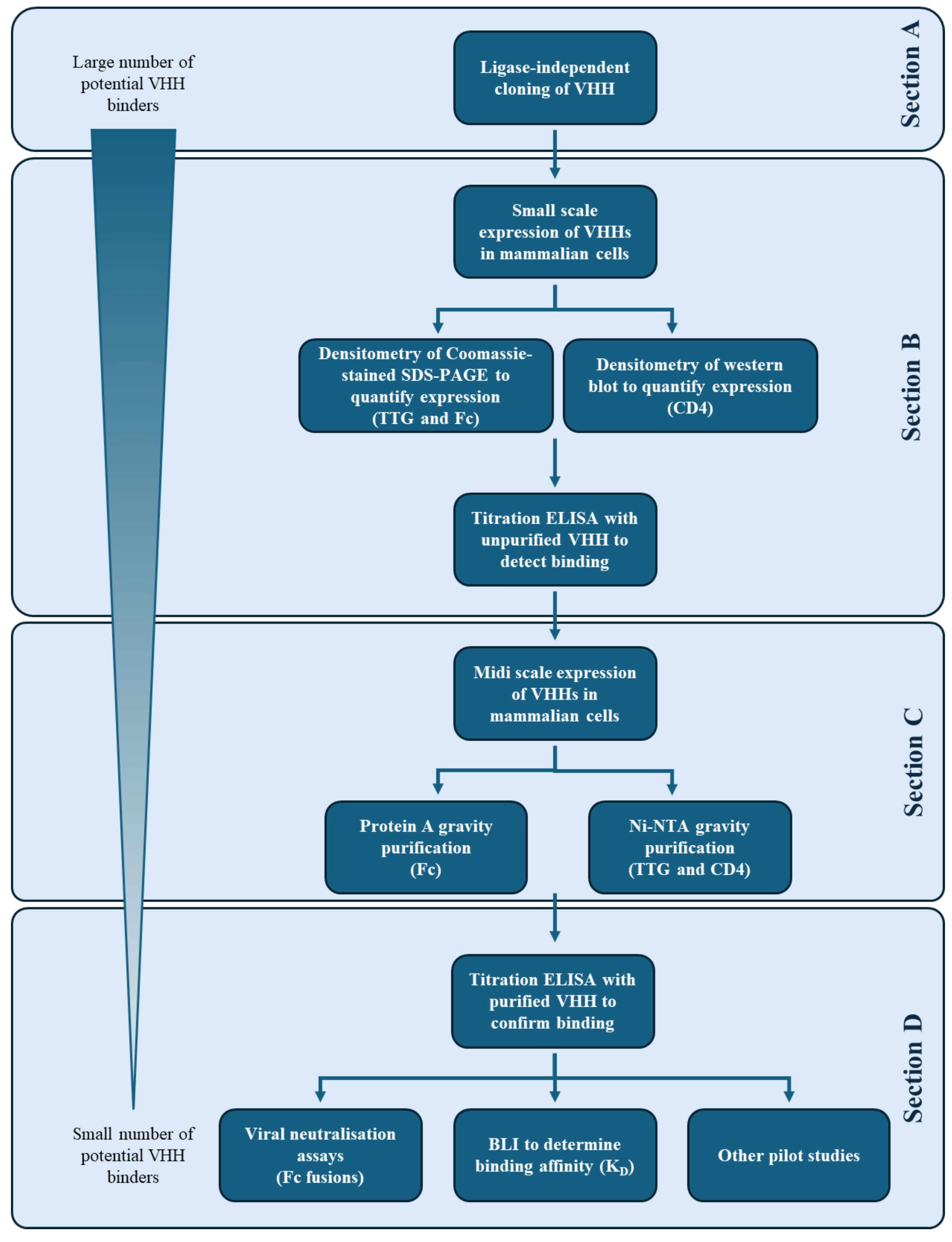
Figure 1. Workflow for the generation of single-domain antibodies (VHHs) in different forms using the modular pOPIN vector suite [1] for characterisation and various downstream applications. Sections A, B, and C each take roughly one week to complete. The modular nature of the workflow has many possible pause points to enable researchers to work flexibly. The timeframe for section D depends on how many downstream tests are carried out.
A. Construction of vectors for expression of VHHs in mammalian cells
Potential antigen-binding VHHs identified by screening a phage display library from either an immunised or non-immunised llama or synthetic VHH library are inserted into mammalian expression vectors in three different formats (IgGFc-His6, CD4-His6, and carboxy-terminal His6) using ligation independent cloning [8]. Confirmation of correct insertion of VHH into the selected vector is verified by colony PCR (Figure 2). Any inserts that were not amplified at the expected sizes were excluded at this point.
A1. Preparation of the digested plasmid
1. In a 1.2 mL cluster tube, add 1 μL of vector (pOPINTTGneo_Fc, pOPINTTGneo_CD4, or pOPINTTGneo) at 100 ng/mL (0.1 ng total) to 10 μL of StellarTM competent cells.
2. Incubate for 10 min on ice.
3. Incubate for 45 s at 42 °C.
4. Incubate for 2 min on ice.
5. Add 200 μL of LB medium and incubate for 1 h at 37 °C without shaking.
6. Spread 50 μL of the resulting culture onto 1% LB agar supplemented with ampicillin (100 μg/mL).
7. Incubate for 16 h at 37 °C.
8. Pick a single colony from each transformation and inoculate 4 mL of LB supplemented with ampicillin (100 μg/mL) in a 24-deep-well block.
9. Cover with a breathable membrane and incubate for 16 h at 37 °C with shaking at 250 rpm.
10. Isolate the plasmid DNA using QIAprep Spin Miniprep kit as per the manufacturer’s instructions.
11. Prepare the digestion mixture of the plasmids in a PCR tube as shown in Table 1.
Note: The three exemplified vectors (pOPINTTGneo_Fc, pOPINTTGneo_CD4, or pOPINTTGneo) are all cut with the same pair of restriction enzymes (KpnI-HF and PmeI)
Table 1. Reaction setup for the preparation of digested plasmid
| Reagent | Final concentration | Volume |
|---|---|---|
| Nuclease-free water | n/a | 80.5 μL |
| 10× rCutSmartTM buffer | 1× | 10 μL |
| KpnI-HF | 25 units | 2.5 μL |
| PmeI | 25 units | 2.5 μL |
| 400 ng/μL vector DNA | 1.8 μg | 4.5 μL |
| Total | n/a | 100 μL |
12. Briefly vortex and centrifuge the reaction tubes and incubate for 1 h at 37 °C.
13. Heat-inactivate the restriction enzymes for 20 min at 60 °C.
14. Clean the resulting linearised plasmid using the PureLink PCR Purification kit as per the manufacturer’s instructions and quantify using a NanoDropTM spectrophotometer.
A2. Preparation of the VHH inserts
1. The DNA template of the target VHHs for PCR can be obtained from a variety of sources: the PCR master plate prepared during the VHH discovery process [4], commercially synthesised double-stranded DNA (dsDNA) fragments, diluted to 10 ng/μL in TE buffer, and plasmid DNA diluted to 10 ng/μL in TE buffer.
2. Prepare dsDNA and plasmid DNA dilutions in a rigid 96-well PCR plate to match the PCR layout.
3. Prepare the PCR master mixes as detailed in Table 2.
4. Add the prepared master mix (22.5 or 24 μL) to the wells of a rigid 96-well PCR plate using a multipipette.
5. Use a multichannel pipette to transfer either 2.5 μL of PCR master plate or 1 μL of dsDNA/plasmid DNA of template into the filled wells of the PCR plate.
6. Seal plate with PCR foil seal, set up the thermal cycler as per Table 3, and perform the PCR.
7. Add 45 μL of HighPrepTM PCR magnetic beads to each well using a multichannel pipette and place on a 96-well DynaMagTM rack for 1 min or until the solution is clear and the beads have been pulled against the wall of the PCR tube. Pipette off the supernatant (SN).
8. Add 100 μL of 70% EtOH and mix by multichannel pipette. Place back on the DynaMagTM rack for 1 min or until the solution is clear and the beads have been pulled against the wall of the PCR tube. Pipette off the SN.
9. Repeat step A2.8 for a total of three times.
10. Leave beads to dry at RT for at least 10 min, add 25 μL of EB buffer, and mix by multichannel pipette. Place back on the DynaMagTM rack for 1 min or until the solution is clear and the beads have been pulled against the wall of the PCR tube.
11. Transfer 20 μL of the purified PCR product containing SN to a new 96-well skirted PCR plate.
12. Use an 8- or 12-channel NanoPhotometer together with a multichannel pipette to quantify the amplified VHH inserts.
13. Store the purified amplified VHH inserts at -20 °C
Note: These can be stored indefinitely at -20 °C.
Table 2. Reaction mixtures for the PCR amplification of VHH clones
| Volume (μL) | |||||||
| Reagent | Final concentration | Fc | CD4 | TTG | |||
| Single reaction | 50× master mix | Single reaction | 50× master mix | Single reaction | 50× master mix | ||
| Nuclease-free water* | n/a | 7.5 | 375 | 9 | 450 | 9 | 450 |
| 10 μM Nb_Fc_Forward primer* | 0.5 μM | 1.25 | 62.5 | 1.25 | 62.5 | 1.25 | 62.5 |
| 10 μM Nb_Fc_Reverse primer* | 0.5 μM | 1.25 | 62.5 | 1.25 | 62.5 | 1.25 | 62.5 |
| 2× Phusion flash PCR master mix* | 1× | 12.5 | 625 | 12.5 | 625 | 12.5 | 625 |
| PCR Master plate | n/a | 2.5 | n/a | n/a | n/a | n/a | n/a |
| 10 ng/μL dsDNA Fragment | 0.4 ng/μL | n/a | n/a | 1 | n/a | n/a | n/a |
| 10 ng/μL plasmid DNA | 0.4 ng/μL | n/a | n/a | n/a | n/a | 1 | n/a |
| Total | 25 | 25 | 25 | ||||
*Indicates reagents that can be scaled up to make a master mix.
Note: Primer sequences can be found in Table S1.
Table 3. PCR amplification conditions
| Step | Temperature (°C) | Duration | No. of cycles |
|---|---|---|---|
| Denaturation | 98 | 60 s | 1 |
| Annealing | 98 | 1 s | |
| Extension | 60 | 1 s | 30 |
| Final extension | 72 | 5 s | |
| Hold | 72 | 1 min | 1 |
| Hold | 12 | Infinite hold | - |
A3. Cloning of the VHH inserts into the prepared plasmid
1. Prepare the InFusion Cloning reaction mixtures of the amplified VHH inserts (step A2.13) and the cut pOPINTTGneo vectors (step A1.14) as shown in Table 4.
2. Transfer the prepared master mix (4 μL) to the wells of a 96-well plate using a multipipette.
3. Add the amplified VHH insert (1 μL) to the wells using a multichannel pipette.
Table 4. Preparation of a 5 μL cloning reaction
| Reagent | Final concentration | Volume |
|---|---|---|
| Nuclease-free water* | n/a | 1.5 μL |
| 5× CEII buffer* | 1× | 1 μL |
| 10 ng/μL VHH insert | 10 ng | 1 μL |
| 85 ng/μL digested vector* | 85 ng | 1 μL |
| Exnase II* | 10% | 0.5 μL |
| Total | 5 μL |
*Indicates reagents that can be scaled up to make a master mix
Note: In cases where VHHs are poorly amplified (less than 10 ng/μL), refer to the Troubleshooting section, problem 3, for alternative reaction mixtures.
4. Briefly vortex and spin down the sample before incubating at 42 °C for 30 min using a thermocycler.
5. Add 20 μL of TE buffer using a multichannel pipette to quench each reaction.
6. Add 10 μL of StellarTM competent cells to 1.2 mL cluster tubes on ice.
7. Transfer 2.5 μL of the quenched infusion reaction to the 1.2 mL cluster tubes and mix gently by pipetting.
8. Incubate on ice for 10 min.
9. Heat shock at 42 °C for 45 s.
10. Incubate on ice for 2 min.
11. Using a multichannel pipette, add 200 μL of LB medium to each well.
12. Incubate for 1 h at 37 °C, without shaking.
13. For blue-white screening, spread 50 μL of the subsequent culture onto 24-well plates containing 1 mL per well of 1% LB agar supplemented with ampicillin (100 μg/mL), IPTG (2 mM), and Xgal (40 μg/mL).
Note: Do not resuspend the cells before plating to avoid growth that is too dense.
14. Leave to dry for 15 min at RT before incubating at 37 °C for 16 h.
15. Pick a single white colony from each well to inoculate 1.2 mL of TB supplemented with ampicillin (100 μg/mL) in a 96-well 2 mL deep-well block.
16. Cover with a breathable membrane and incubate for 16 h at 37 °C and 400 rpm.
17. Pellet cells by centrifugation (4,000× g, 10 min, 4 °C) and isolate the plasmids using the Wizard® SV 96 plasmid DNA purification system as per the manufacturer’s instructions.
18. Quantify plasmid DNA using a NanoPhotometer® N120 and a multichannel.
Note: A minimum of 4 μg of DNA is required for the transfection of 4 mL small-scale mammalian cultures. DNA is eluted from the Wizard® SV 96 plasmid DNA purification system in 100 μL. Taking this into account, any clones with yields lower than 40 ng/μL can be disregarded.
19. Prepare the colony PCR reaction mixture as per Table 5.
20. Distribute the master mix (24 μL per well) to a 96-well PCR plate using a multipipette.
21. Use a multichannel pipette to add the isolated plasmid DNA (1 μL) to the wells of the 96-well plate.
Table 5. Reaction mixture for the colony PCR
| Reagent | Final concentration | Volumes for single reaction (μL) | Volumes for 50× master mix (μL) |
|---|---|---|---|
| Nuclease-free water * | n/a | 19.75 | 987.5 |
| 10 μM TTG_Forward * | 0.2 μM | 0.5 | 25 |
| 10 μM TTG_Reverse* | 0.2 μM | 0.5 | 25 |
| 10× Taq buffer* | 1× | 2.5 | 125 |
| 25 mM dNTP* | 0.25 mM | 0.25 | 12.5 |
| Taq polymerase* | 1 unit | 0.5 | 25 |
| 10 ng/μL miniprep DNA | 0.4 ng/μL | 1 | n/a |
| Total | n/a | 25 |
*Indicates reagents that can be scaled up to make a master mix.
Note: Primer sequences can be found in Table S1.
22. Cover the plate with a foil seal and set up the thermocycler as indicated in Table 6.
Table 6. Colony PCR conditions
| Step | Temperature (°C) | Duration | No. of cycles |
|---|---|---|---|
| Denaturation | 95 | 7 min | 1 |
| Annealing | 95 | 15 s | |
| Extension | 55 | 30 s | 35 |
| Final extension | 68 | 1 min 40 s | |
| Hold | 68 | 5 min | 1 |
| Hold | 12 | Infinite hold | - |
23. In a new 96-well plate, add 5× GelPilot DNA loading dye (5 μL) to the wells using a multipipette.
24. Use a multichannel pipette to add PCR product (5 μL) to the prepared plate.
25. Load sample (10 μL) and HyperLadderTM 1 kb (5 μL) on a 1% TBE agarose gel, supplemented with 1× SYBR safe DNA gel stain.
26. Run at 80 V in 1× TBE buffer for 40 min.
27. Image on a ChemiDoc Imaging System and identify which colonies possess the correctly sized VHH insert in their respective vector (Figure 2).
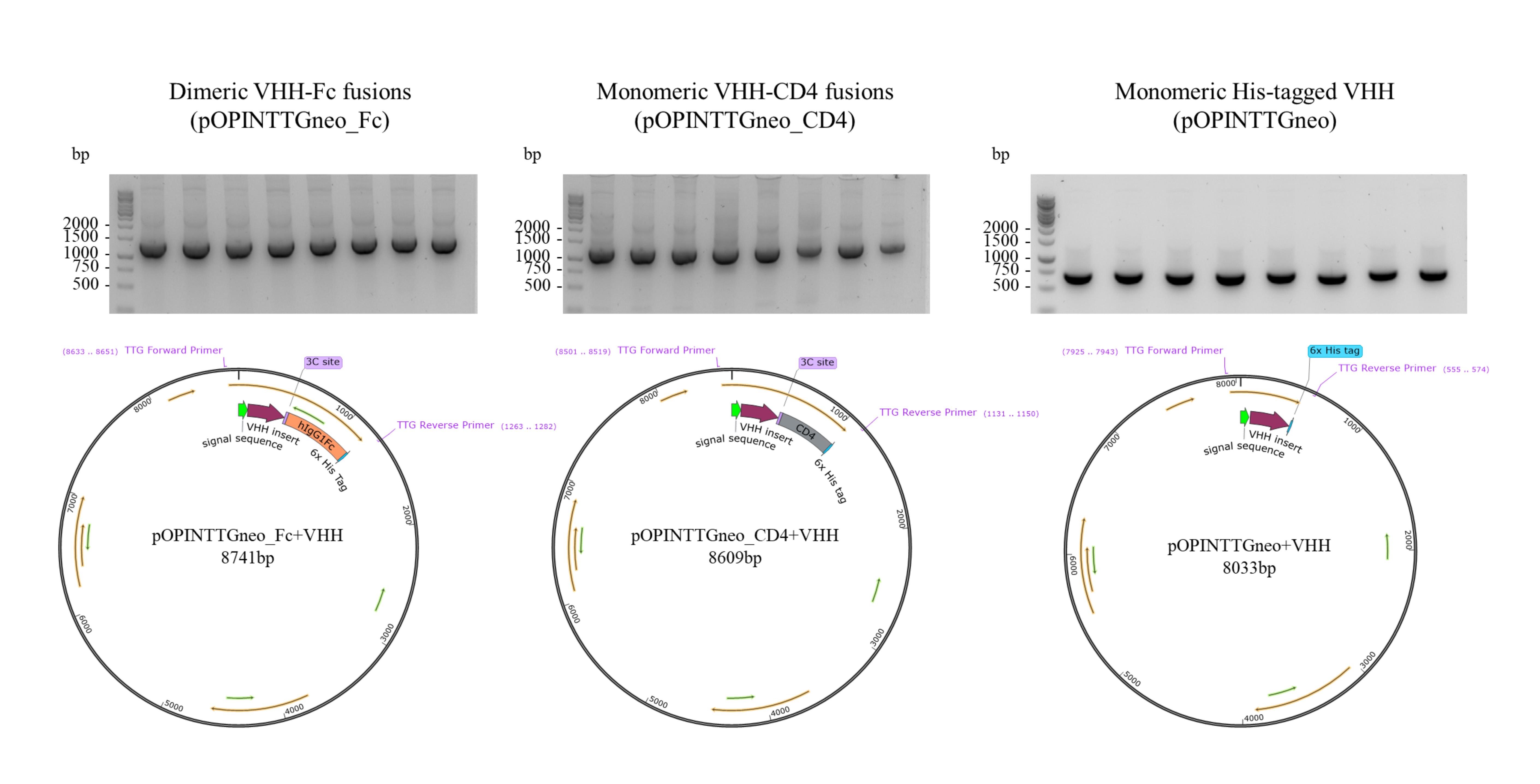
Figure 2. Validation of successful ligation of single-domain antibody (VHH) inserts into pOPINTTG vector suite. Shown by products from colony PCR of eight different VHHs from three different antigens cloned into pOPINTTGneo_Fc (~1,400 bp), pOPINTTGneo_CD4 (~1,200 bp), and pOPINTTGneo (~700 bp), respectively, on a 1% TBE agarose gel and representative plasmid maps generated in SnapGene 8.2.
B. Analysis of VHH production in mammalian cells and their binding capacity using unpurified material
Successfully cloned VHHs are used for the small-scale transfection of mammalian culture (4 mL) to confirm expression by analysing levels of secreted VHH in the culture medium. This can be carried out using Coomassie-stained gels or anti-His western blot for densitometric analysis. Each expressed VHH is subsequently tested for binding to its respective antigen in a titration ELISA using this same unpurified culture medium. VHHs giving a titratable signal by ELISA signal are selected for the next step of the workflow.
B1. Small-scale expression of VHHs in mammalian cells
1. Seed Expi293 cells at 0.5× 10⁶ cells/mL and grow at 37 °C with 5% CO2 and 120 rpm shaking for 48 h.
Note: This cell type can reliably be used up to passage 20 for robust protein expression.
2. Begin the transfection procedure only when cells reach between 1.5 and 2× 10⁶ cells/mL and are at least 95% viable when using an automated cell counter and Trypan Blue solution (diluted 1:1 with cell suspension).
3. Make a transfection master mix consisting of Opti-MEM and polyethylenimine (PEI) MAX 40K transfection reagent as shown in Table 7 for all samples and distribute into each well of 24-deep-well plates.
Table 7. Reaction mixture for transfection
| Reagent | Final concentration | Volume per 4 mL culture (μL) | Volume for 24-well block mastermix (μL) |
|---|---|---|---|
| Opti-MEM | 10% v/v | 400 | 10,000 |
| 1 mg/mL PEI MAX 40K | 5.4 μg/mL | 21 | 525 |
4. Add the transfection master mix into the wells of a 24-deep-well round-bottom plate (421 μL per well).
5. Add 4 μg of DNA of each of the cloned VHH plasmids (step A3.17) to each filled well and incubate at RT for 10 min.
6. Add 4 mL of cell suspension to each well and cover with a breathable membrane.
7. Grow cells for 16–18 h at 37 °C with 5% CO2 and 220 rpm shaking.
8. Create a master mix of enhancers for the required number of wells as per Table 8.
Table 8. Enhancer master mix for small-scale mammalian culture
| Reagent | Final concentration | Volume per 4 mL culture (μL) | Volume for 24-well block master mix (μL) |
|---|---|---|---|
| Glucose (45% w/v in HBSS) | 0.82% | 73 | 1825 |
| Valproic acid (300 mM in HBSS) | 5 mM | 67 | 1675 |
| Sodium propionate (1 M in HBSS) | 6.75 mM | 27 | 675 |
9. Add 167 μL of enhancer master mix to each well.
10. Grow cells for a further 72 h at 37 °C, 5% CO2, 220 rpm.
11. Centrifuge the plate (4,000× g, 10 min, 4 °C). The culture medium contains the secreted VHH and will be referred to as the supernatant (SN) in future steps.
12. Transfer the SN to individual 15 mL conical centrifuge tubes and store at 4 °C for short-term use.
B2. Analysis of expression of VHH in small-scale mammalian culture
The Expi293 cells are cultured in protein-free media; therefore, expression of VHHs can be assessed by SDS-PAGE analysis and Coomassie staining of media samples 96 h after transfection. Under reducing conditions, expect to observe bands at ~45 kDa for dimeric VHH-Fc fusions, ~35 kDa for monomeric VHH-CD4 fusions, and ~15 kDa for monomeric His-tagged VHHs. By running known amounts of a reference protein, the level of expression can be estimated by densitometry of the stained gel.
1. Prepare densitometry control samples
a. For Coomassie-stained gels: include two control wells containing 1 and 2 μg of BSA.
b. For western blots: include two control wells containing 500 and 250 ng of a 14 kDa His-tagged control.
2. Use a multipipette to add 2× Laemmli buffer (10 μL) to each well of a 96-well plate.
3. Add SN and control samples (10 μL) to the wells.
4. Cover plate with a PCR foil lid and heat at 95 °C for 5 min.
5. Load ladder, sample (10 μL), and controls (10 μL) onto a 4%–12% NuPAGE Bis-Tris midi gel.
a. For Coomassie-stained gels, use Mark12 protein ladder (10 μL).
b. For western blots, use PageRulerTM pre-stained protein ladder (1.5 μL).
6. Run gels in 1× MES buffer at 180 V for 40 min.
7. For Coomassie-stained gels:
a. Stain gels with Coomassie for 15–20 min.
b. Rinse the gels with Milli-Q water.
c. Image on a ChemiDoc Imaging System.
8. For western blots:
a. Use the iBlotTM Gel Transfer device to transfer the proteins from the SDS-PAGE gel onto an iBlotTM Regular Transfer Stack as per the manufacturer’s instructions (20 V for 7 min).
b. Move the nitrocellulose membrane to a western blot box and add Ponceau S for 2 min to visualise proteins
Note: To save costs, when running western blots, a Ponceau S staining step eliminates the need to run one gel for visualisation by Coomassie staining and a second gel for the western blot.
c. Rinse nitrocellulose membranes with Milli-Q water.
d. Image on a ChemiDoc Imaging System.
e. Add NaOH (1 M) dropwise onto the membrane until all colour has disappeared.
f. Rinse nitrocellulose membranes with Milli-Q water.
g. Add 5% milk PBS to nitrocellulose membranes for 1 h at RT on a rocking platform.
h. Wash 3× with PBS-T over 15 min.
i. Make a 1:5,000 dilution of mouse monoclonal anti-polyhistidine-peroxidase antibody in antibody dilution buffer and incubate with the membrane for 1 h at RT on a rocking platform.
Note: Prepare the minimum volume of antibody dilution required to cover the nitrocellulose membrane to save costs while using no less than 1 μL of antibody in 5 mL of dilution buffer to ensure pipetting accuracy.
j. Wash 3× with PBS-T over 15 min.
k. Add prepared ECL western blotting substrate
Note: Prepare the minimum volume of ECL western blotting substrate to cover the nitrocellulose membrane to save costs.
l. Image immediately using auto-rapid exposure, merging images captured using the Chemiluminescence, Cy5, and Cy3 channels on a ChemiDoc Imaging System.
9. Use the 68 kDa BSA or 14 kDa His-tagged VHH densitometry controls to perform densitometric analysis using ImageLab or ImageJ (details in Software section) to provide an estimated concentration of the expressed VHHs (Image Lab Software User Guide).
10. Sequence the plasmids of the clones that have successfully expressed the correctly sized VHH with the TTG_Forward primer (Table S1) to validate the sequence.
B3. Confirmation of binding activity of unpurified VHH by titration ELISA
Note: Antigen-binding ELISAs of cell supernatants are carried out in 384-well plates. Volumes for 96-well ELISA can be found in previously published work [4]. Any VHH that showed low signal (absorbance or fluorescence) and no evident titration was excluded at this point.
1. Coat wells of a 384-well high-binding plate with 50 nM target antigen (25 μL per well) and incubate overnight at 4 °C.
Note: While an antigen coating concentration of 50 nM is generally optimal, this can be adjusted if required, and protein buffer can be used in place of PBS if the protein is very sensitive.
2. Wash with 3× PBS (80 μL per well) using a plate washer.
3. Add 2% milk PBS to each well (75 μL per well) using a multichannel pipette and incubate at RT for 1 h, 500 rpm on a microplate shaker.
4. Wash with 3× PBS-T (80 μL per well) using a plate washer.
5. Prepare a serial dilution of crude VHH SN in 2% milk PBS (25 μL per well) and incubate at RT for 1 h, 500 rpm on a microplate shaker.
a. For VHH-Fc, a 4-step 1:10 dilution series is set up from 1000 to 10 ng/mL (as determined by densitometry using Coomassie-stained gels; Figure 3).
b. For VHH-CD4, an 8-step 1:10 dilution series is set up from the undiluted SN (as determined by densitometry using anti-His western blot; Figure 3).
c. For VHH-TTG, a 4-step 1:2 dilution series is set up (without any quantitation using densitometry).
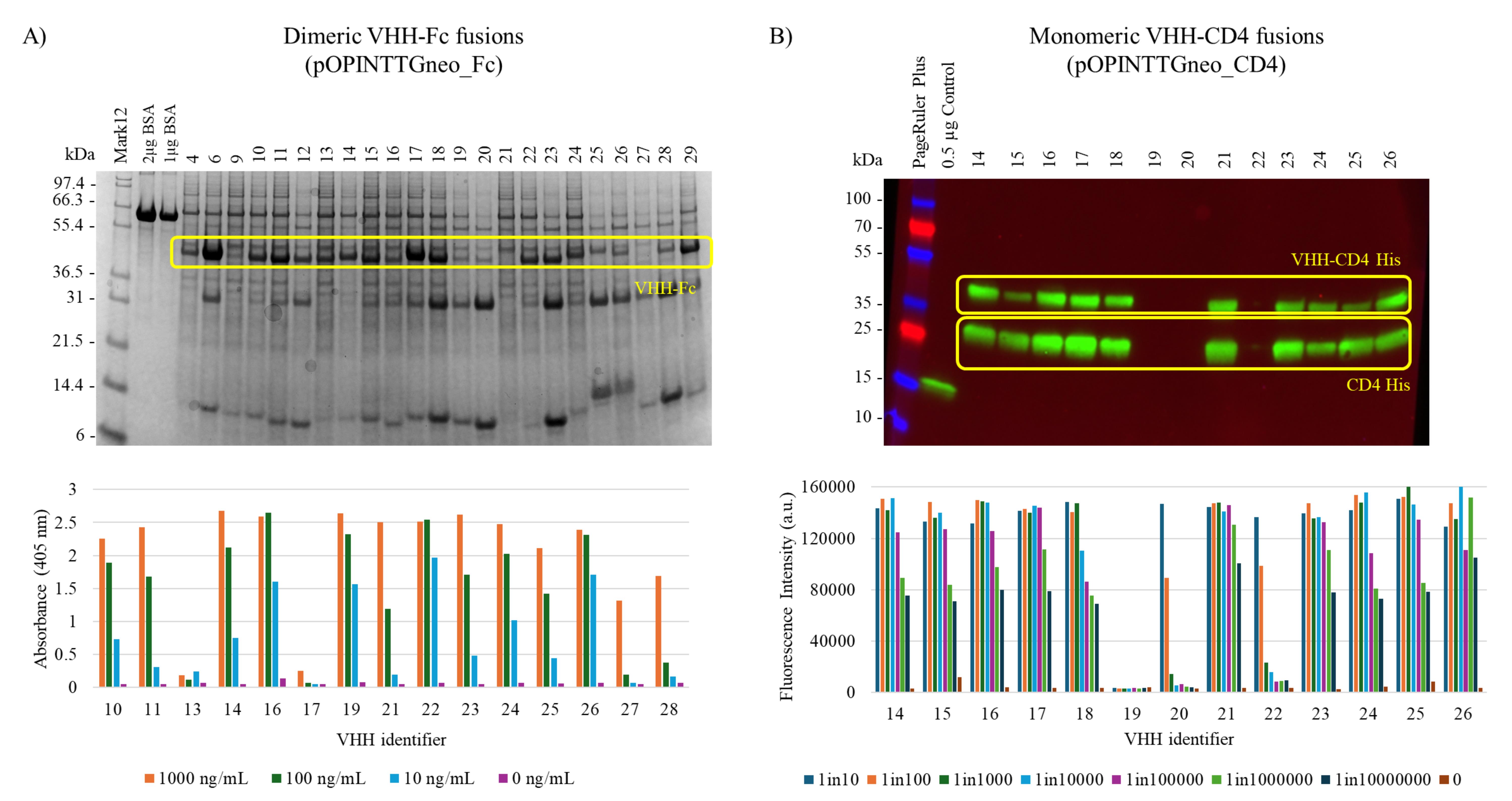
Figure 3. Identification of single-domain antibody (VHH) binders from a small-scale expression in mammalian cells. Analysis of the VHH secreted into the culture medium by Expi293 cells is visualised either by (A) Coomassie staining of 4%–12% Bis-Tris SDS-PAGE gels for VHH-Fc or by (B) anti-His western blot using anti-polyhistidine-peroxidase antibody for VHH-CD4. Expressed VHHs were quantified using densitometry. The ability of these crude VHH preparations to bind their respective antigens is determined by a titration ELISA (n = 1 for VHH-Fc fusions and n = 2 for VHH-CD4 fusions). Western blot loading control is a previously purified His-tagged VHH.
6. Wash with 3× PBS-T (80 μL per well) using a plate washer.
7. Add antibody dilutions as follows (25 μL per well) and incubate for 1 h at RT on a microplate shaker:
a. For VHH-Fc: anti-Fc HRP at 1:1,000 dilution in 0.1% BSA PBS.
b. For VHH-CD4 and VHH-TTG: mouse monoclonal anti-polyhistidine-peroxidase antibody at 1:5,000 dilution in 0.1% BSA PBS.
8. Wash with 3× PBS-T (80 μL per well) using a plate washer.
9. Add QuantaBlu (25 μL per well) and incubate for 15 min in the dark at RT without shaking.
10. Measure the plate for fluorescence using Ex 345 nm/Em 455 nm on a plate reader such as the CLARIOstar Plus.
C. Parallelised purification of midi-scale expressed VHHs using affinity chromatography
Having identified VHH clones that have antigen-binding activity, the next step of the workflow is to prepare material for further characterisation. To produce approximately 100–500 μg of purified protein, midi-scale expression in 30 mL of Expi293 cultures grown in specialised TPP® TubeSpin bioreactor tubes is carried out. These tubes offer a space-saving solution. The footprint of the 50 mL centrifuge tube storage box in which 9 midi scale expressions can be carried out is 12.5 cm × 12.5 cm (approximately equivalent to just 3–4× 125 mL conical flasks usually used for a single 30 mL culture). They also provide a cost-saving solution as the culture does not require transferring to another piece of plasticware for centrifugation. To facilitate the parallel purification of many VHHs without the use of relatively expensive chromatography equipment, a custom multi-column purification apparatus (Figure 4) was designed and built. The components of this apparatus were cut using a laser cutter and assembled using superglue. Detailed assembly instructions can be found in Supplementary file 1.
Focusing on gravity purification, different resins can be loaded into the Poly-Prep® chromatography columns for purification of VHHs with varying tags. Unbound and eluted fractions can easily be collected in 24-well and 2 mL 96-deep-well plates, respectively. Using this setup, it is possible to purify eight VHHs simultaneously, and two such purification runs can easily be performed by a single person in a day. For the dimeric VHH-Fc fusion proteins, Protein A Plus Agarose is used for purification, whilst for the monomeric VHH-CD4 fusions and simple His-tagged VHHs (pOPINTTGneo), Ni-NTA resin is used. All purified VHHs are desalted into PBS, and binding to their respective antigens is confirmed by titration ELISA. In general, VHHs made in a fusion format provide a higher yield than those produced as simple His-tagged monomers: roughly a 5-fold increase in yield as an Fc fusion and nearly a 10-fold increase for CD4 fusions (see Validation).
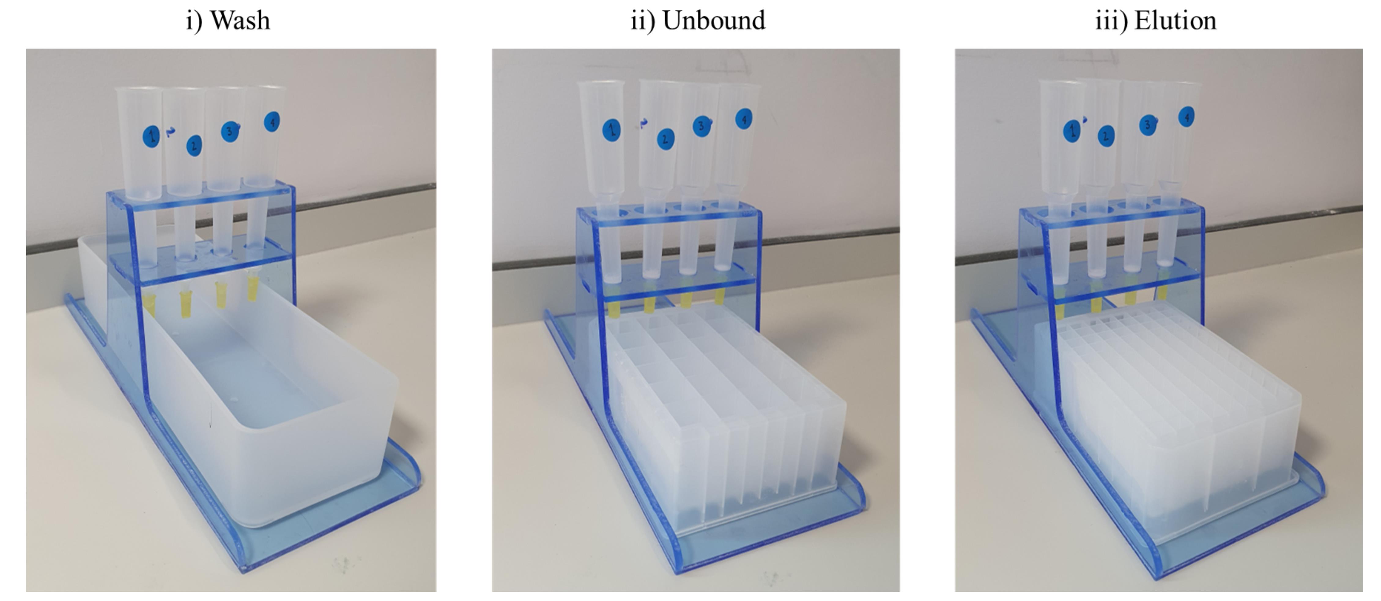
Figure 4. Custom multi-column purification apparatus containing Poly-Prep® chromatography columns. i) over a waste trough to collect washes and regeneration reagents, ii) a 24-deep-well block for collection of unbound protein, and iii) a 96-well 2 mL deep-well block for collection of eluted, purified single-domain antibodies (VHHs). Detailed instructions for the construction of the custom multi-column purification apparatus can be found in Supplementary File 1.
C1. Scaled preparation of plasmid DNA
1. Use plasmid DNA from step A3.17 to transform into StellarTM competent cells as detailed in section A1, but with the following modifications:
a. Add 200 μL of LB media, instead of 400 μL, for the 1-h incubation at 37 °C.
b. Do not resuspend the culture after the 1-h incubation. Spread 50 μL of the resulting culture onto 24-well plates containing 1 mL per well of 1% LB agar supplemented with ampicillin (100 μg/mL), instead of standard-sized Petri dishes.
2. Leave plates to dry for 15 min at RT before incubation at 37 °C for 16 h.
3. Take a single colony for each of the VHH clones to inoculate 35 mL of TB supplemented with ampicillin (100 μg/mL) in 50 mL conical centrifuge tubes.
4. Incubate at 37 °C for 16 h at 250 rpm.
5. Isolate and quantitate the plasmid DNA using the Plasmid Plus Midi kit as per the manufacturer’s instructions.
C2. Midi-scale expression of VHHs in mammalian cells
1. Seed Expi293 cells at 0.5 × 10⁶ cells/mL and grow at 37 °C with 5% CO2 and 120 rpm shaking for 48 h.
2. Begin the transfection procedure only when cells reach between 1.5 and 2 × 10⁶ cells/mL and are at least 95% viable using an automated cell counter and Trypan Blue solution (diluted 1:1 with cell suspension).
3. Make a transfection master mix consisting of Opti-MEM and PEI MAX 40K transfection reagent as shown in Table 9 for all samples.
Table 9. Reaction mixture for transfection
| Reagent | Final concentration | Volume per 30 mL culture (μL) |
|---|---|---|
| Opti-MEM | 10% v/v | 3,000 |
| PEI MAX 40K | 5.4 μg/mL | 160 |
4. Distribute the master mix into 50 mL TubeSpin bioreactor tubes.
5. Add 30 μg of each plasmid (step C1.5) to each tube and incubate for 10 min.
6. Add 30 mL of cell suspension to each tube and place in a 50 mL centrifuge tube storage box (do not use the box lid).
7. Grow cells for 16 h at 37 °C with 5% CO2 and 220 rpm shaking.
8. Create a master mix of enhancers for the required number of tubes as per Table 10.
Table 10. Reaction mixture for enhancers
| Reagent | Final concentration | Volume per 30 mL culture (μL) |
|---|---|---|
| Glucose (45% w/v in HBSS) | 0.82% | 550 |
| Valproic acid (300 mM in HBSS) | 5 mM | 500 |
| Sodium propionate (1 M in HBSS) | 6.75 mM | 200 |
9. Add 1.25 mL of master mix per tube.
10. Grow cells for a further 72 h at 37 °C, 5% CO2, 220 rpm.
11. Centrifuge the tubes (4,000× g, 30 min, 4 °C).
12. Transfer SN to individual 50 mL conical centrifuge tubes and store at 4 °C for short-term use.
Note: Whilst storage of SN at 4 °C is adequate for the short term, it can also be stored at –20 °C and can withstand multiple freeze-thaw cycles without any noticeable impact on protein binding in a titration ELISA.
C3. Parallelised gravity purification using protein A (for dimeric VHH-Fc fusions)
1. Add 1 mL of PierceTM Protein A Plus agarose resin to Poly-Prep® chromatography columns.
2. Drain storage buffer, leaving a bed volume of 0.5 mL, and wash with 10 mL of PBS (Note II).
3. Add 3 mL of each SN (step C2.12) to each column and mix with resin to form a resin–SN slurry.
4. Return the slurry to the remaining SN in the 50 mL conical centrifuge tube and agitate on a roller mixer for 1 h at RT.
5. Pour the resin mixture back into the Poly-Prep® chromatography columns and collect unbound fractions in 24-deep-well plates.
6. Wash the resin with PBS (3 × 10 mL).
7. During this time, add 220 μL of protein A neutralisation buffer into 12 wells of a 2 mL deep-well 96-well block. If purifying four VHHs, add this buffer to the 12 wells in rows A, C, F, and H.
8. Add 12 mL of protein A elution buffer to the resin and collect eluted VH-Fcs into the prepared 2 mL deep-well block.
9. If precipitation is present, centrifuge the fraction at 17,000× g for 2 min at 4 °C and transfer the SN to a fresh 2 mL microcentrifuge tube.
10. Use a multipipette to add 2× Laemmli buffer (10 μL) to each well of a 96-well plate.
11. Add samples from unbound and eluted fractions (10 μL) to the wells.
12. Cover plate with a PCR foil lid and heat at 95 °C for 5 min.
13. Load Mark12 unstained standard and samples (10 μL) onto a 4%–12% NuPAGE Bis-Tris mini gel.
14. Run gels in 1× MES buffer at 200 V for 30 min.
15. Stain gel with Coomassie for 15–20 min.
16. Rinse the gel with Milli-Q water.
17. Image on a ChemiDoc imaging system.
18. Regenerate the protein A resin after each use by placing the columns over a waste trough and running 10 mL of the following buffers over the resin in sequence: 1× PBS, 0.5 M NaOH, and 1× PBS. Resins are stored in PBS at 4 °C until the next use.
C4. Parallelised purification using Ni-NTA resin (for His-tagged monomeric VHH-CD4 fusions and simple His-tagged monomer)
1. In Poly-Prep® chromatography columns, drain and wash Ni-NTA Superflow resin (1 mL) with Milli-Q water (10 mL).
2. Wash resin with 10 mL of wash buffer and collect waste in a trough.
3. Add 3 mL of SN to each column and mix with resin to form a resin–SN slurry.
4. Return the slurry to the remaining SN in the centrifuge tube and agitate on a roller mixer for 1 h at 4 °C.
5. Pour the resin mixture back into the Poly-Prep® chromatography columns and collect unbound fractions in 24-deep-well plates.
6. Wash resin with 3 × 10 mL of wash buffer and continue to collect the wash flowthrough (FT) in 24-deep-well plates.
7. Add 5 mL of elution buffer to the resin and collect 0.5 mL fractions of eluted VHHs into a 0.5 mL deep-well 96-well block.
8. Use a multipipette to add 2× Laemmli buffer (10 μL) to each well of a 96-well plate.
9. Add samples from unbound, FT, and eluted fractions (10 μL) to the wells.
10. Cover plate with a PCR foil lid and heat at 95 °C for 5 min.
11. Load Mark12 unstained standard and samples (10 μL) onto a 4%–12% NuPAGE Bis-Tris mini gel.
12. Run gels in 1× MES buffer at 200 V for 30 min.
13. Stain gel with Coomassie for 15–20 min.
14. Rinse the gel with Milli-Q water.
15. Image on a ChemiDoc imaging system.
16. Regenerate the Ni-NTA Superflow resin after each use by placing the columns over a waste through and running 10 mL of the following buffers over the resin in sequence: Milli-Q, stripping buffer*, Milli-Q, 1 M NaOH, Milli-Q, regeneration buffer*, Milli-Q. Resins are stored in 20% (v/v) ethanol.
Note: Waste from buffers marked with a * are toxic and should be treated as nickel waste.
C5. Desalting
Note: For dimeric VHH-Fc fusions purified using protein A, there was no difference in antigen binding ability observed between desalted and non-desalted products (Figure S1). However, since Ni-NTA-purified proteins required the removal of imidazole, all VHHs were desalted into PBS.
1. From step C4.15, identify elution fractions containing purified VHH and pool into a single 15 mL conical centrifuge tube per VHH.
2. Desalt 1 mL of pooled purified VHH using a PD Miditrap desalting column as per the manufacturer’s instructions.
Note: PD Miditrap desalting columns were used for ease of running multiple columns simultaneously and without considerable preparation. We found that other commercial desalting columns that facilitate larger sample volumes were not consistent in performance. Alternatively, a 5 mL ÄKTA desalting column could be used with a syringe as a low-throughput alternative.
3. Measure the absorbance of the desalted nanobodies using a NanoDropTM spectrophotometer at 280 nm.
4. Use the VHH sequence (starting from the signal sequence cleavage site and including the relevant fusion proteins and tags) in the ProtParam tool on Expasy (https://web.expasy.org/protparam/) [9] to determine the size (kDa) and extinction coefficient (ε) of each VHH. Signal sequences are cleaved during expression and, as such, are not included in the sequence for computing molecular parameters.
5. Use the absorbance value, extinction coefficient (ε), and size (kDa) to calculate the concentration of the purified and desalted VHHs.
a. (A280/ε) × 1,000,000 = μM
b. μM × kDa = μg/mL
D. Confirmation of binding activity of purified VHHs
After successfully purifying and desalting VHHs, a titration ELISA is carried out to confirm binding to the target antigen and to assess relative affinities. Further analysis by either biolayer interferometry (BLI) or surface plasmon resonance (SPR) is carried out on the strongest binders to measure binding affinities and is not covered by this protocol.
Validation of protocol
The protocol described in this article was designed and implemented for a VHH discovery campaign, targeting the S2 domain of the spike protein of SARS-CoV-1 and SARS-CoV-2 viruses. Potential binders (n = 75) identified through screening of an immunised VHH phage library were expressed as Fc fusions at small-scale and unpurified cell supernatants tested for antigen-binding by ELISA (Figure 3A). This identified potential binders (n = 40) that were subsequently expressed at midi scale, purified (Figure 5), and binding-validated by ELISA against the S2 protein of SARS-CoV-2 variant BA.1 (n = 38/40 as two of the clones selected from the SN screening failed to bind as purified proteins) (Figure 6). Purified products were used for viral neutralisation assays.
A pilot study using a protein large language model (pLLM) [10] to predict residue changes that improve binding activity was carried out on one of the VHHs from the SARS-CoV-2 S2 binder project, described above. Mutant VHHs (n = 88) containing one or more residue changes relative to the parent sequence were expressed as CD4 fusions so that monomeric binding activity could be assessed. Antigen-binding ELISAs of unpurified cell supernatants were used to identify expressible binders (n = 74) (Figure 5B). Eight of these were pursued in midi-scale expressions to produce VHH-CD4 fusions (Figure 5) for further binding characterisation studies (Figure 6).
The numbers of VHHs retained at each stage of the workflow are indicated in Table 11, highlighting the filtering nature of the methodology described in this paper. For the SARS-CoV-2 S2 targeting nanobodies, from the initial identification of 75 potential binders to the expression of 38 genuine binders, it is evident that time and money can be saved by implementing the small-scale filtering stage in the workflow and identifying the clones that need prioritisation for scaled expression.
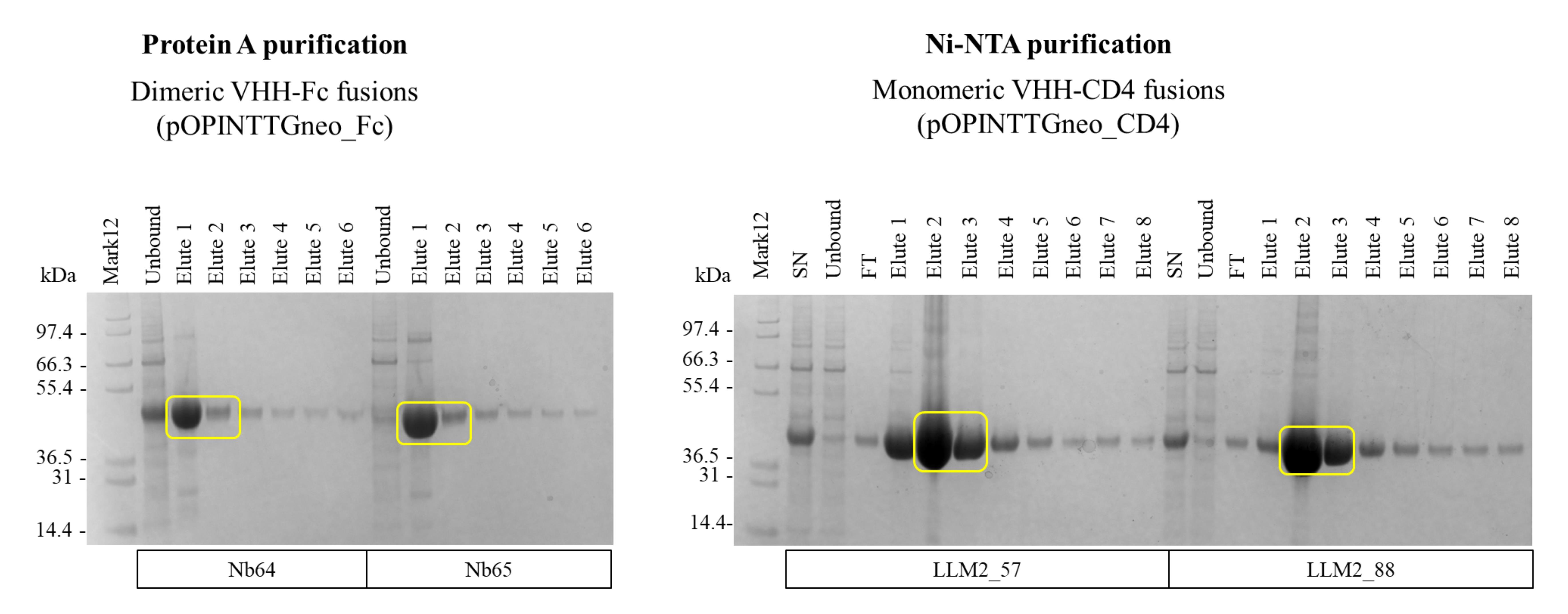
Figure 5. Simultaneous purification of single-domain antibodies (VHHs) expressed at midi-scale using custom multi-column purification apparatus. Unbound and sequentially eluted VHH fractions from either protein A (VHH-Fc fusion) or Ni-NTA (VHH-CD4 fusion) resins were analysed on SDS-PAGE gels as described in sections C3 and C4, respectively. Only a representative number of nanobodies that were purified are shown.
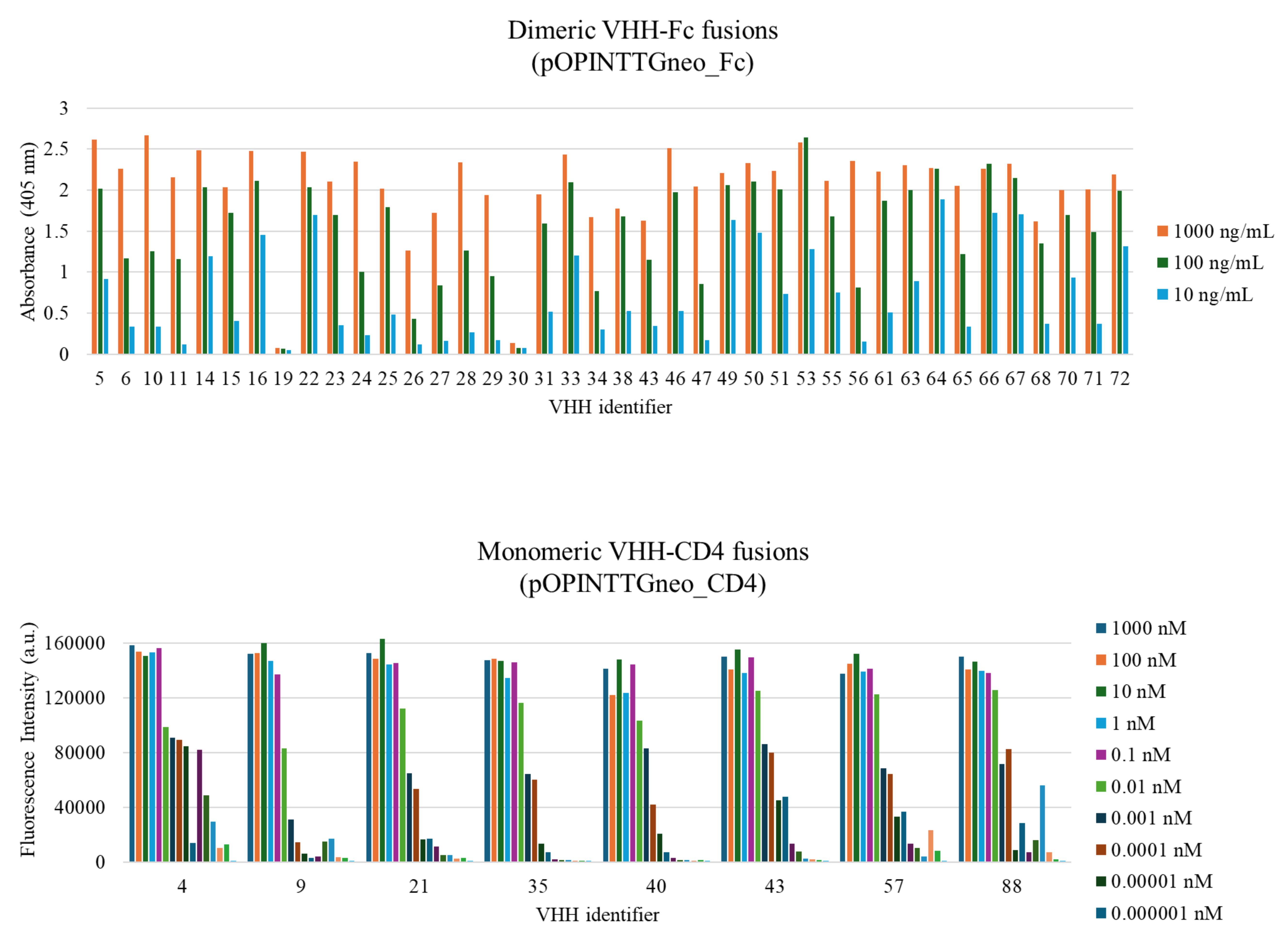
Figure 6. Confirmation of binding of purified single-domain antibodies (VHHs) to their respective antigens by titration ELISA. n = 1 for VHH-Fc fusions and n = 2 for VHH-CD4 fusions.
Table 11. Summary of the number of VHH clones retained at each stage of the workflow
| Stage of workflow | pOPINTTG neo_Fc | pOPINTTG neo_CD4 |
|---|---|---|
| Identified VHH sequences targeting the specific antigen | 75 | 88 |
| Colonies obtained for InFusion cloning into the vector | 68 | 85 |
| Plasmid successfully isolated (>40 ng/μL) | 64 | 85 |
| Correct insert size as determined by colony PCR | 59 | 85 |
| Successful expression: small scale | 46 | 74 |
Successful binder: unpurified SN ELISA (Absorbance >0.5 or fluorescence >50,000 with visible titration across dilutions) | 40 | 74 |
Successful binder: purified VHH ELISA (Absorbance >0.5 or fluorescence >50,000 with visible titration across dilutions) | 38 | 8 pursued |
General notes and troubleshooting
General notes
1. This protocol is intended to be high throughput and low cost. We make use of multichannel pipettes, multipipettes, and 12-channel spectrophotometers wherever possible to make the process more efficient and reduce the risk of repetitive strain injury. However, if these are not available, single-channel pipettes and single-channel spectrophotometers can be used instead. This workflow can be further optimised with the use of lab automation; however, that is not a low-cost strategy.
2. Due to the nature of working with high numbers of samples and different plate formats, we strongly encourage the creation of plate maps to visualise sample location and tables to confirm volumes that need to be added per sample.
3. While we describe the usage of commercially available pre-cast gels and pre-made transfer stacks, homemade alternatives are suitable for use and will have a reduced associated cost.
4. When working with resins, ensure that the resin bed does not dry out throughout the purification and recharging processes.
5. It is recommended that users carry out technical replicates for the various titration ELISAs, duplicates for the crude supernatant, and triplicates for the purified VHHs.
Troubleshooting
Problem 1: No colonies present after transformation.
Possible causes: Low concentration of DNA, poor transformation efficiency, or not enough culture to yield growth.
Solutions: Increase the DNA concentration or volume of competent cells used in the transformation, or plate a larger volume onto the agar.
Problem 2: Colonies are too dense after transformation.
Possible cause: High transformation efficiency.
Solution: Dilute the culture 1:2 with LB medium and re-plate.
Problem 3: VHH inserts made from PCR master plates (section A2) have a low yield.
Possible cause: Poor amplification of the template.
Solution: If the yield of VHH insert is particularly low after Phusion PCR amplification, carry out bead cleanup of the DNA insert and re-run the same reaction using the cleaned DNA as the template (1 μL at 10 ng/μL).
Problem 4: VHH inserts (section A2) with a yield too low for cloning into the pOPIN vector suite as per Table 4.
Possible cause: Poor amplification of the template.
Solution: Adapt the cloning reaction (section A3) as shown in Table 12.
Table 12. Cloning reaction mixtures adapted for low-yield VHH inserts
| Reagent | Volumes for inserts at <5 ng/μL (μL) | Volumes for inserts at <1 ng/μL (μL) |
|---|---|---|
| Nuclease-free water | - | 2 |
| 5x CEII buffer | 1 | 3 |
| VHH insert | 2.5 | 8 |
| 85 ng/μL digested vector | 1 | 1 |
| Exnase II | 0.5 | 1 |
| Total | 5 | 15 |
Problem 5: Poor yields from the Wizard® SV 96 plasmid DNA purification system (section A3).
Possible causes: Incomplete drying of the membrane after washing or incomplete elution of the final product.
Solutions: To ensure complete removal of excess EtOH after washing steps and prior to elution, the binding plate can be left for longer at RT to dry out or can be centrifuged over a waste plate to further dry out the membrane. If incomplete elution occurs, it is possible to carry out a centrifugation step with the binding plate placed over the elution plate to ensure collection of the full elution volume.
Problem 6: No expression observed (section C2).
Possible cause: Incorrect sequence in scaled DNA preparation.
Solution: Verify the sequence of the insert after scaled preparation of plasmid DNA.
Problem 7: No or low signal on titration ELISA or signal drops off very quickly.
Possible cause: Weak binder.
Solution: Try a higher starting concentration or a smaller fold dilution to address low signal.
Supplementary information
The following supporting information can be downloaded here:
1. Table S1: Primers used in this workflow.
2. File S1: Assembly of the custom multi-column purification apparatus.
3. Figure S1: Comparison of non-desalted and desalted VHH-Fc after protein A purification on a titration ELISA.
Acknowledgments
This work was supported by the Rosalind Franklin Institute, with funding from the Engineering and Physical Sciences Research Council UK (EPSRC), and grants from the Biotechnology and Biological Sciences Research Council UK (BBSRC) for nanobody discovery (ref. BB/V018523/1) and the Wellcome Trust for technology development (ref. 223733/Z/21/Z).
References
- Nettleship J. E., Rada H., Owens R. J., (2019). A High-Throughput Pipeline for Protein Production. In: Vincentelli R., (Ed). High-throughput protein production and purification: methods and protocols. Humana Press: 33–49.
- Pardon, E., Laeremans, T., Triest, S., Rasmussen, S. G. F., Wohlkönig, A., Ruf, A., Muyldermans, S., Hol, W. G. J., Kobilka, B. K., Steyaert, J., et al. (2014). A general protocol for the generation of Nanobodies for structural biology. Nat Protoc. 9(3): 674–693. https://doi.org/10.1038/nprot.2014.039
- Zimmermann, I., Egloff, P., Hutter, C. A. J., Kuhn, B. T., Bräuer, P., Newstead, S., Dawson, R. J. P., Geertsma, E. R. and Seeger, M. A. (2020). Generation of synthetic nanobodies against delicate proteins. Nat Protoc. 15(5): 1707–1741. https://doi.org/10.1038/s41596-020-0304-x
- Eyssen, L., Ramadurai, S., Abdelkarim, S., Buckle, I., Cornish, K., Lin, H., Jones, A., Stephens, G. and Owens, R. (2025). From Llama to Nanobody: A Streamlined Workflow for the Generation of Functionalised VHHs. Bio Protoc. 14(1341): e4962. https://doi.org/10.21769/bioprotoc.4962
- Le Bas, A., Mikolajek, H., Huo, J., Norman, C., Dormon, J., Naismith, J. and Owens, R. (2022). Production and Crystallization of Nanobodies in Complex with the Receptor Binding Domain of the SARS-CoV-2 Spike Protein. Bio Protoc. 12(9): e4406. https://doi.org/10.21769/bioprotoc.4406
- Huo, J., Mikolajek, H., Le Bas, A., Clark, J. J., Sharma, P., Kipar, A., Dormon, J., Norman, C., Weckener, M., Clare, D. K., et al. (2021). A potent SARS-CoV-2 neutralising nanobody shows therapeutic efficacy in the Syrian golden hamster model of COVID-19. Nat Commun. 12(1): e1038/s41467–021–25480–z. https://doi.org/10.1038/s41467-021-25480-z
- Djender, S., Schneider, A., Beugnet, A., Crepin, R., Desrumeaux, K., Romani, C., Moutel, S., Perez, F. and de Marco, A. (2014). Bacterial cytoplasm as an effective cell compartment for producing functional VHH-based affinity reagents and Camelidae IgG-like recombinant antibodies. Microb Cell Fact. 13(1): 140. https://doi.org/10.1186/preaccept-1444588381355050
- Berrow, N. S., Alderton, D., Sainsbury, S., Nettleship, J., Assenberg, R., Rahman, N., Stuart, D. I. and Owens, R. J. (2007). A versatile ligation-independent cloning method suitable for high-throughput expression screening applications. Nucleic Acids Res. 35(6): e45–e45. https://doi.org/10.1093/nar/gkm047
- Gasteiger, E., Hoogland, C., Gattiker, A., Duvaud, S., Wilkins, M. R., Appel, R. D. and Bairoch, A. (2005). Protein Identification and Analysis Tools on the ExPASy Server. The Proteomics Protocols Handbook. Humana Press. 571–607. https://doi.org/10.1385/1-59259-890-0:571
- Hie, B. L., Shanker, V. R., Xu, D., Bruun, T. U. J., Weidenbacher, P. A., Tang, S., Wu, W., Pak, J. E. and Kim, P. S. (2023). Efficient evolution of human antibodies from general protein language models. Nat Biotechnol. 42(2): 275–283. https://doi.org/10.1038/s41587-023-01763-2
Article Information
Publication history
Received: Feb 13, 2026
Accepted: Mar 27, 2026
Available online: Apr 12, 2026
Published: May 5, 2026
Copyright
© 2026 The Author(s); This is an open access article under the CC BY-NC license (https://creativecommons.org/licenses/by-nc/4.0/).
How to cite
Campaigne, H. A., Parker, K., Owens, R. J. and Eyssen, L. E. A. (2026). Parallelised Cloning, Mammalian Cell Expression, and Purification of Nanobodies Identified by Phage Display. Bio-protocol 16(9): e5682. DOI: 10.21769/BioProtoc.5682.

 Download Citation in RIS Format
Category
Molecular Biology > DNA > DNA cloning
Molecular Biology > Protein > Expression
Biochemistry > Protein > Expression
Do you have any questions about this protocol?
Post your question to gather feedback from the community. We will also invite the authors of this article to respond.
 Post a Question


