- Protocols

- Articles and Issues
- For Authors
- About
- Become a Reviewer

Past Issue in 2025
Volume: 15, Issue: 23
Biochemistry
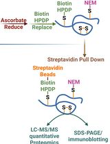
Quantitative Proteomics of Nitrosylated Proteins in Melanoma Using the Biotin-Switch Technique Combined With Tandem Mass Tag Labeling
Bioinformatics and Computational Biology
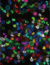
Detailed Protocol for Segmentation and Quantification of Overlapping Prospore Membranes using DeMemSeg
Biological Sciences
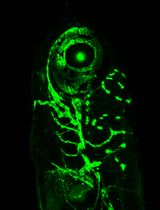
Room-Temperature Storage of Zebrafish and Medaka Sperm Using Lactic Acid-Stabilized L-15 Medium
Biophysics
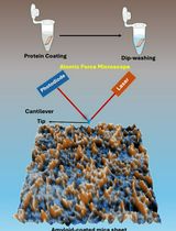
An Optimized Protocol for High-Quality AFM Imaging of Amyloid Fibrils
Immunology
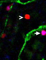
Utilizing EdU to Track Leukocyte Recruitment to the Brain
Microbiology
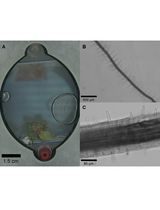
Imaging the Entire Sexual Life Cycle of the Budding Yeast Saccharomyces cerevisiae Using a Microfluidic Platform
Molecular Biology
Analyzing the Translatome of Lymphatic and Venous Endothelial Cells In Vivo via Translating Ribosome Affinity Purification (TRAP)
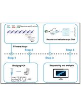
Bridging PCR-Based Genome-Walking Protocol
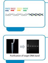
Implementation of Fusion Primer-Driven Racket PCR Protocol for Genome Walking
Neuroscience
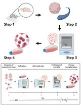
Revisiting Primary Microglia Isolation Protocol: An Improved Method for Microglia Extraction
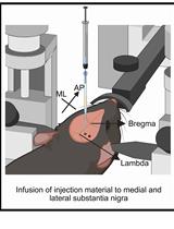
A One-Step Mouse Model of Parkinson’s Disease Combining rAAV-α-Synuclein and Preformed Fibrils of α-Synuclein
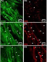
Whole-Mount Immunostaining for the Visual Separation of A- and C-Fibers in the Study of the Sciatic Nerve
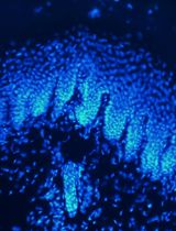
Intraepidermal Nerve Fiber Quantification of the Mouse Hind Paw Footpads: A Detailed and Simplified Protocol
Plant Science

Highly Efficient Agrobacterium-Mediated Transformation of Tomato cv Micro-Tom From Cotyledon Explants
Synchronizing Germination Rates Across Plant Species for Fabricated Ecosystems EcoFAB 2.0
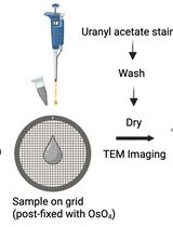
Preparation and Negative Staining for Visualization of Cyanoglobule Lipid Droplets Using Transmission Electron Microscopy
Stem Cell
A Protocol to Induce Brown and Beige Adipocyte Differentiation From Murine and Human Adipose-Derived SVF
A Simplified 3D-Plasma Culture Method for Generating Minimally Manipulated Autologous Equine Muscle-Derived Progenitor Cells