Past Issue in 2024

Volume: 14, Issue: 8

left

Dec

20

Dec

5

Nov

20

Nov

5

Oct

20

Oct

5

Sep

20

Sep

5

Aug

20

Aug

5

Jul

20

Jul

5

Jun

20

Jun

5

May

20

May

5

Apr

20

Apr

5

Mar

20

Mar

5

Feb

20

Feb

5

Jan

20

Jan

5

right

Biochemistry

Preparing and Evaluating the Stability of Therapeutically Relevant Oligonucleotide Duplexes

Preparing and Evaluating the Stability of Therapeutically Relevant Oligonucleotide Duplexes

SI Shreyas G. Iyer
AK Andrea L. Kasinski
3565 Views
Apr 20, 2024
The field of oligonucleotide therapeutics is rapidly advancing, particularly for combating orphan diseases and cancer. However, the intrinsic instability of oligonucleotides, especially RNA, poses a substantial challenge in the face of the harsh conditions encountered intracellularly and in circulation. Therefore, evaluating the stability of oligos in serum is of great significance when developing oligonucleotide therapeutics. This protocol outlines a dependable and reproducible method for preparing oligonucleotide duplexes, coupled with confirmation by gel electrophoresis. Subsequently, the protocol defines a mechanism to assess the stability of the oligo duplexes in serum. This protocol seeks to establish a standardized reference for researchers, enabling them to compare the impact of various modifications on oligo stability and assess the degradation kinetics effectively.

Cancer Biology

Conditional Depletion of STN1 in Mouse Embryonic Fibroblasts

Conditional Depletion of STN1 in Mouse Embryonic Fibroblasts

SK Sara Knowles
WC Weihang Chai
3391 Views
Apr 20, 2024
The CTC1-STN1-TEN1 (CST) complex is a single-strand DNA-binding protein complex that plays an important role in genome maintenance in various model eukaryotes. Dysfunction of CST is the underlying cause of the rare genetic disorder known as Coats plus disease. In addition, down regulation of STN1 promotes colorectal cancer development in mice. While prior studies have utilized RNAi to knock down CST components in mammalian cells, this approach is associated with off-target effects. Attempts to employ CRISPR/Cas9-based knockout of CST components in somatic cell lines have been unsuccessful due to CST's indispensable role in DNA replication and cell proliferation. To address these challenges, we outline a novel approach utilizing a Cre-loxP-based conditional knockout in mouse embryonic fibroblasts (MEFs). This method offers an alternative means to investigate the function and characteristics of the CST complex in mammalian systems, potentially shedding new light on its roles in genome maintenance.

Cell Biology

Automated Layer Analysis (ALAn): An Image Analysis Tool for the Unbiased Characterization of Mammalian Epithelial Architecture in Culture

Automated Layer Analysis (ALAn): An Image Analysis Tool for the Unbiased Characterization of Mammalian Epithelial Architecture in Culture

CC Christian Cammarota
DB Dan T. Bergstralh
TF Tara M. Finegan
3546 Views
Apr 20, 2024
Cultured mammalian cells are a common model system for the study of epithelial biology and mechanics. Epithelia are often considered as pseudo–two dimensional and thus imaged and analyzed with respect to the apical tissue surface. We found that the three-dimensional architecture of epithelial monolayers can vary widely even within small culture wells, and that layers that appear organized in the plane of the tissue can show gross disorganization in the apical-basal plane. Epithelial cell shapes should be analyzed in 3D to understand the architecture and maturity of the cultured tissue to accurately compare between experiments. Here, we present a detailed protocol for the use of our image analysis pipeline, Automated Layer Analysis (ALAn), developed to quantitatively characterize the architecture of cultured epithelial layers. ALAn is based on a set of rules that are applied to the spatial distributions of DNA and actin signals in the apical-basal (depth) dimension of cultured layers obtained from imaging cultured cell layers using a confocal microscope. ALAn facilitates reproducibility across experiments, investigations, and labs, providing users with quantitative, unbiased characterization of epithelial architecture and maturity.Key features• This protocol was developed to spatially analyze epithelial monolayers in an automated and unbiased fashion.• ALAn requires two inputs: the spatial distributions of nuclei and actin in cultured cells obtained using confocal fluorescence microscopy. • ALAn code is written in Python3 using the Jupyter Notebook interactive format.• Optimized for use in Marbin-Darby Canine Kidney (MDCK) cells and successfully applied to characterize human MCF-7 mammary gland–derived and Caco-2 colon carcinoma cells.• This protocol utilizes Imaris software to segment nuclei but may be adapted for an alternative method. ALAn requires the centroid coordinates and volume of nuclei.Graphical overview

Immunology

T-Cell-Based Platform for Functional Screening of T-Cell Receptors Identified in Single-Cell RNA Sequencing Data Sets of Tumor-Infiltrating T-Cells

T-Cell-Based Platform for Functional Screening of T-Cell Receptors Identified in Single-Cell RNA Sequencing Data Sets of Tumor-Infiltrating T-Cells

AE Aaron Rodriguez Ehrenfried
SZ Stefan Zens
LS Laura K. Steffens
HK Hannes Kehm
AP Alina Paul
CL Claudia Lauenstein
MV Michael Volkmar
IP Isabel Poschke
ZM Zibo Meng
RO Rienk Offringa
5074 Views
Apr 20, 2024
The advent of single-cell RNA sequencing (scRNAseq) has enabled in-depth gene expression analysis of several thousand cells isolated from tissues. We recently reported the application of scRNAseq toward the dissection of the tumor-infiltrating T-cell repertoire in human pancreatic cancer samples. In this study, we demonstrated that combined whole transcriptome and T-cell receptor (TCR) sequencing provides an effective way to identify tumor-reactive TCR clonotypes on the basis of gene expression signatures. An important aspect in this respect was the experimental validation of TCR-mediated anti-tumor reactivity by means of an in vitro functional assay, which is the subject of the present protocol. This assay involves the transient transfection of mRNA gene constructs encoding TCRα/β pairs into a well-defined human T-cell line, followed by co-cultivation with the tumor cells of interest and detection of T-cell activation by flow cytometry. Due to the high transfectability and the low background reactivity of the mock-transfected T-cell line to a wide variety of tumor cells, this assay offers a highly robust and versatile platform for the functional screening of large numbers of TCR clonotypes as identified in scRNAseq data sets. Whereas the assay was initially developed to test TCRs of human origin, it was more recently also applied successfully for the screening of TCRs of murine origin.

Microbiology

CRISPR/Cas9 Ribonucleoprotein-Mediated Mutagenesis in Sporisorium reilianum

CRISPR/Cas9 Ribonucleoprotein-Mediated Mutagenesis in Sporisorium reilianum

JW Janina Werner
WZ Weiliang Zuo
Gunther Doehlemann Gunther Doehlemann
1573 Views
Apr 20, 2024
Clustered regularly interspaced short palindromic repeats/CRISPR-associated protein 9 (CRISPR/Cas9) has become the state of the art for mutagenesis in filamentous fungi. Here, we describe a ribonucleoprotein complex (RNP)-mediated CRISPR/Cas9 for mutagenesis in Sporisorium reilianum. The efficiency of the method was tested in vitro with a cleavage assay as well as in vivo with a GFP-expressing S. reilianum strain. We applied this method to generate frameshift- and knock-out mutants in S. reilianum without a resistance marker by using an auto-replicating plasmid for selection. The RNP-mediated CRISPR/Cas9 increased the mutagenesis efficiency, can be applied for all kinds of mutations, and enables a marker-free genome editing in S. reilianum.

Molecular Biology

CRISPR/dCas9-Tet1-Mediated DNA Methylation Editing

CRISPR/dCas9-Tet1-Mediated DNA Methylation Editing

JQ Junming Qian
SL Shawn X. Liu
2537 Views
Apr 20, 2024
DNA methylation is a key epigenetic mechanism underlying many biological processes, and its aberrant regulation has been tightly associated with various human diseases. Precise manipulation of DNA methylation holds the promise to advance our understanding of this critical mechanism and to develop novel therapeutic methods. Previously, we were only able to alter genome-wide DNA methylation by treating with small molecules (e.g., 5-Aza-2-deoxycytidine) or perturbing relevant genes (e.g., DNA methyltransferase) targetlessly, which makes it challenging to investigate the functional significance of this epigenetic mark at specific genomic loci. By fusing the catalytic domain of a key enzyme in the DNA demethylation process (Ten-eleven translocation dioxygenases 1, Tet1) with a reprogrammable sequence-specific DNA-targeting molecular protein, dCas9, we developed a DNA methylation editing tool (dCas9-Tet1) to demethylate specific genomic loci in a targeted manner. This dCas9-Tet1 system allows us to study the role of DNA methylation at almost any given loci with only the replacement of a single-guide RNA. Here, we describe a protocol that enables modular and scalable manipulation of DNA methylation at specific genomic loci in various cell cultures with high efficiency and specificity using the dCas9-Tet1 system.
A Novel Method for Floxed Gene Manipulation Using TAT-Cre Recombinase in Ex Vivo Precision-Cut Lung Slices (PCLS)

A Novel Method for Floxed Gene Manipulation Using TAT-Cre Recombinase in Ex Vivo Precision-Cut Lung Slices (PCLS)

SC Sek-Shir Cheong
TL Tiago C. Luis
MH Matthew Hind
CD Charlotte H. Dean
1215 Views
Apr 20, 2024
Precision-cut lung slices (PCLS), ex vivo 3D lung tissue models, have been widely used for various applications in lung research. PCLS serve as an excellent intermediary between in vitro and in vivo models because they retain all resident cell types within their natural niche while preserving the extracellular matrix environment. This protocol describes the TReATS (TAT-Cre recombinase-mediated floxed allele modification in tissue slices) method that enables rapid and efficient gene modification in PCLS derived from adult floxed animals. Here, we present detailed protocols for the TReATS method, consisting of two simple steps: PCLS generation and incubation in a TAT-Cre recombinase solution. Subsequent validation of gene modification involves live staining and imaging of PCLS, quantitative real-time PCR, and cell viability assessment. This four-day protocol eliminates the need for complex Cre-breeding, circumvents issues with premature lethality related to gene mutation, and significantly reduces the use of animals. The TReATS method offers a simple and reproducible solution for gene modification in complex ex vivo tissue-based models, accelerating the study of gene function, disease mechanisms, and the discovery of drug targets.

Neuroscience

Imaging Single-Cell Ca2+ Dynamics of Brainstem Neurons and Glia in Freely Behaving Mice

Imaging Single-Cell Ca2+ Dynamics of Brainstem Neurons and Glia in Freely Behaving Mice

AB Amol M. Bhandare
ND Nicholas Dale
RH Robert T. R. Huckstepp
1829 Views
Apr 20, 2024
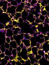
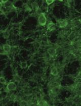
In vivo brain imaging, using a combination of genetically encoded Ca2+ indicators and gradient refractive index (GRIN) lens, is a transformative technology that has become an increasingly potent research tool over the last decade. It allows direct visualisation of the dynamic cellular activity of deep brain neurons and glia in conscious animals and avoids the effect of anaesthesia on the network. This technique provides a step change in brain imaging where fibre photometry combines the whole ensemble of cellular activity, and multiphoton microscopy is limited to imaging superficial brain structures either under anaesthesia or in head-restrained conditions. We have refined the intravital imaging technique to image deep brain nuclei in the ventral medulla oblongata, one of the most difficult brain structures to image due to the movement of brainstem structures outside the cranial cavity during free behaviour (head and neck movement), whose targeting requires GRIN lens insertion through the cerebellum—a key structure for balance and movement. Our protocol refines the implantation method of GRIN lenses, giving the best possible approach to image deep extracranial brainstem structures in awake rodents with improved cell rejection/acceptance criteria during analysis. We have recently reported this method for imaging the activity of retrotrapezoid nucleus and raphe neurons to outline their chemosensitive characteristics. This revised method paves the way to image challenging brainstem structures to investigate their role in complex behaviours such as breathing, circulation, sleep, digestion, and swallowing, and could be extended to image and study the role of cerebellum in balance, movement, motor learning, and beyond.Key features• We developed a protocol that allows imaging from brainstem neurons and glia in freely behaving rodents.• Our refined method of GRIN lenses implantation and cell sorting approach gives the highest number of cells with the least postoperative complications.• The revised deep brainstem imaging method paves way to understand complex behaviours such as cardiorespiratory regulation, sleep, swallowing, and digestion.• Our protocol can be implemented to image cerebellar structures to understand their role in key functions such as balance, movement, motor learning, and more.Graphical overview