Past Issue in 2024

Volume: 14, Issue: 2

left

Dec

20

Dec

5

Nov

20

Nov

5

Oct

20

Oct

5

Sep

20

Sep

5

Aug

20

Aug

5

Jul

20

Jul

5

Jun

20

Jun

5

May

20

May

5

Apr

20

Apr

5

Mar

20

Mar

5

Feb

20

Feb

5

Jan

20

Jan

5

right

Biochemistry

Quantitative Determination of Cholesterol Hydroxylase Specificities by GC–MS/MS in Living Mammalian Cells

Quantitative Determination of Cholesterol Hydroxylase Specificities by GC–MS/MS in Living Mammalian Cells

HS Hodaka Saito
MN Mizuki Nishimura
RS Ryuichiro Sato
YY Yoshio Yamauchi
1538 Views
Jan 20, 2024
Cholesterol is oxygenated by a variety of cholesterol hydroxylases; oxysterols play diverse important roles in physiological and pathophysiological conditions by regulating several transcription factors and cell-surface receptors. Each oxysterol has distinct and overlapping functions. The expression of cholesterol hydroxylases is highly regulated, but their physiological and pathophysiological roles are not fully understood. Although the activity of cholesterol hydroxylases has been characterized biochemically using radiolabeled cholesterol as the substrate, their specificities remain to be comprehensively determined quantitatively. To better understand their roles, a highly sensitive method to measure the amount of various oxysterols synthesized by cholesterol hydroxylases in living mammalian cells is required. Our method described here, with gas chromatography coupled with tandem mass spectrometry (GC–MS/MS), can quantitatively determine a series of oxysterols endogenously synthesized by forced expression of one of the four major cholesterol hydroxylases—CH25H, CYP7A1, CYP27A1, and CYP46A1—or induction of CH25H expression by a physiological stimulus. This protocol can also simultaneously measure the amount of intermediate sterols, which serve as markers for cellular cholesterol synthesis activity.Key features• Allows measuring the amount of a variety of oxysterols synthesized endogenously by cholesterol hydroxylases using GC–MS/MS.• Comprehensive and quantitative analysis of cholesterol hydroxylase specificities in living mammalian cells.• Simultaneous quantification of intermediate sterols to assess cholesterol synthesis activity.Graphical overview

Biological Engineering

The Development of an Advanced Model for Multilayer Human Skin Reconstruction In Vivo

The Development of an Advanced Model for Multilayer Human Skin Reconstruction In Vivo

MP Maryna Pavlova
VB Velmurugan Balaiya
JF Jocelyn C. Flores
MF Michael Ferreyros
KB Katie Bush
AH Amy Hopkin
IK Igor Kogut
DR Dennis R. Roop
GB Ganna Bilousova
1765 Views
Jan 20, 2024
Human skin reconstruction on immune-deficient mice has become indispensable for in vivo studies performed in basic research and translational laboratories. Further advancements in making sustainable, prolonged skin equivalents to study new therapeutic interventions rely on reproducible models utilizing patient-derived cells and natural three-dimensional culture conditions mimicking the structure of living skin. Here, we present a novel step-by-step protocol for grafting human skin cells onto immunocompromised mice that requires low starting cell numbers, which is essential when primary patient cells are limited for modeling skin conditions. The core elements of our method are the sequential transplantation of fibroblasts followed by keratinocytes seeded into a fibrin-based hydrogel in a silicone chamber. We optimized the fibrin gel formulation, timing for gel polymerization in vivo, cell culture conditions, and seeding density to make a robust and efficient grafting protocol. Using this approach, we can successfully engraft as few as 1.0 × 10⁶ fresh and 2.0 × 10⁶ frozen-then-thawed keratinocytes per 1.4 cm2 of the wound area. Additionally, it was concluded that a successful layer-by-layer engraftment of skin cells in vivo could be obtained without labor-intensive and costly methodologies such as bioprinting or engineering complex skin equivalents.Key features• Expands upon the conventional skin chamber assay method (Wang et al., 2000) to generate high-quality skin grafts using a minimal number of cultured skin cells.• The proposed approach allows the use of frozen-then-thawed keratinocytes and fibroblasts in surgical procedures.• This system holds promise for evaluating the functionality of skin cells derived from induced pluripotent stem cells and replicating various skin phenotypes.• The entire process, from thawing skin cells to establishing the graft, requires 54 days.Graphical overviewGeneration of a human skin equivalent on an immunodeficient mouse using a fibrin-based grafting system. A schematic of the protocol is shown. Cultured keratinocytes and fibroblasts resuspended in a fibrin-based gel are delivered as layers into a silicon chamber inserted underneath the skin of an immunocompromised mouse. First, a fibrin gel containing encapsulated fibroblasts (up to 2 × 10⁶ per 1.4 cm2 wound) is delivered into the chamber and allowed to solidify for 15 minutes. Second, a fibrin gel containing 1.0–2.0 × 10⁶ keratinocytes is applied on top of the fibroblast layer. On day 7 post-grafting, the chamber is removed, and the wound with the graft is allowed to heal for 4–5 weeks. During healing, a scab forms and eventually falls off. By day 54, the graft is fully established.
Mesenteric Parametrial Fat Pad Surgery for in vivo Implantation of Hepatocytes in Nude Mice

Mesenteric Parametrial Fat Pad Surgery for in vivo Implantation of Hepatocytes in Nude Mice

SS Saloni Sinha
DN Duc-Huy T. Nguyen
NH Nora Hassan
QA Qazi Ali
JS Jason Sethiadi
ST Sergey Tsoy
RS Robert E. Schwartz
1200 Views
Jan 20, 2024
Cell-based liver therapies utilizing functionally stabilized engineered hepatic tissue hold promise in improving host liver functions and are emerging as a potential alternative to whole-organ transplantation. Owing to the ability to accommodate a large ex vivo engineered hepatocyte mass and dense vascularization, the mesenteric parametrial fat pad in female nude mice forms an ideal anatomic microenvironment for ectopic hepatocyte transplantation. However, the lack of any reported protocol detailing the presurgical preparation and construction of the engineered hepatic hydrogel, fat pad surgery, and postsurgical care and bioluminescence imaging to confirm in vivo hepatocyte implantation makes it challenging to reliably perform and test engraftment and integration with the host. In this report, we provide a step-by-step protocol for in vivo hepatocyte implantation, including preparation of hepatic tissue for implantation, the surgery process, and bioluminescence imaging to assess survival of functional hepatocytes. This will be a valuable protocol for researchers in the fields of tissue engineering, transplantation, and regenerative medicine.Key features• Primary human hepatocytes transduced ex vivo with a lentiviral vector carrying firefly luciferase are surgically implanted onto the fat pad.• Bioluminescence helps monitor survival of transplanted hepatic tissue over time.• Applicable for assessment of graft survival, graft-host integration, and liver regeneration.Graphical overview

Developmental Biology

Live Imaging and Analysis of Meiotic Cytokinesis in Drosophila Testes

Live Imaging and Analysis of Meiotic Cytokinesis in Drosophila Testes

GK Govind Kunduri
JA Jairaj K. Acharya
1880 Views
Jan 20, 2024
All living organisms require the division of a cell into daughter cells for their growth and maintenance. During cell division, both genetic and cytoplasmic contents are equally distributed between the two daughter cells. At the end of cell division, cytoplasmic contents and the plasma membrane are physically separated between the two daughter cells via a process known as cytokinesis. Hundreds of proteins and lipids involved in the cytokinetic process have been identified; however, much less is known about the mechanisms by which these molecules regulate cytokinesis, being therefore an intense area of current research. Male meiotic cytokinesis in Drosophila melanogaster testes has been shown to be an excellent model to study cytokinesis in vivo. Currently, several excellent protocols are available to study cytokinesis in Drosophila testes. However, improved methods are required to study cytokinesis under in vitro and ex vivo conditions. Here, we demonstrate a simple method to perform live imaging on individual spermatocyte cysts isolated from adult testes. We evaluate amenability of this in vitro method for treatment with pharmacological agents. We show that cytokinesis is strongly inhibited upon treatment with Dynasore, a dynamin inhibitor known to block clathrin-mediated endocytosis. In addition, we also demonstrate an ex vivo method to perform live imaging on whole mount adult testes on gas permeable membrane chambers. We believe the protocols described here are valuable tools to study cytokinetic mechanisms under various genetic and treatment conditions.Key features• In vitro method to study male meiotic cytokinesis in dissected spermatocyte cysts.• In vitro method allows acute treatment with various pharmacological agents to study cytokinesis.• Ex vivo method to image male meiosis cytokinesis in intact adult testes.• Requires 15–60 min to set up and could be imaged up to 6–12 h.Graphical overviewIn vitro and ex vivo live imaging of male meiotic cytokinesis in adult Drosophila testes

Molecular Biology

Methodology to Create Auxin-Inducible Degron Tagging System to Control Expression of a Target Protein in Mammalian Cell Lines

Methodology to Create Auxin-Inducible Degron Tagging System to Control Expression of a Target Protein in Mammalian Cell Lines

AR Amit Rahi
DS Deepika K. Sodhi
CM Christine B. Magdongon
RS Rajina Shakya
DV Dileep Varma
2474 Views
Jan 20, 2024
The auxin-inducible degron (AID) system is a versatile tool in cell biology and genetics, enabling conditional protein regulation through auxin-induced degradation. Integrating CRISPR/Cas9 with AID expedites tagging and depletion of a required protein in human and mouse cells. The mechanism of AID involves interactions between receptors like TIR1 and the AID tag fused to the target protein. The presence of auxin triggers protein ubiquitination, leading to proteasome-mediated degradation. We have used AID to explore the mitotic functions of the replication licensing protein CDT1. Swift CDT1 degradation via AID upon auxin addition achieves precise mitotic inhibition, revealing defects in mitotic spindle structure and chromosome misalignment. Using live imaging, we found that mitosis-specific degradation of CDT1 delayed progression and chromosome mis-segregation. AID-mediated CDT1 inhibition surpasses siRNA-based methods, offering a robust approach to probe CDT1’s mitotic roles. The advantages of AID include targeted degradation and temporal control, facilitating rapid induction and reversal of degradation—contrasting siRNA’s delayed RNA degradation and protein turnover. In summary, the AID technique enhances precision, control, and efficiency in studying protein function and regulation across diverse cellular contexts. In this article, we provide a step-by-step methodology for generating an efficient AID-tagging system, keeping in mind the important considerations that need to be adopted to use it for investigating or characterizing protein function in a temporally controlled manner.Key features• The auxin-inducible degron (AID) system serves as a versatile tool, enabling conditional protein regulation through auxin-induced degradation in cell biology and genetics.• Integration of CRISPR/Cas9 knock-in technology with AID expedites the tagging and depletion of essential proteins in mammalian cells.• AID’s application extends to exploring the mitotic functions of the replication licensing protein CDT1, achieving precise mitotic inhibition and revealing spindle defects and chromosome misalignment.• The AID system and its diverse applications advance the understanding of protein function and cellular processes, contributing to the study of protein regulation and function.Graphical overview Cdt1–auxin-inducible degron (AID) tagging workflow. (A) Schematic of the cloned Cdt1 gRNA vector and the repair template generated to endogenously tag the Cdt1 genomic locus with YFP and AID at the C-terminal using CRISPR/CAS9-based genome editing. The two plasmids are transfected into DLD1-TIR1 stable cells, followed by sorting and scaling up of YFP-positive single cells. (B) The molecular mechanism of auxin-induced proteasome-mediated degradation of the target protein (CDT1) shown at the bottom of the figure is well worked out.

Neuroscience

Isolation and Enrichment of Major Primary Neuroglial Cells from Neonatal Mouse Brain

Isolation and Enrichment of Major Primary Neuroglial Cells from Neonatal Mouse Brain

SK Santosh Kumar Samal
MS Madhav Sharma
JD Jayasri Das Sarma
2882 Views
Jan 20, 2024
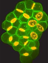
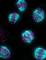
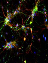
The central nervous system (CNS) relies on the complex interaction of neuroglial cells to carry out vital physiological functions. To comprehensively understand the structural and functional interplay between these neuroglial cells, it is essential to establish an appropriate in vitro system that can be utilized for thorough investigation. Traditional protocols for establishing primary neuronal and mixed glial cultures from prenatal mice or neural stem cells require sacrificing pregnant mice and have the drawback of yielding only specific types of cells. Our current protocol overcomes these drawbacks by utilizing the brain from day-0 pups to isolate CNS resident neuroglial cells including astrocytes, microglia, oligodendrocytes [oligodendrocyte precursor cells (OPCs) and differentiated oligodendrocytes], and meningeal fibroblasts, as well as hippocampal neurons, avoiding sacrificing pregnant mice, which makes this procedure efficient and cost effective. Furthermore, through this protocol, we aim to provide step-by-step instructions for isolating and establishing different primary neuroglial cells and their characterization using cell-specific markers. This study presents an opportunity to isolate, culture, and establish all major CNS resident cells individually. These cells can be utilized in various cell-based and biochemical assays to comprehensively investigate the cell-specific roles and behaviors of brain resident cells in a reductionist approach.Key features• Efficient isolation of major neuroglial cells like meningeal fibroblasts, neurons, astrocytes, oligodendrocytes, and microglia from a single day-0 neonatal mouse pup’s brain.• Circumvents the sacrifice of pregnant female mice.• Acts as a bridging experimental method between secondary cell lines and in vivo systems.• Isolated cells can be used for performing various cell-based and biochemical assays.Graphical overviewSteps for isolation of meningeal fibroblast and neuroglial cells from day 0 pups of mice (Created using BioRender.com)
Optical Modulation of the Blood-Brain Barrier for Glioblastoma Treatment

Optical Modulation of the Blood-Brain Barrier for Glioblastoma Treatment

QC Qi Cai
HF Hanwen Fan
XL Xiaoqing Li
MG Monica Giannotta
RB Robert Bachoo
ZQ Zhenpeng Qin
1503 Views
Jan 20, 2024
The blood–brain barrier (BBB) is a major obstacle to the diagnostics and treatment of many central nervous system (CNS) diseases. A prime example of this challenge is seen in glioblastoma (GBM), the most aggressive and malignant primary brain tumor. The BBB in brain tumors, or the blood–brain–tumor barrier (BBTB), prevents the efficient delivery of most therapeutics to brain tumors. Current strategies to overcome the BBB for therapeutic delivery, such as using hyperosmotic agents (mannitol), have impeded progress in clinical translation limited by the lack of spatial resolution, high incidences of complications, and potential for toxicity. Focused ultrasound combined with intravenously administered microbubbles enables the transient disruption of the BBB and has progressed to early-phase clinical trials. However, the poor survival with currently approved treatments for GBM highlights the compelling need to develop and validate treatment strategies as well as the screening for more potent anticancer drugs. In this protocol, we introduce an optical method to open the BBTB (OptoBBTB) for therapeutic delivery via ultrashort pulse laser stimulation of vascular targeting plasmonic gold nanoparticles (AuNPs). Specifically, the protocol includes the synthesis and characterization of vascular-targeting AuNPs and a detailed procedure of optoBBTB. We also report the downstream characterization of the drug delivery and tumor treatment efficacy after BBB modulation. Compared with other barrier modulation methods, our optical approach has advantages in high spatial resolution and minimally invasive access to tissues. Overall, optoBBTB allows for the delivery of a variety of therapeutics into the brain and will accelerate drug delivery and screening for CNS disease treatment.Key features• Pulsed laser excitation of vascular-targeting gold nanoparticles non-invasively and reversibly modulates the blood–brain barrier permeability.• OptoBBTB enhances drug delivery in clinically relevant glioblastoma models.• OptoBBTB has the potential for drug screening and evaluation for superficial brain tumor treatment.Graphical overview
High-throughput Analysis of Capillary Density in Skeletal Muscle Cross Sections

High-throughput Analysis of Capillary Density in Skeletal Muscle Cross Sections

TA Tooba Abbassi-Daloii
SM Sander D. Mallon
Se Salma el Abdellaoui
LV Lenard M. Voortman
VR Vered Raz
1362 Views
Jan 20, 2024
Capillary density in skeletal muscles is key to estimate exercise capacity in healthy individuals, athletes, and those with muscle-related pathologies. Here, we present a step-by-step, high-throughput semi-automated method for quantifying capillary density from whole human skeletal muscle cross-sections, in areas of the muscle occupied by myofibers. We provide a detailed protocol for immunofluorescence staining, image acquisition, processing, and quantification. Image processing is performed in ImageJ, and data analysis is conducted in R. The provided protocol allows high-throughput quantification of capillary density.Key features• This protocol builds upon the method and results described in Abbassi-Daloii et al. (2023b).• It includes step-by-step details on image acquisition and image processing of the entire muscle section.• It enables high-throughput and semi-automated image quantification of capillary density.• It provides a robust analysis for determining capillary density over the entire muscle cross section.Graphical overview

Correction

Correction Notice: Isolation and Culture of Murine Hepatic Stellate Cells

Correction Notice: Isolation and Culture of Murine Hepatic Stellate Cells

RM Rucha V. Modak
DZ Dietmar M. Zaiss
529 Views
Jan 20, 2024