Past Issue in 2023

Volume: 13, Issue: 6

left

Dec

20

Dec

5

Nov

20

Nov

5

Oct

20

Oct

5

Sep

20

Sep

5

Aug

20

Aug

5

Jul

20

Jul

5

Jun

20

Jun

5

May

20

May

5

Apr

20

Apr

5

Mar

20

Mar

5

Feb

20

Feb

5

Jan

20

Jan

5

right

Biochemistry

Protein Pull-down Assay Using HiBiT-tag-dependent Luciferase Activity Measurement

Protein Pull-down Assay Using HiBiT-tag-dependent Luciferase Activity Measurement

MA Masashi Arakawa
EM Eiji Morita
1815 Views
Mar 20, 2023
Co-immunoprecipitation or pull-down assays are frequently used to analyze protein–protein interactions. In these experiments, western blotting is commonly used to detect prey proteins. However, sensitivity and quantification problems remain in this detection system. Recently, the HiBiT-tag-dependent NanoLuc luciferase system was developed as a highly sensitive detection system for small amounts of proteins. In this report, we introduce the method of using HiBiT technology for the detection of prey protein in a pull-down assay. Using this protocol, we demonstrate the formation of a ternary complex consisting of Japanese encephalitis virus NS4B and two host factors, namely valosin-containing protein, and nuclear protein localization protein 4, which is a critical biological event during flavivirus replication in cells.
Quantification of Ethylene Production in Leaf and Bud Tissue of the Subtropical Tree Crop Litchi (Litchi chinensis Sonn.) Using Gas Chromatography and Flame Ionization Detection

Quantification of Ethylene Production in Leaf and Bud Tissue of the Subtropical Tree Crop Litchi (Litchi chinensis Sonn.) Using Gas Chromatography and Flame Ionization Detection

RC Regina B. Cronje
AJ Arnoldus J. Jonker
1064 Views
Mar 20, 2023
Ethylene is an important plant hormone that is involved in the regulation of numerous processes in plant development. It also acts as a signaling molecule in response to biotic and abiotic stress conditions. Most studies have investigated ethylene evolution of harvested fruit or small herbaceous plants under controlled conditions, but only a few explored ethylene release in other plant tissues, such as leaves and buds, particularly those of subtropical crops. However, in light of increasing environmental challenges in agriculture (such as temperature extremes, droughts, floods, and high solar radiation), studies on these challenges and on potential chemical treatments for mitigating their effects on plant physiology have become more and more important. Thus, adequate techniques for the sampling and analysis of tree crops are needed to ensure accurate ethylene quantification. As part of a study on ethephon as a mitigating agent to improve litchi flowering under warm winter conditions, a protocol was developed for ethylene quantification in leaf and bud tissue of litchi following ethephon application, taking into account that these plant organs release lower ethylene concentrations than fruit. At sampling, leaves and buds were placed in glass vials of appropriate sizes for the respective plant tissue volumes and allowed to equilibrate for 10 min to release possible wound ethylene before incubating the samples for 3 h at ambient temperature. Thereafter, ethylene samples were aspirated from the vials and analyzed using a gas chromatograph with flame ionization detection, the TG-BOND Q+ column for separation of ethylene, and helium as the carrier gas. Quantification was achieved based on a standard curve derived from an external standard gas calibration with certified ethylene gas. This protocol will also be appropriate for other tree crops with similar plant materials as study foci. It will enable researchers to accurately determine ethylene production in various studies investigating the role of ethylene in general plant physiology or stress-induced plant responses following a range of treatment conditions.

Cancer Biology

Multiphoton Microscopy of FITC-labelled Fusobacterium nucleatum in a Mouse in vivo Model of Breast Cancer

Multiphoton Microscopy of FITC-labelled Fusobacterium nucleatum in a Mouse in vivo Model of Breast Cancer

LP Lishay Parhi
AS Amjad Shhadeh
NM Naseem Maalouf
TA Tamar Alon-Maimon
VS Viviana Scaiewicz
GB Gilad Bachrach
995 Views
Mar 20, 2023
Over the past decades, the main techniques used to visualize bacteria in tissue have improved but are still mainly based on indirect recognition of bacteria. Both microscopy and molecular recognition are being improved, but most procedures for bacteria detection in tissue involve extensive damage. Here, we describe a method to visualize bacteria in tissue slices from an in vivo model of breast cancer. This method allows examining trafficking and colonization of fluorescein-5-isothiocyanate (FITC)-stained bacteria in various tissues. The protocol provides direct visualization of fusobacterial colonization in breast cancer tissue. Rather than processing the tissue or confirming bacterial colonization by PCR or culture, the tissue is directly imaged using multiphoton microscopy. This direct visualization protocol causes no damage to the tissue; therefore, all structures can be identified. This method can be combined with others to co-visualize bacteria, types of cells, or protein expression in cells.

Cell Biology

Live Imaging of Phagoptosis in ex vivo Drosophila Testis

Live Imaging of Phagoptosis in ex vivo Drosophila Testis

DK Diana Kanaan
BS Boris Shklyar
LP Lilach Porat-Kuperstein
HT Hila Toledano
1285 Views
Mar 20, 2023
Phagoptosis is a prevalent type of programmed cell death (PCD) in adult tissues in which phagocytes non-autonomously eliminate viable cells. Therefore, phagoptosis can only be studied in the context of the entire tissue that includes both the phagocyte executors and the targeted cells doomed to die. Here, we describe an ex vivo live imaging protocol of Drosophila testis to study the dynamics of phagoptosis of germ cell progenitors that are spontaneously removed by neighboring cyst cells. Using this approach, we followed the pattern of exogenous fluorophores with endogenously expressed fluorescent proteins and revealed the sequence of events in germ cell phagoptosis. Although optimized for Drosophila testis, this easy-to-use protocol can be adapted to a wide variety of organisms, tissues, and probes, thus providing a reliable and simple means to study phagoptosis.

Developmental Biology

The Assessment of Beta Cell Mass during Gestational Life in the Mouse

The Assessment of Beta Cell Mass during Gestational Life in the Mouse

YK Yury Kryvalap
JC Jan Czyzyk
912 Views
Mar 20, 2023
Successful advancement in the treatment of diabetes mellitus is not possible without well-established methodology for beta cell mass calculation. Here, we offer the protocol to assess beta cell mass during embryonic development in the mouse. The described protocol has detailed steps on how to process extremely small embryonic pancreatic tissue, cut it on the cryostat, and stain tissue slides for microscopic analysis. The method does not require usage of confocal microscopy and takes advantage of enhanced automated image analysis with proprietary as well as open-source software packages.

Microbiology

Separating Inner and Outer Membranes of Escherichia coli by EDTA-free Sucrose Gradient Centrifugation

Separating Inner and Outer Membranes of Escherichia coli by EDTA-free Sucrose Gradient Centrifugation

SS Sheng Shu
WM Wei Mi
2106 Views
Mar 20, 2023
The envelope of Gram-negative bacteria consists of an outer membrane (OM), a peptidoglycan cell wall, and an inner membrane (IM). The OM and IM have different components of proteins and lipids. Separating the IM and OM is a basic biochemical procedure to further study lipids and membrane proteins in different locations. Sucrose gradient ultracentrifugation of lysozyme/EDTA-treated total membrane is the most widely used method to separate the IM and OM of Gram-negative bacteria. However, EDTA is often harmful to protein structure and function. Here, we describe a relatively simple sucrose gradient ultracentrifugation method to separate the IM and OM of Escherichia coli. In this method, the cells are broken by a high-pressure microfluidizer, and the total cell membrane is collected by ultracentrifugation. The IM and OM are then separated on a sucrose gradient. Because EDTA is not used, this method is beneficial for subsequent membrane protein purification and functional study.

Molecular Biology

Isolation of RNA from the Murine Colonic Tissue and qRT-PCR for Inflammatory Cytokines

Isolation of RNA from the Murine Colonic Tissue and qRT-PCR for Inflammatory Cytokines

IS Ibrahim M. Sayed
KI Kaili Inouye
SD Soumita Das
LA Laura Crotty Alexander
2795 Views
Mar 20, 2023
E-cigarette (E-cig) inhalation affects health status by modulating inflammation profiles in several organs, including the brain, lung, heart, and colon. The effect of flavored fourth-generation pod-based E-cigs (JUUL) on murine gut inflammation is modulated by both flavor and exposure period. Exposure of mice to JUUL mango and JUUL mint for one month upregulated inflammatory cytokines, particularly TNF-α, IL-6, and Cxcl-1 (IL-8). JUUL Mango effects were more prominent than those incurred by JUUL Mint after one month of exposure. However, JUUL Mango reduced the expression of colonic inflammatory cytokines after three months of exposure. In this protocol, we detail the process of RNA isolation from the mouse colon and the use of extracted RNA in profiling the inflammatory milieu. Efficient RNA extraction from the murine colon is the most important step in the evaluation of inflammatory transcripts in the colon.

Neuroscience

Fast Detection and Quantification of  Interictal Spikes and Seizures in a  Rodent Model of Epilepsy Using an  Automated Algorithm

Fast Detection and Quantification of Interictal Spikes and Seizures in a Rodent Model of Epilepsy Using an Automated Algorithm

KJ Kyle J. Jackson
SS Shaunik Sharma
GT Grant Tiarks
SR Saul Rodriguez
AB Alexander G. Bassuk
1391 Views
Mar 20, 2023
The electroencephalogram (EEG) is a powerful tool for analyzing neural activity in various neurological disorders, both in animals and in humans. This technology has enabled researchers to record the brain’s abrupt changes in electrical activity with high resolution, thus facilitating efforts to understand the brain’s response to internal and external stimuli. The EEG signal acquired from implanted electrodes can be used to precisely study the spiking patterns that occur during abnormal neural discharges. These patterns can be analyzed in conjunction with behavioral observations and serve as an important means for accurate assessment and quantification of behavioral and electrographic seizures. Numerous algorithms have been developed for the automated quantification of EEG data; however, many of these algorithms were developed with outdated programming languages and require robust computational hardware to run effectively. Additionally, some of these programs require substantial computation time, reducing the relative benefits of automation. Thus, we sought to develop an automated EEG algorithm that was programmed using a familiar programming language (MATLAB), and that could run efficiently without extensive computational demands. This algorithm was developed to quantify interictal spikes and seizures in mice that were subjected to traumatic brain injury. Although the algorithm was designed to be fully automated, it can be operated manually, and all the parameters for EEG activity detection can be easily modified for broad data analysis. Additionally, the algorithm is capable of processing months of lengthy EEG datasets in the order of minutes to hours, reducing both analysis time and errors introduced through manual-based processing.

Plant Science

A Miniature Sucrose Gradient for Polysome Profiling

A Miniature Sucrose Gradient for Polysome Profiling

Ansul Lokdarshi Ansul Lokdarshi
Av Albrecht G. von Arnim
3738 Views
Mar 20, 2023
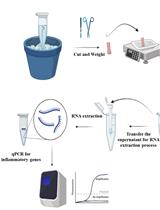
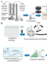
Polysome profiling by sucrose density gradient centrifugation is commonly used to study the overall degree of translation (messenger RNA to protein synthesis). Traditionally, the method begins with synthesis of a 5–10 mL sucrose gradient onto which 0.5–1 mL of cell extract is layered and centrifuged at high speed for 3–4 h in a floor-model ultracentrifuge. After centrifugation, the gradient solution is passed through an absorbance recorder to generate a polysome profile. Ten to twelve fractions (0.8–1 mL each) are collected for isolating different RNA and protein populations. The overall method is tedious and lengthy (6–9 h), requires access to a suitable ultracentrifuge rotor and centrifuge, and requires a substantial amount of tissue material, which can be a limiting factor. Moreover, there is often a dilemma over the quality of RNA and protein populations in the individual fractions due to the extended experiment times. To overcome these challenges, here we describe a miniature sucrose gradient for polysome profiling using Arabidopsis thaliana seedlings that takes ~1 h centrifugation time in a tabletop ultracentrifuge, reduced gradient synthesis time, and also less tissue material. The protocol described here can be easily adapted to a wide variety of organisms and polysome profiling of organelles, such as chloroplasts and mitochondria.Key Features• Mini sucrose gradient for polysome profiling that requires less than half the processing time vs. traditional methods.• Reduced starting tissue material and sample volume for sucrose gradients.• Feasibility of RNA and protein isolation from polysome fractions.• Protocol can be easily modified to a wide variety of organisms (and even polysome profiling of organelles, such as chloroplast and mitochondria).Graphical OverviewFigure 1. Graphical overview of polysome profiling using mini sucrose gradient. A. One milliliter each of 15% (w/v) and 50% (w/v) sucrose gradient solution is added to the individual chambers of the gradient maker. While mixing with a small magnetic stirrer in the 50% solution chamber, base station knob is turned to open position, allowing sucrose gradient solution to slowly flow through the outlet into a 2.2 mL gradient tube. After centrifugation at 50,000 rpm (213,626.2 × g) in a swinging bucket rotor for 70 min at 4 °C, the gradient tube is stored at 4 °C for the next steps. B. Cell extract from 12-day-old vertically grown Arabidopsis thaliana seedlings is centrifuged twice and 100 µL of supernatant is gently layered on the pre-made sucrose gradient from step A. After centrifugation as described in step A, polysome profile is obtained by feeding the gradient solution through an absorbance recorder (A254 nm). Eight (200 µL) fractions are collected for RNA and protein isolation.

Stem Cell

Determining Bone-forming Ability and Frequency of Skeletal Stem Cells by Kidney Capsule Transplantation and Limiting Dilution Assay

Determining Bone-forming Ability and Frequency of Skeletal Stem Cells by Kidney Capsule Transplantation and Limiting Dilution Assay

HU Hitoshi Uchida
TM Takamitsu Maruyama
WH Wei Hsu
903 Views
Mar 20, 2023
Adult stem cells not only maintain tissue homeostasis but are also critical for tissue regeneration during injury. Skeletal stem cells are multipotent stem cells that can even generate bones and cartilage upon transplantation to an ectopic site. This tissue generation process requires essential stem cell characteristics including self-renewal, engraftment, proliferation, and differentiation in the microenvironment. Our research team has successfully characterized and isolated skeletal stem cells (SSCs) from the cranial suture called suture stem cells (SuSCs), which are responsible for craniofacial bone development, homeostasis, and injury-induced repair. To assess their stemness features, we have demonstrated the use of kidney capsule transplantation for an in vivo clonal expansion study. The results show bone formation at a single-cell level, thus permitting a faithful assessment of stem cell numbers at the ectopic site. The sensitivity in assessing stem cell presence permits using kidney capsule transplantation to determine stem cell frequency by limiting dilution assay. Here, we described detailed protocols for kidney capsule transplantation and limiting dilution assay. These methods are extremely valuable both for the evaluation of skeletogenic ability and the determination of stem cell frequency.