Past Issue in 2022

Volume: 12, Issue: 20

left

Dec

20

Dec

5

Nov

20

Nov

5

Oct

20

Oct

5

Sep

20

Sep

5

Aug

20

Aug

5

Jul

20

Jul

5

Jun

20

Jun

5

May

20

May

5

Apr

20

Apr

5

Mar

20

Mar

5

Feb

20

Feb

5

Jan

20

Jan

5

right

Biochemistry

Staphylococcus aureus 30S Ribosomal Subunit Purification and Its Biochemical and Cryo-EM Analysis

Staphylococcus aureus 30S Ribosomal Subunit Purification and Its Biochemical and Cryo-EM Analysis

MB Margarita Belinite
IK Iskander Khusainov
SM Stefano Marzi
1957 Views
Oct 20, 2022
The ribosome is a complex cellular machinery whose solved structure allowed for an incredible leap in structural biology research. Different ions bind to the ribosome, stabilizing inter-subunit interfaces and structurally linking rRNAs, proteins, and ligands. Besides cations such as K+ and Mg2+, polyamines are known to stabilize the folding of RNA and overall structure. The bacterial ribosome is composed of a small (30S) subunit containing the decoding center and a large (50S) subunit devoted to peptide bond formation. We have previously shown that the small ribosomal subunit of Staphylococcus aureus is sensitive to changes in ionic conditions and polyamines concentration. In particular, its decoding center, where mRNA codons and tRNA anticodons interact, is prone to structural deformations in the absence of spermidine. Here, we report a detailed protocol for the purification of the intact and functional 30S, achieved through specific ionic conditions and the addition of spermidine. Using this protocol, we obtained the cryo-electron microscopy (cryo-EM) structure of the 30S–mRNA complex from S. aureus at 3.6 Å resolution. The 30S–mRNA complex formation was verified by a toeprinting assay. In this article, we also include a description of toeprinting and cryo-EM protocols. The described protocols can be further used to study the process of translation regulation.Graphical abstract:

Biological Engineering

A Novel Method of Inducible Directed Evolution to Evolve Complex Phenotypes

A Novel Method of Inducible Directed Evolution to Evolve Complex Phenotypes

IA Ibrahim S. Al’Abri
ZL Zidan Li
DH Daniel J. Haller
NC Nathan Crook
2532 Views
Oct 20, 2022
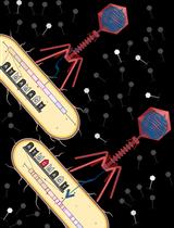
Directed evolution is a powerful technique for identifying beneficial mutations in defined DNA sequences with the goal of improving desired phenotypes. Recent methodological advances have made the evolution of short DNA sequences quick and easy. However, the evolution of DNA sequences >5kb in length, notably gene clusters, is still a challenge for most existing methods. Since many important microbial phenotypes are encoded by multigene pathways, they are usually improved via adaptive laboratory evolution (ALE), which while straightforward to implement can suffer from off-target and hitchhiker mutations that can adversely affect the fitness of the evolved strain. We have therefore developed a new directed evolution method (Inducible Directed Evolution, IDE) that combines the specificity and throughput of recent continuous directed evolution methods with the ease of ALE. Here, we present detailed methods for operating Inducible Directed Evolution (IDE), which enables long (up to 85kb) DNA sequences to be mutated in a high throughput manner via a simple series of incubation steps. In IDE, an intracellular mutagenesis plasmid (MP) tunably mutagenizes the pathway of interest, located on the phagemid (PM). MP contains a mutagenic operon (danQ926, dam, seqA, emrR, ugi, and cda1) that can be expressed via the addition of a chemical inducer. Expression of the mutagenic operon during a cell cycle represses DNA repair mechanisms such as proofreading, translesion synthesis, mismatch repair, and base excision and selection, which leads to a higher mutation rate. Induction of the P1 lytic cycle results in packaging of the mutagenized phagemid, and the pathway-bearing phage particles infect naïve cells, generating a mutant library that can be screened or selected for improved variants. Successive rounds of IDE enable optimization of complex phenotypes encoded by large pathways (as of this writing up to 36 kb), without requiring inefficient transformation steps. Additionally, IDE avoids off-target genomic mutations and enables decoupling of mutagenesis and screening steps, establishing it as a powerful tool for optimizing complex phenotypes in E. coli.Graphical abstract: Figure 1. Overview of Inducible Directed Evolution (IDE). Pathways of interest are cloned into a P1 phagemid (PM) backbone and transformed into a strain of E. coli containing MP (diversification strain). The mutagenesis plasmid is induced to generate mutations. Phage lysate is produced and used to infect a strain that expresses the phenotype of interest (screening/selection strain). The resulting strain library is screened to identify those with improved properties. Narrowed-down libraries can then go through another IDE cycle by infecting a fresh diversification strain.

Biophysics

Single-molecule Force Spectroscopy on Biomembrane Force Probe to Characterize Force-dependent Bond Lifetimes of Receptor–ligand Interactions on Living Cells

Single-molecule Force Spectroscopy on Biomembrane Force Probe to Characterize Force-dependent Bond Lifetimes of Receptor–ligand Interactions on Living Cells

TZ Tongtong Zhang
CA Chenyi An
WH Wei Hu
WC Wei Chen
1857 Views
Oct 20, 2022
The transmembrane receptor–ligand interactions play a vital role in the physiological and pathological processes of living cells, such as immune cell activation, neural synapse formation, or viral invasion into host cells. Mounting evidence suggests that these processes involve mechanosensing and mechanotransduction, which are directly mediated by the force-dependent transmembrane receptor–ligand interactions. Some single-molecule force spectroscopy techniques have been applied to investigate force-dependent kinetics of receptor–ligand interactions. Among these, the biomembrane force probe (BFP), a unique and powerful technique, can quantitatively and accurately determine the force-dependent parameters of transmembrane receptor–ligand interactions at the single-molecule level on living cells. The stiffness, spatial resolution, force, and bond lifetime range of BFP are 0.1–3 pN/nm, 2–3 nm, 1–103 pN, and 5 × 10⁻⁴–200 s, respectively. Therefore, this technique is very suitable for studying transient and weak interactions between transmembrane receptors and their ligands. Here, we share in detail the in situ characterization of the single-molecule force-dependent bond lifetime of transmembrane receptor–ligand interactions, based on a force-clamp assay with BFP.
Single Protein Detection and Imaging with Evanescent Scattering Microscopy

Single Protein Detection and Imaging with Evanescent Scattering Microscopy

PZ Pengfei Zhang
LZ Lei Zhou
RW Rui Wang
XZ Xinyu Zhou
JJ Jiapei Jiang
ZW Zijian Wan
SW Shaopeng Wang
1372 Views
Oct 20, 2022
Single-molecule measurements provide statistical distributions of molecular properties, in addition to the ensemble averages. Evanescent detection approaches have been widely used for single-molecule detection because the evanescent field can significantly enhance the light-analyte interaction and reduce the background noise. However, current evanescent single-molecule detection systems mostly require specially designed sensing components. Here, we show that single proteins can be imaged on a plain cover glass surface by detecting the evanescent waves scattered by the target molecules. This allows us to quantify the protein–antibody interactions at the single-molecule level. This protocol describes a label-free single-molecule imaging approach with conventional consumables and may pave the road for detecting single molecules with commercial optical microscopy.

Cancer Biology

Analysis of the Ubiquitination and Phosphorylation of Vangl Proteins

Analysis of the Ubiquitination and Phosphorylation of Vangl Proteins

DF Di Feng
ZH Ziwei He
BG Bo Gao
2750 Views
Oct 20, 2022
The core planar cell polarity (PCP) protein Vang/Vangl, including Vangl1 and Vangl2 in vertebrates, is indispensable during development. Our previous studies showed that the activity of Vangl is tightly controlled by two important posttranslational modifications, ubiquitination and phosphorylation. Vangl is ubiquitinated through an endoplasmic reticulum-associated degradation (ERAD) pathway and is phosphorylated by casein kinase 1 (CK1) in response to Wnt. Here, we present step-by-step procedures to analyze Vangl ubiquitination and phosphorylation, including cell culture, transfection, sample preparation, and signal detection, as well as the use of newly available phospho-specific antibodies to detect Wnt-induced Vangl2 phosphorylation. The protocol described here can be applicable to the analysis of posttranslational modifications of other membrane proteins.

Cell Biology

Measuring Intracellular H2O2 in Intact Human Cells Using the Genetically Encoded Fluorescent Sensor HyPer7

Measuring Intracellular H2O2 in Intact Human Cells Using the Genetically Encoded Fluorescent Sensor HyPer7

LJ Lianne J. H. C. Jacobs
MH Michaela N. Hoehne
JR Jan Riemer
2569 Views
Oct 20, 2022
Depending on its local concentration, hydrogen peroxide (H2O2) can serve as a cellular signaling molecule but can also cause damage to biomolecules. The levels of H2O2 are influenced by the activity of its generator sites, local antioxidative systems, and the metabolic state of the cell. To study and understand the role of H2O2 in cellular signaling, it is crucial to assess its dynamics with high spatiotemporal resolution. Measuring these subcellular H2O2 dynamics has been challenging. However, with the introduction of the super sensitive pH-independent genetically encoded fluorescent H2O2 sensor HyPer7, many limitations of previous measurement approaches could be overcome. Here, we describe a method to measure local H2O2 dynamics in intact human cells, utilizing the HyPer7 sensor in combination with a microscopic multi-mode microplate reader. Graphical abstract: Overview of HyPer7 sensor function and measurement results.

Developmental Biology

First-line Evaluation of Sperm Parameters in Mice (Mus musculus)

First-line Evaluation of Sperm Parameters in Mice (Mus musculus)

GM Guillaume Martinez
2403 Views
Oct 20, 2022
Infertility has become a major public health problem, with a male factor involved in about half the cases. Mice are the most widely used animal model in reproductive biology research laboratories, but changes in sperm parameters in mice can be subtle and, in the absence of official guidelines, it is important that analyses are carried out in a strict and reproductive manner. This protocol successively details the different steps required to obtain spermatozoa under good conditions, the measurement of sperm motility using a Computer Assisted Sperm Analysis System (CASA) device, the calculation of sperm concentration in the epididymides using a sperm counting cell, and the examination of sperm morphology. The combination of these assays provides an overview of the basic sperm parameters in mice. This is both a diagnostic and a decision-making tool for researchers to orient their scientific strategy according to the observed abnormalities.

Microbiology

Negative Staining Transmission Electron Microscopy of HIV Viral Particles Permeabilized with PFO and Capsid Stabilized with IP6

Negative Staining Transmission Electron Microscopy of HIV Viral Particles Permeabilized with PFO and Capsid Stabilized with IP6

DL Derrick Lau
CM Chantal L. Márquez
MP Michael W. Parker
TB Till Böcking
1636 Views
Oct 20, 2022
The human immunodeficiency virus 1 (HIV-1) consists of a viral membrane surrounding the conical capsid. The capsid is a protein container assembled from approximately 1,500 copies of the viral capsid protein (CA), functioning as a reaction and transport chamber for the viral genome after cell entry. Transmission electron microscopy (TEM) is a widely used technique for characterizing the ultrastructure of isolated viral capsids after removal of the viral membrane, which otherwise hinders negative staining of structures inside the viral particle for TEM. Here, we provide a protocol to permeabilize the membrane of HIV-1 particles using a pore-forming toxin for negative staining of capsids, which are stabilized with inositol hexakisphosphate to prevent premature capsid disassembly. This approach revealed the pleomorphic nature of capsids with a partially intact membrane surrounding them. The permeabilization strategy using pore-forming toxins can be readily applied to visualize the internal architecture of other enveloped viruses using TEM.Graphical abstract:

Neuroscience

MiniSOG2-mediated Specific Photoablation of Motor Neurons in Ascidian Embryos

MiniSOG2-mediated Specific Photoablation of Motor Neurons in Ascidian Embryos

MU Madoka K. Utsumi
TA Taichi Akahoshi
KO Kotaro Oka
KH Kohji Hotta
1358 Views
Oct 20, 2022
When understanding the neuronal function of a specific neural circuit, single-cell level photoablation of a targeted cell is one of the useful experimental approaches. This protocol describes a method to photoablate specific motor neurons via the mini singlet oxygen generator (miniSOG2), a light–oxygen–voltage (LOV)-based optogenetic tool used for ablating targeted cells in arbitrary areas. MiniSOG2 could induce the cell death pathway by generating reactive oxygen species (ROS) upon blue light illumination. Photoablation of a specific cell using the miniSOG2 was performed to show that, in Ciona intestinalis type A (Ciona robusta), a single pair of motor neurons, MN2/A10.64, is necessary to drive their tail muscle contraction. The membrane targeted miniSOG2 combined with neuron-specific promoter (pSP-Neurog::miniSOG2-CAAX) was electroplated into the Ciona egg and transiently expressed at specific neurons of the embryo. MN2 labeled with pSP-Neurog:mCherry-CAAX was irradiated using a 440-nm laser from the lateral side for 10 min to ablate its neural function. The behavior of the embryo before and after the irradiation was recorded with a high-speed camera.Graphical abstract:

Plant Science

Measurement of Ascorbate Peroxidase Activity in Sorghum

Measurement of Ascorbate Peroxidase Activity in Sorghum

PK Praveen Kumar
2931 Views
Oct 20, 2022
The ascorbate peroxidase (APX) is a widely distributed antioxidant enzyme. It differs from catalase and other peroxidases in that it scavenges/reduces reactive oxygen species (ROS) such as hydrogen peroxide (H2O2) to water using reduced ascorbate as the electron donor. It is advantageous over other similar antioxidant enzymes in scavenging ROS since ascorbate may react with superoxide, singlet oxygen, and hydroxyl radical, in addition to reacting with H2O2. The estimation of its activity is helpful to analyze the level of oxidative stress in living systems under stressful conditions. The present protocol was performed to analyze the impact of heavy metal chromium (Cr) toxicity on sorghum plants in the form of APX enzyme activity under the application of glycine betaine (GB) and arbuscular mycorrhizal fungi (AMF) as stress ameliorators. Plant defense strategies against heavy metals toxicity involve the utilization of APX and the instigation of AMF symbiotic system, as well as their possible collaboration with one another or with the plant antioxidant system; this has been examined and discussed in literature. In this protocol, an increased APX activity was observed on underlying functions and detoxification capabilities of GB and AMF that are typically used by plants to enhance tolerance to Cr toxicity.Graphical abstract: Flow chart of standardized or calibrated enzyme assay with leaf samples of sorghum