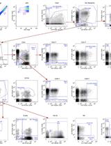
Cytokine-Stimulated Phosphoflow of Whole Blood Using CyTOF Mass Cytometry
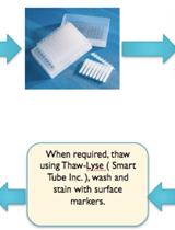
The ability to assess the function of a range of cytokine, antigen receptor, and Toll-like receptor (TLR) signaling pathways in a range of immune cells could provide a kind of fingerprint of the state of the human immune system. The mass cytometry or CyTOF, platform allows for the parallel application of about 40 labeled antibodies to a single sample, creating the possibility to read out many cell types and signaling pathways in a single small blood sample. We developed such a mass cytometry panel, consisting of 22 antibodies to cell surface lineage markers and 8 antibodies to phospho-specific epitopes of signaling proteins. These antibodies were chosen to discriminate all major white blood cell lineages, to a level of detail that includes subsets such as naïve, central memory, effector memory, and late effector CD4+ and CD8+T cells, naïve, transitional, and switched memory B cells, plasmablasts, myeloid and plasmacytoid dendritic cells, CD16+ and CD16+CD56+ NK cells, CD16+ and classical monocytes etc. 32 such cell subsets are defined in our standard gating scheme. The eight phospho-specific antibodies were chosen to represent major signaling nodes responsive to cytokine, TLR, and antigen receptor signaling. This antibody panel is used with 8 standard stimulation conditions (unstimulated, IFNa, IL-6, IL-7, IL-10, IL-21, LPS, PMA+ ionomycin), although other stimuli can be added. Comparison of healthy controls to subjects with immune deficiencies of unknown etiology may help elucidate the mechanisms of such deficiencies. Phosphorylation of tyrosine, serine, and threonine residues is critical for the control of protein activity involved in various cellular events. An assortment of kinases and phosphatases regulate intracellular protein phosphorylation in many different cell signaling pathways, such as T and B cell signaling, those regulating apoptosis, growth and cell cycle control, plus those involved with cytokine, chemokine, and stress responses. Phosphoflow assays combine phospho-specific antibodies with the power of flow cytometry to enhance phospho protein study. In our assay, peripheral blood mononuclear cells are stimulated by cytokines, fixed, surface-stained with a cocktail of antibodies labeled with MAXPAR (Brand Name) metal-chelating polymers and permeabilized with methanol. They are then stained with intracellular phospho-specific antibodies. We use a CyTOFTM mass cytometer to acquire the ICP-MS data. The current mass window selected is approximately AW 103-203, which includes the lanthanides used for most antibody labeling, as well as iridium and rhodium for DNA intercalators. Subsequent analysis of the dual count signal data using FlowJo software allows for cell types to be analyzed based on the dual count signal in each mass channel. The percentage of each cell type is determined and reported as a percent of the parent cell type. Median values are reported to quantitate the level of phosphorylation of each protein in response to stimulation. Comparing the level of phosphorylation between samples can offer insight to the status of the immune system. Whole blood stimulation is the closest to the in vivo condition and it allows for assessment of granulocyte population as well as lymphocytes and monocytes.