Technologies for RNA Detection

Technologies for RNA Detection

Ribonucleic acid (RNA) molecules are essential for multiple cellular processes, including protein synthesis, regulation of gene expression, and maintenance of chromosomal structure. To fulfill these key roles, RNAs interact directly and/or indirectly with other cellular biomolecules, such as proteins and additional RNAs, often forming complex structures. Identifying and characterizing the roles and structures of RNAs within these subcellular complexes and processes requires the application of highly advanced spatial and temporal techniques. Indeed, recent years have seen a plethora of single-cell-based RNA sequencing (RNA-seq) approaches, single-molecule RNA visualization methodologies, and artificial intelligence–based prediction models for RNA localization and structure. This special issue highlights some of the latest technological advances in RNA detection and visualization, RNA-seq methodologies, structural elucidation, and characterization of RNA–protein binding.


专辑委员会 实验方案目录
Enhancement of RNA Imaging Platforms by the Use of Peptide Nucleic Acid-Based Linkers

Enhancement of RNA Imaging Platforms by the Use of Peptide Nucleic Acid-Based Linkers

基于肽核酸连接臂提升 RNA 成像平台性能

0 Favorites
1701 Views
Jun 20, 2026

RNA imaging techniques enable researchers to monitor RNA localization, dynamics, and regulation in live or fixed cells. While the MS2-MCP system—comprising the MS2 RNA hairpin and its binding partner, the MS2 coat protein (MCP)—remains the most widely used approach, it relies on a tag containing multiple fluorescent proteins and has several limitations, including the potential to perturb RNA function due to the tag’s large mass. Alternative methods using small-molecule binding aptamers have been developed to address these challenges. This protocol describes the synthesis and characterization of RNA-targeting probes incorporating a peptide nucleic acid (PNA)-based linker within the cobalamin (Cbl)-based probe of the Riboglow platform. Characterization in vitro involves a fluorescence turn-on assay to determine binding affinity (KD) and selective 2′-hydroxyl acylation analyzed by primer extension (SHAPE) footprinting analysis to assess RNA-probe interactions at a single nucleotide resolution. To show the advancement of PNA probes in live cells, we present a detailed approach to perform both stress granule (SG) and U-body assays. By combining sequence-specific hybridization with structure-based recognition, our approach enhances probe affinity and specificity while minimizing disruption to native RNA behavior, offering a robust alternative to protein-based RNA imaging systems.

Amplification-Free Detection of Highly Structured RNA Molecules Using SCas12aV2

Amplification-Free Detection of Highly Structured RNA Molecules Using SCas12aV2

利用 SCas12aV2 实现高度结构化 RNA 分子的免扩增检测

0 Favorites
1150 Views
Jun 20, 2026

The CRISPR/Cas12a system has revolutionized molecular diagnostics; however, conventional Cas12a-based methods for RNA detection typically require transcription and pre-amplification steps. Our group has recently developed a diagnostic technique known as the SCas12a assay, which combines Cas12a with a split crRNA, achieving amplification-free detection of miRNA. However, this method still encounters challenges in accurately quantifying long RNA molecules with complex secondary structures. Here, we report an enhanced version termed SCas12aV2 (split-crRNA Cas12a version 2 system), which enables direct detection of RNA molecules without sequence limitation while demonstrating high specificity in single-nucleotide polymorphism (SNP) applications. We describe the general procedure for preparing the SCas12a system and its application in detecting RNA targets from clinical samples.

Visualizing Diverse RNA Functions in Living Cells With Spinach<sup>TM</sup> Family of Fluorogenic Aptamers

Visualizing Diverse RNA Functions in Living Cells With SpinachTM Family of Fluorogenic Aptamers

利用SpinachTM 系列荧光适配体可视化活细胞中多种RNA功能

0 Favorites
1558 Views
Jun 20, 2026

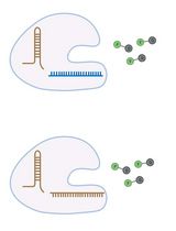
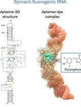
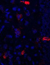
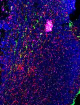
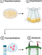
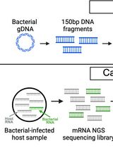
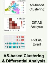
RNA is now recognized as a highly diverse and dynamic class of molecules whose localization, processing, and turnover are central to cell function and disease. Live-cell RNA imaging is therefore essential for linking RNA behavior to mechanism. Existing approaches include quenched hybridization probes that directly target endogenous transcripts but face delivery and sequestration issues, protein-recruitment tags such as MS2/PP7 that add large payloads and can perturb localization or decay, and CRISPR–dCas13 imaging that requires substantial protein cargo and careful control of background and off-target effects. Here, we present a protocol for live-cell RNA imaging using the SpinachTM family of fluorogenic RNA aptamers. The method details the design and cloning of SpinachTM-tagged RNA constructs, selection and handling of cognate small-molecule fluorophores, expression in mammalian cell lines, dye loading, and image acquisition on standard fluorescence microscopes, followed by quantitative analysis of localization and dynamics. We include controls to verify aptamer expression and signal specificity, guidance for multiplexing with related variants (e.g., Broccoli, Corn, Squash, Beetroot), and troubleshooting for dye permeability and signal optimization. Application examples illustrate use in tracking cellular delivery of mRNA therapeutics, monitoring transcription and decay in response to perturbations, and the forming of toxic RNA aggregates. Compared with prior methods, SpinachTM tags are compact, genetically encodable, and fluorogenic, providing high-contrast imaging in both the nucleus and cytoplasm with single-vector simplicity and multiplexing capability. The protocol standardizes key steps to improve robustness and reproducibility across cell types and laboratories.

Enhanced RNA-Seq Expression Profiling and Functional Enrichment in Non-model Organisms Using Custom Annotations

Enhanced RNA-Seq Expression Profiling and Functional Enrichment in Non-model Organisms Using Custom Annotations

基于自定义注释的非模式生物 RNA-seq 表达谱与功能富集分析优化

0 Favorites
747 Views
Jun 20, 2026

Functional enrichment analysis is essential for understanding the biological significance of differentially expressed genes. Commonly used tools such as g:Profiler, DAVID, and GOrilla are effective when applied to well-annotated model organisms. However, for non-model organisms, particularly for bacteria and other microorganisms, curated functional annotations are often scarce. In such cases, researchers often rely on homology-based approaches, using tools like BLAST to transfer annotations from closely related species. Although this strategy can yield some insights, it often introduces annotation errors and overlooks unique species-specific functions. To address this limitation, we present a user-friendly and adaptable method for creating custom annotation R packages using genomic data retrieved from NCBI. These packages can be directly imported as libraries into the R environment and are compatible with the clusterProfiler package, enabling effective gene ontology and pathway enrichment analysis. We demonstrate this approach by constructing an R annotation package for Mycobacterium tuberculosis H37Rv, as an example. The annotation package is then utilized to analyze differentially expressed genes from a subset of RNA-seq dataset (GSE292409), which investigates the transcriptional response of M. tuberculosis H37Rv to rifampicin treatment. The chosen dataset includes six samples, with three serving as untreated controls and three exposed to rifampicin for 1 h. Further, enrichment analysis was performed on genes to demonstrate changes in response to the treatment. This workflow provides a reliable and scalable solution for functional enrichment analysis in organisms with limited annotation resources. It also enhances the accuracy and biological relevance of gene expression interpretation in microbial genomics research.

High-Resolution Mapping of RNA–RNA Interactions Across the HIV-1 Genome With HicapR

High-Resolution Mapping of RNA–RNA Interactions Across the HIV-1 Genome With HicapR

基于 HiCapR 的 HIV-1 全基因组 RNA–RNA 相互作用高分辨率图谱构建

0 Favorites
267 Views
Jun 20, 2026

The genomes of RNA viruses can fold into dynamic structures that regulate their own infection and immune evasion processes. Proximity ligation methods (e.g., SPLASH) enable genome-wide interaction mapping but lack specificity when dealing with low-abundance targets in complex samples. Here, we describe HiCapR, a protocol integrating in vivo psoralen crosslinking, RNA fragmentation, proximity ligation, and hybridization capture to specifically enrich viral RNA–RNA interactions. Captured libraries are sequenced, and chimeric reads are analyzed via a customized computational pipeline to generate constrained secondary structures. HiCapR generates high-resolution RNA interaction maps for viral genomes. We applied it to resolve the in vivo structure of the complete HIV-1 RNA genome, identifying functional domains, homodimers, and long-range interactions. The protocol's robustness has been previously validated on the SARS-CoV-2 genome. HiCapR combines proximity ligation with targeted enrichment, providing an efficient and specific tool for studying RNA architecture in viruses, with broad applications in virology and antiviral development.

Electrophoretic Mobility Shift Assay (EMSA) for Assessing RNA–Protein Binding and Complex Formation Using Recombinant RNA-Binding Proteins and In Vitro–Transcribed RNA

Electrophoretic Mobility Shift Assay (EMSA) for Assessing RNA–Protein Binding and Complex Formation Using Recombinant RNA-Binding Proteins and In Vitro–Transcribed RNA

利用重组 RNA 结合蛋白和体外转录 RNA,通过 EMSA 评估 RNA–蛋白质结合及复合物形成

1 Favorites
597 Views
Jun 20, 2026

Evaluating RNA–protein interactions is key to understanding post-transcriptional gene regulation. Electrophoretic mobility shift assays (EMSAs) remain a widely used technique to study these interactions, revealing information about binding affinities and binding modalities, including cooperativity and complex formation. Here, we detail, in a step-by-step protocol, how to perform EMSAs. We describe how to generate, purify, and quantitate 32P-radiolabeled RNA by in vitro transcription, as well as the expression and purification of recombinant RNA-binding proteins in E. coli using ELAV as an example. We then describe how to set up binding reactions using serial dilutions in a microtiter plate format of recombinant ELAV and in vitro–transcribed RNA and how to perform EMSAs using native low-crosslinked acrylamide gels, with detailed graphically supported instructions and troubleshooting guides.

Using Combined Fluorescent In Situ Hybridization With Immunohistochemistry to Co-localize mRNA in Diverse Neuronal Cell Types

Using Combined Fluorescent In Situ Hybridization With Immunohistochemistry to Co-localize mRNA in Diverse Neuronal Cell Types

结合荧光原位杂交与免疫组织化学分析不同神经元细胞类型中的 mRNA 共定位

0 Favorites
271 Views
Jun 20, 2026

Understanding gene expression within defined neuronal populations is essential for dissecting the cellular and molecular diversity of the brain. mRNA assays provide a direct readout of gene expression, capturing transcriptional changes that may precede or occur independently of protein abundance, whereas protein assays reflect the cumulative effects of translation, modification, and degradation. Moreover, in histological analysis, immunohistochemical protein detection results in visually diffuse labeling, which makes it difficult to quantitatively assess levels and locations of expression at high resolution. Here, we present a protocol that allows for mRNA detection in single neuronal cell types with a high degree of sensitivity and anatomical resolution. This protocol combines fluorescent in situ hybridization (FISH) with immunohistochemistry (IHC) on the same tissue section. Briefly, FISH is carried out by ACDBio RNAscope® fluorescent in situ hybridization technology, which involves processing the tissue sections, followed by signal amplification. This involves target retrieval, probe hybridization, and signal enhancement. Then, the tissue section is processed for IHC, which involves blocking nonspecific sites and incubation with primary antibodies, followed by development of a fluorescent signal with secondary antibodies. Typically, visual mRNA detection with FISH can be seen as individual puncta, whereas targeting the protein with an antibody results in filled cells or processes. The variation in staining pattern allows for the quantification of distinct mRNA transcripts within different neuronal populations, which renders co-localization analyses easy and efficient.

Simultaneous Immunofluorescence-Based In Situ mRNA Expression and Protein Detection in Bone Marrow Biopsy Samples

Simultaneous Immunofluorescence-Based In Situ mRNA Expression and Protein Detection in Bone Marrow Biopsy Samples

基于免疫荧光的骨髓活检样本原位 mRNA 表达与蛋白同步检测

0 Favorites
162 Views
Jun 20, 2026

Fluorescence in situ hybridization (FISH) can be employed to study the expression and subcellular localization of nucleic acids by using labeled antisense strands that hybridize with the target RNA or DNA molecules. Likewise, immunofluorescence antibody staining (IF) takes advantage of the specific interaction between a fluorophore-labeled antibody and its corresponding antigen. This protocol reports the combination of RNA-FISH and IF antibody staining for simultaneous detection of both RNA transcripts and proteins of interest in routine formalin-fixed paraffin-embedded (FFPE) bone marrow biopsy samples. Herein, we provide a detailed description of the methodology that we have developed and optimized to study the spatial expression of two transcripts—TGFB1 and PDGFA1—in human hematopoietic (CD45+) and non-hematopoietic (CD271+) cells in the bone marrow of patients with acute lymphoblastic leukemia (ALL).

One-Step Affinity Purification of MarathonRT Reverse Transcriptase for RNA Sequencing Applications

One-Step Affinity Purification of MarathonRT Reverse Transcriptase for RNA Sequencing Applications

用于 RNA 测序的 MarathonRT 逆转录酶一步亲和纯化方法

0 Favorites
143 Views
Jun 20, 2026

Transfer RNAs (tRNAs) are important regulators of translation and cellular function. Several high-throughput sequencing methods have been developed to quantitatively analyze tRNA isoacceptors in cells. However, the strong secondary structures and extensive post-transcriptional modification of most tRNA molecules present significant challenges for many reverse transcriptases, negatively impacting sequencing library preparation and causing quantification biases. Currently, the field utilizes processive next-generation reverse transcriptases (ngRTs), such as Induro (New England Biolabs) and UltraMarathonRT (RNAConnect), to address these issues. Despite being used in multiple protocols, these commercial products face little competition and remain costly. However, non-commercial alternatives, such as the original MarathonRT (MRT), are available from gene repositories. MRT is a next-generation reverse transcriptase derived from the Eubacterium rectale group II intron maturase, which can read through RNA secondary structures and chemical modifications. Here, we present a simplified expression and purification protocol for producing highly active MRT that is stable over 1 year. This cost-effective protocol yields a heterogeneous protein preparation with no discernible competing enzymatic activities; it mitigates previously reported precipitation issues, saving one day of laboratory work and eliminating two chromatography-based purification steps. Moreover, the use of the resulting protein preparation has been verified in the mim-tRNAseq pipeline, where it was shown to perform equally to the commercial alternatives Induro and UltraMarathonRT. In addition, we have developed a simple and cost-effective assay for measuring the enzymatic activity of MRT, allowing for batch comparison.

Enriching Bacteria-Specific RNA From Host Samples Before NGS With Transcript-Capture

Enriching Bacteria-Specific RNA From Host Samples Before NGS With Transcript-Capture

基于转录本捕获的宿主样本细菌特异性 RNA 富集方法

0 Favorites
62 Views
Jun 20, 2026

Pathogen gene expression from host samples is often challenging to study due to low signal and high host RNA background. PCR probes have been recently used to hybridize and extract bacterial sequences from next-generation sequencing (NGS) libraries generated from in vitro and animal models of infection; however, these strategies require purchasing commercially synthesized probes that often do not capture the entire transcriptome. Transcript-capture sequencing is a novel capture approach for extracting RNA of a target bacterial species from samples in which there is substantial contamination by the host or other microbes. Biotinylated 150-base-pair DNA probes are generated in-house from bacterial DNA spanning the entire bacterial genome. Probes are hybridized to the cDNA of NGS sequencing libraries prepared from host samples to capture and enrich for bacterial-specific RNA reads before sequencing. This method results in a >200-fold increase in bacterial RNA reads from infected host samples (including in vitro, animal, and human samples) and generates complete bacterial transcriptomes with high gene coverage (>80%). Use of this protocol on infected host samples reveals a snapshot of bacterial activity during disease that may improve understanding of the physiological state of pathogens within their hosts.

Stepwise Protocol for Alternative Splicing Analysis in Single-Cell SMART-Seq2 RNA-Seq Data

Stepwise Protocol for Alternative Splicing Analysis in Single-Cell SMART-Seq2 RNA-Seq Data

单细胞 SMART-Seq2 RNA-seq 数据中可变剪接分析的分步流程

0 Favorites
82 Views
Jun 20, 2026

RNA alternative splicing (AS) is an essential process that expands transcriptomic and proteomic diversity in eukaryotic cells and contributes to cellular heterogeneity across physiological and pathological conditions in humans. With the advent of single-cell RNA sequencing (scRNA-seq), it has become possible to study AS at cellular resolution, although robust and standardized analytical workflows remain to be developed. Here, we present a stepwise protocol for analyzing AS in single cells from pediatric high-grade gliomas (pHGGs) harboring the histone H3.3 lysine 27-to-methionine (H3.3K27M) mutation using SMART-Seq2 scRNA-seq data. Starting from raw sequencing reads, the workflow includes read alignment, gene-level quantification, splice junction and intron quantification, and single-nucleotide variant-based mutation detection. Gene expression–based clustering and cell-type annotation are performed by using the Seurat R package. AS analysis in tumor cells is then conducted using the MARVEL R package in combination with customized scripts to calculate percent spliced-in (PSI) values, identify variable AS events, perform dimensionality reduction, cluster cells, conduct differential AS analysis, and visualize splicing patterns. This protocol provides a reproducible and comprehensive framework for dissecting AS dynamics at single-cell resolution. It is readily adaptable to other SMART-Seq2 datasets and facilitates systematic investigation of splicing heterogeneity in diverse biological contexts.

PrimeFlow<sup>TM</sup> Assay for Cell Type–Specific Co-detection of Transgene RNA and Protein in Mouse Spleens From Preclinical Studies

PrimeFlowTM Assay for Cell Type–Specific Co-detection of Transgene RNA and Protein in Mouse Spleens From Preclinical Studies

基于 PrimeFlowTM 的临床前小鼠脾脏样本转基因 RNA 与蛋白细胞类型特异性同步检测

0 Favorites
40 Views
Jun 20, 2026

The PrimeFlowTM assay is a flow cytometry–based method for the co-detection of RNAs and proteins in cells. When combined with cell characterization by immunophenotyping, PrimeFlowTM can be used to simultaneously detect RNA and proteins in a cell type–specific manner in complex heterogeneous samples, offering an advantage over bulk tissue analysis methods. Here, we describe the implementation of the PrimeFlowTM assay protocol for the detection of transgene mRNA and protein expression in spleen samples from mice treated in vivo with luciferase mRNA-lipid nanoparticles (LNPs). This protocol involves spleen tissue dissociation for cell isolation, followed by cell fixation and permeabilization to allow immunolabeling of intracellular luciferase protein. The immunophenotyping strategy is based on immunolabeling with mouse CD marker antibodies for the identification of T cells, B cells, monocytes, granulocytes/macrophages, NK cells, and non-hematopoietic cells. The RNAs of luciferase and a housekeeping gene, β-actin, are detected with sequence-specific probe sets by employing sequential oligonucleotide annealing steps and fluorescent labeling using a branched DNA (bDNA) technology. Samples are analyzed by flow cytometry. Based on our analysis, we conclude it is feasible to apply the PrimeFlowTM approach for evaluating successful drug targeting to the cell types of interest and any potential differences in the kinetics of RNA delivery and protein expression in various tissue cells, supporting the discovery and development of RNA therapeutics.

Editorial: Technologies for RNA Detection

Editorial: Technologies for RNA Detection

社论:RNA 检测技术

0 Favorites
30 Views
Jun 20, 2026
DiRT v2.0: An Optimized Pipeline for Detecting Dicistronic tRNA-mRNA Transcripts in Plants

DiRT v2.0: An Optimized Pipeline for Detecting Dicistronic tRNA-mRNA Transcripts in Plants

DiRT v2.0:用于检测植物双顺反子 tRNA-mRNA 转录本的优化流程

0 Favorites
38 Views
Jun 20, 2026

The canonical role of transfer RNAs (tRNAs) in protein synthesis has been extensively characterized; however, recent studies have uncovered novel functions for tRNA as a mediator of long-distance signaling in plants. Several studies have identified dicistronic tRNA-mRNA transcripts that contain a tRNA gene and an adjacent protein-coding gene (PCG) that are transcribed as a single unit. These transcripts are associated with RNA systemic mobility through the plant’s vascular tissues, potentially acting as non-cell-autonomous signaling messengers in coordinating development and stress responses. Here, we report a computational pipeline to detect dicistronic tRNA-mRNA transcripts from short-read next-generation RNA-sequencing datasets; to our knowledge, this is the only established pipeline for the systematic identification of such candidates in plants. The dicistronic RNA transcript version 2 (v2) described here improves on the earlier version DiRT v1 by expanding the repertoire of dicistronic transcripts detected to include tRNA-like structures (TLS) as well as functional tRNAs, which were already supported in the pipeline. The updated protocol also includes detection of dicistronic tRNA or TLS sequences within genomic features such as untranslated regions (UTRs). The accurate detection of both tRNAs and UTR-embedded tRNA-like sequences (TLS) is critical, as these RNA structures have been reported to function as mediators of long-distance RNA mobility. Furthermore, as NGS datasets are prone to sequencing artifacts and potential DNA contamination, we improved the pipeline’s statistical robustness by including read coverage of flanking intronic regions as a baseline control. To account for potential DNA contamination during RNA-seq library preparation, detected tRNA-mRNA transcripts are deemed as putatively dicistronic only if the coverage of their intergenic region is significantly higher (Student’s t-test, FDR Vitis spp. Ruggeri 140) and Arabidopsis thaliana datasets and validated in vitro using RT-PCR. We provide a fast and reliable method to detect dicistronic transcripts that can be applied to any short-read RNA-sequencing dataset, fast-tracking the functional characterization of these newly emerging transcripts.

RNA Detection Technologies: A Method‑Centric Guide to Principles and Reproducibility

RNA Detection Technologies: A Method‑Centric Guide to Principles and Reproducibility

RNA检测技术:原理解析与可重复性实践

0 Favorites
31 Views
Jun 20, 2026

RNA detection techniques have expanded into a diverse methodological landscape spanning hybridization, amplification, imaging, and sequencing. In this review, we provide a method‑centric synthesis of the major technologies that define this landscape, emphasizing how each method’s core principle, practical strengths, and sources of variability shape its reproducibility. Beginning with foundational approaches, we trace the development of isothermal amplification, quantitative and digital PCR, microarrays, single‑molecule imaging, multiplexed spatial methods, and amplification‑free digital quantification. We then examine the transformative impact of bulk, single‑cell, long‑read, direct‑RNA, and spatial transcriptomics, as well as CRISPR‑based detection and metabolic labeling for RNA dynamics. Across these technologies, we focus on reproducibility as a defining dimension of evaluation: mature methods benefit from established standards, whereas newer approaches remain pre‑standardization and require careful, experiment‑specific controls. Rigorous method selection must be guided by the biological question, required resolution, sample constraints, and the maturity of each method’s reproducibility framework. We conclude that RNA detection methods form interconnected methodological paths of problem‑solving rather than simple replacements.