- Protocols

- Articles and Issues
- For Authors
- About
- Become a Reviewer

Past Issue in 2026
Volume: 16, Issue: 1
Biophysics
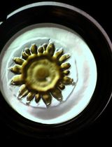
A Compact Schlieren Optics Device for Imaging Biological Samples
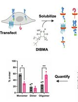
SiMPull-POP: Quantification of Membrane Protein Assembly via Single Molecule Photobleaching
Cell Biology
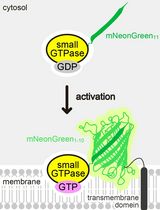
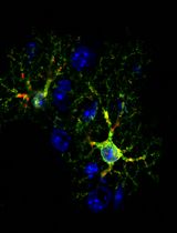
Detecting the Activation of Endogenous Small GTPases via Fluorescent Signals Utilizing a Split mNeonGreen: Small GTPase ActIvitY ANalyzing (SAIYAN) System
Generating ER-TRG and CA-ER-TRG Knock-in Mice and Quantitative In Vivo Imaging of ER-phagy
Microbiology
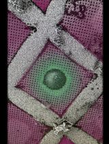
FLARE: A Flow Cytometry–Based Fluorescent Assay for Measuring HSV-1 Nuclear Egress


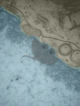
Reproducible Sample Preparation of Virus-Infected Cells for Cryo-FIB/ET Using Manual Plunge Freezing
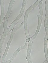
Creating Loss-of-Function Mutants of Neurospora crassa Using a Novel CRISPR/Cas9 System
Molecular Biology
Optimized Method for High-Quality Isolation of Single-Nuclei From Mosquito Fat Body for RNA Sequencing
Neuroscience
Simultaneous Non-Invasive Electrocardiogram and Respiration Rate Recordings in Head-Fixed Awake Mice
Plant Science
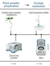
Quantification of Protochlorophyllide (Pchlide) Content in Arabidopsis Seedlings Using a High-Performance Liquid Chromatography (HPLC) System
Stem Cell
Efficient Fluorescent Labeling of Human Trophoblast Stem Cells via a CRISPR/Cas9-Mediated Knock-In Approach in a Safe Harbor Locus