
- Home
- Protocols

-

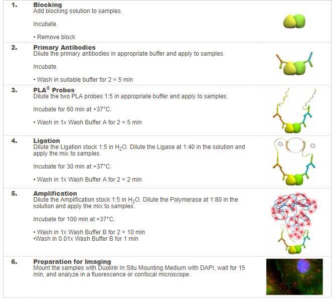
Proximity ligation assay
Last updated date: Dec 2, 2019 Views: 819 Forks: 0
*When remove coverslips from a solution, always pat liquid drops off onto a kimwipe before move into 24-well plate. Do all wash steps on a slow shaker.
Day 1
- Permeabilize and block the cells with blocking solution at RT for 15 min.
- blocking solution:
- 5 mL PBS w/o Ca
- 100 uL 10% NP40
- 100 uL donkey serum
- blocking solution:
- Stain the cells in a mix of the two primary antibodies at 4°C overnight.
- We do not use the diluent and blocking solution come with the kit, as we don’t know what they are inside, and our homemade blocking solution and antibody buffer work fine. However the diluent and blocking solution included in the kit should work very well in most of cases.
- Make antibody buffer (for both primary and secondary antibodies).
- 8 mL PBS w/o Ca
- 40 uL donkey serum
- Add primary antibodies at the concentration you determined when doing regular IF.
- The two primary antibodies are from different species (e.g. mouse and rabbit).
- Make a humidified chamber using a large Petri dish, paper towels, water, and parafilm.
- Place 20-40 uL drops of the antibody mix on the parafilm.
- Invert coverslips into drops of antibody mix so that the cells are face down in the antibody. Place at 4°C overnight.
- Make IF Wash Buffer (You will need 3 mL per coverslip)
- 50 mL PBS w/o Ca
- 50 uL Tween 20 (final conc. is 0.1%)
- 333 uL 30% BSA (final conc. is 0.2%)
- Make Wash Buffer A (You will need 9 mL per coverslip)
- 1000 mL miliQ water
- 8.8 g NaCl
- 1.2 g Tris base
- 0.5 mL Tween-20
- Adjust pH to 7.4 with HCl
- Filter. Store at 4 C. Bring to RT before use
- Make Wash Buffer B (You will need 4.5 mL per coverslip.)
- 1000 mL milliQ water
- 5.84 g NaCl
- 4.24 g Tris base
- 26.0 g Tris-HCl
- Adjust pH to 7.5 with HCl (or NaOH?)
- Filter. Store at 4°C. Bring to RT before use
Day 2
6. Place aliquots of IF wash buffer, Buffer A, and Buffer B at RT so that they will be RT when you use them.
7. Prepare PLA secondary probes (can do step 7 and step 8 at same time)
- You need a PLUS probe of one species and a MINUS probe of the other species, which match your primary antibodies (e.g. donkey anti-mouse-MINUS, and donkey anti-rabbit-PLUS).
- Measure out 20 uL antibody buffer for each coverslip. Per 20 uL, add 4 uL of anti-mouse and 4 uL anti-rabbit. Mix by flicking (do not vortex).
- Let probe mixture sit at room temperature for 20 min.
8. Wash off primary antibody.
- Wash coverslip in 24-well plate 5 mins, 3 times, with 1 mL IF wash buffer.
9. Incubate coverslips in PLUS/MINUS mix in a humidified chamber.
- The humidified chamber will have to go in the incubator, so keep this as sterile as possible. Make the chamber extra-moist!
- Transfer a coverslip from 24-well plate to the parafilm, cells facing up.
- Place a 20 uL drop of the PLUS/MINUS mix on the top of the coverslip. The kit and scientists who have done this before are all adamant about doing the reactions face-up, so don’t try face down.
- Repeat for all coverslips.
- Place the humidified chamber in the 37°C incubator for 1h. While waiting, proceed to next steps.
10. Prepare 24-well plate for next washing steps.
- Aspirate old IF wash buffer off.
- Aliquot 1.5 mL of wash buffer A into each well.
11. Dilute ligation stock.
- Make a master mix, where each coverslip will get a volume of 20 uL (19.5 ul):
- 15.5 uL nuclease-free water
- 4 uL ligation 5x stock (thaw on ice, make sure ATP precipitate dissolved)
- Keep at RT in the dark.
- DO NOT add ligase to the ligation dilution until immediately before addition to coverslips.
12. Wash Coverslips
- Wash coverslip in 24-well plate 5 mins, 3 times, with 1.5 mL wash buffer A.
13. Add ligase to the ligation dilution.
- For each coverslip, mix:
- 19.5 uL diluted ligation mix from step 11
- 0.5 uL ligase
- Vortex. Keep in dark at RT until ready to use (use quickly, though).
14. Place coverslips in ligase/ligation solution
- Move a coverslip to the parafilm of humidified chamber and place cells face up.
- Place a 20 uL drop of ligase/ligation solution on the coverslip.
- Repeat for other coverslips.
- Place in incubator for 30 min. While waiting, proceed to next steps.
15. Dilute amplification stock
- Make a master mix, where each coverslip will get a volume of 40 uL (39.5 ul, don’t try 20ul):
- 31.5 uL nuclease-free water
- 8 uL amplification 5X stock (thaw on ice in dark)
- DO NOT add the Polymerase until immediately before addition to coverslips.
- Keep in dark at RT (light sensitive reagent).
16. Prepare 24-well plate for next washing steps.
- Aspirate used wash buffer A off the wells.
- Aliquot 1.5 mL fresh wash buffer A into each well.
17. Wash coverslips
- When the 30mins ligation is over, move coverslips into the awaiting wash buffer A in the 24-well plate. Wash coverslip in the dark for 5 min, 3 times, with 1.5 mL IF wash buffer.
18. Add polymerase to amplification solution
- To the master mix from step 15, add 0.5 uL polymerase per 39.5ul. Vortex mix.
- Leave in dark at RT until ready to use (but use quickly).
19. Place coverslips in Amplification/Polymerase solution
- Move a coverslip to the humidified chamber, and place cells face up.
- Quickly place a 40 uL drop of amplification/polymerase solution onto the coverslip.
- Repeat for all coverslips.
- Place in 37°C incubator for 100 min. Proceed to next steps while waiting.
20. Make a 100x diluted Wash Buffer B
- Make a dilution so that per coverslip you have a volume of 1.5 mL:
- 50 uL Wash Buffer B
- 5 mL milliQ water
21. Prepare 24-well plate for next washing steps.
- Aspirate used wash buffer A off the wells.
- Aliquot 1.5 mL fresh, undiluted wash buffer B into each well.
22. Wash coverslips
- When the 100-min amplification is over,Wash coverslip in the dark for 10 min, 3 times, with 1.5 mL undiluted wash buffer B.
- Aspirate wash buffer B and replace with 1.5 mL 100x diluted wash buffer B. Place on shaker in the dark for 5 min.
23. Mount coverslips
- Pat liquid on coverslipd drops off onto a kimwipe. Place a minimal volume of Duolink In Situ Mounting Medium with DAPI on a clean slide (7-8ul). Cut tip, as mounting medium contains glycerol.
- Place the coverslip in the drop of mounting media. Repeat for other coverslip.
- Let sit for 15 minutes at RT in the dark.
- Gently vacuum around the edge of the coverslip to remove excess mounting medium.
- Seal edges with clear nail polish and let dry at RT in the dark.
- Store slides at -20°C in the dark.
- Xiang, Y K(2019). Proximity ligation assay. Bio-protocol Preprint. bio-protocol.org/prep82.
- Shen, A., Chen, D., Kaur, M., Bartels, P., Xu, B., Shi, Q., Martinez, J. M., Man, K. M., Nieves-Cintron, M., Hell, J. W., Navedo, M. F., Yu, X. and Xiang, Y. K.(2019). β-blockers augment L-type Ca2+ channel activity by targeting spatially restricted β2AR signaling in neurons. eLife. DOI: 10.7554/eLife.49464
Do you have any questions about this protocol?
Post your question to gather feedback from the community. We will also invite the authors of this article to respond.