
- Home
- Protocols

-

Surgical removal of iBAT
Last updated date: Apr 1, 2026 Views: 203 Forks: 0
Surgical Denervation of Interscapular Brown Adipose Tissue (iBAT)
Materials
- Anesthetic agent
- Syringe
- Injection needle
- Sterilized surgical instrument sets
- Straight forceps
- Curved forceps
- Scissors
- Note: To prevent contamination, it is recommended to use separate instrument sets for skin incision and for deeper tissue dissection. Instruments should be sterilized before use in different animals.
- Sutures: Pronova (blue, 4-0, 90 cm, taper-cut needle, 26 mm, strong curvature; 3975H)
- 5% Hibitane (chlorhexidine; dilute 1:30 before use)
- Electric shaver
- Hair removal can also be performed manually; however, shaving is recommended to minimize skin damage.
- Heating pad (optional, for maintaining body temperature during surgery)
Procedure
- After anesthesia, shave the surgical area using an electric shaver.
- Disinfect and sterilize the shaved area with diluted 5% Hibitane (chlorhexidine).
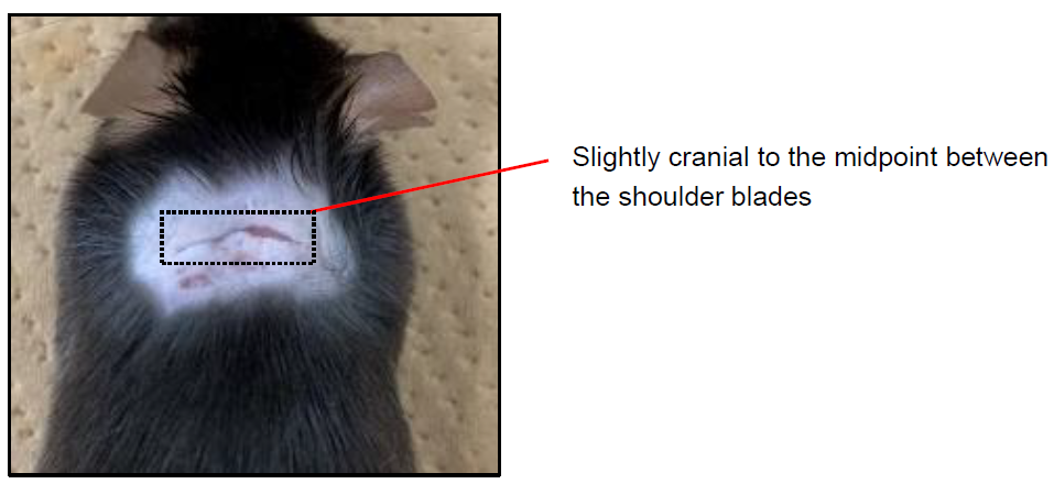
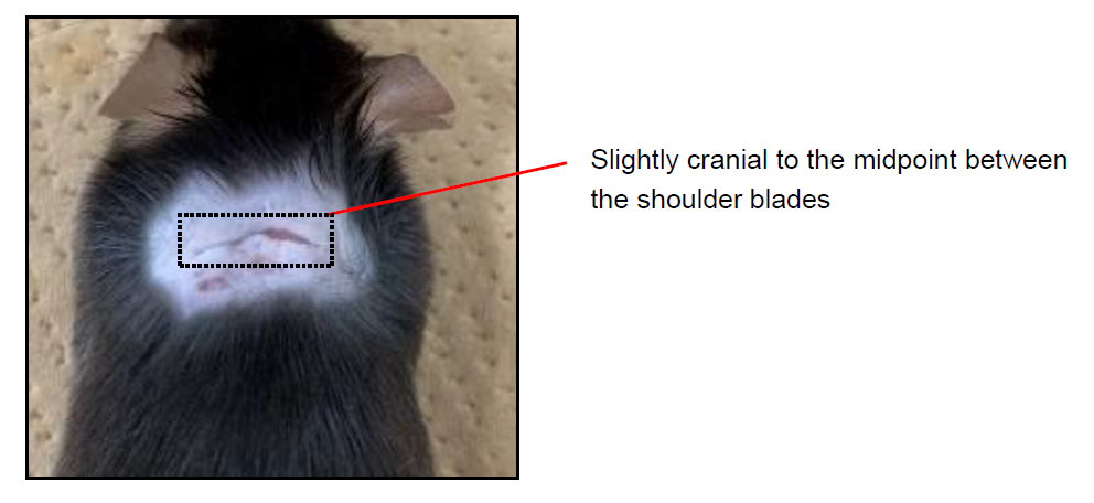
Make a skin incision as shown in the figure, slightly cranial to the midpoint between the shoulder blades.
Make a small incision in the cranial portion of the interscapular white adipose tissue (WAT). Using forceps, gently grasp and lift the WAT upward to visualize the underlying sympathetic nerves.
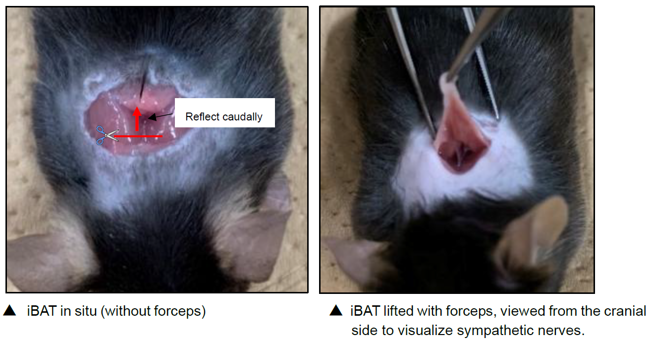
Using scissors, transect the sympathetic nerves (4–5 branches per pad) in each left and right iBAT.
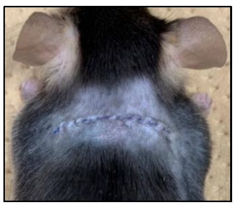
Close the skin incision using sutures.
Notes / Critical Points
- Keep the incision as small as possible, and perform the procedure while viewing from the cranial side to facilitate identification of the nerves.
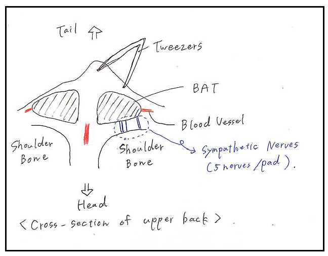
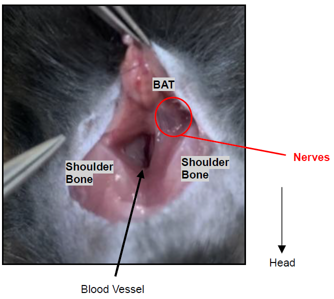
- The sympathetic nerves typically appear as bundled fibers: 3–4 branches located centrally and 1–2 branches positioned more laterally. For beginners, it is recommended to first observe the anatomy under a stereomicroscope.
- A relatively large blood vessel is often present near the center of the surgical field, with additional vessels located more laterally. Accidental injury to these vessels may cause bleeding that can obscure visualization of the sympathetic nerves. Although minor bleeding is usually manageable, these vessels should be preserved whenever possible to maintain a clear surgical field and minimize tissue damage.
Surgical Removal of Interscapular Brown Adipose Tissue (iBAT)
Materials
- Anesthetic agent
- Syringe
- Injection needle
- Sterilized surgical instrument sets
- Straight forceps
- Curved forceps
- Scissors
Note: To prevent contamination, it is recommended to use separate instrument sets for skin incision and for deeper tissue dissection. Instruments should be sterilized before use in different animals.
- Sutures:
- Pronova (blue, 4-0, 90 cm, taper-cut needle, 26 mm, strong curvature; 3975H)
- 5% Hibitane (chlorhexidine; dilute 1:30 before use)
- Electric shaver
- Hair removal can also be performed manually; however, shaving is recommended to minimize skin damage.
- Heating pad (optional, for maintaining body temperature during surgery)
Procedure
- After anesthesia, shave the surgical area using an electric shaver.
- Disinfect and sterilize the shaved area with diluted 5% Hibitane (chlorhexidine).
Make a skin incision as shown in the figure, slightly cranial to the midpoint between the shoulder blades.
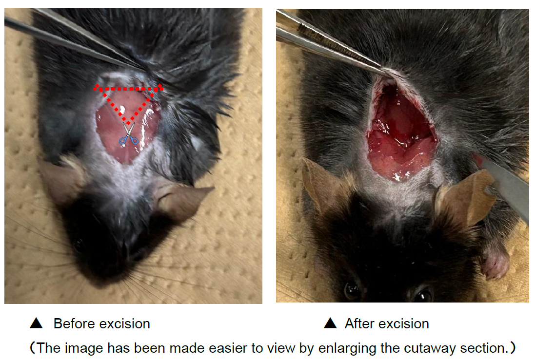
Make a small incision in the cranial portion of the interscapular white adipose tissue (WAT). Using forceps, gently grasp and lift the WAT upward to identify the location of the iBAT. Then, as shown in the figure, carefully dissect along the surrounding WAT using scissors and remove the iBAT completely.
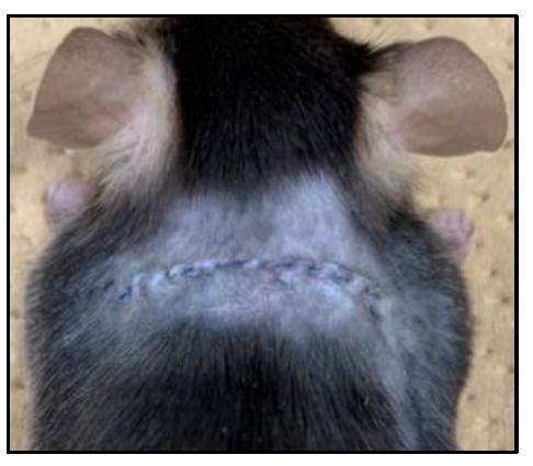
Close the skin incision using sutures.
Notes
- Keep the incision as small as possible.
Additional Information
- No obvious wound-directed behaviors (e.g., scratching with hind limbs) were observed after surgery.
- The incision typically heals within approximately one week.
- Animals can be used for experiments 1–2 weeks after surgery.
Reference
Vaughan, C.H., Zarebidaki, E., Ehlen, J.C., Bartness, T.J. (2014) Analysis and measurement of the sympathetic and sensory innervation of white and brown adipose tissue. Methods Enzymol. 537: 199-225 https://doi.org/10.1016/b978-0-12-411619-1.00011-2
Related files
 Protocol_Surgical Denervation or Removal of BAT.pdf
 - Izumi-Mishima, Y and Sakaue, H(2026). Surgical removal of iBAT. Bio-protocol Preprint. bio-protocol.org/prep2921.
- Izumi-Mishima, Y., Tsutsumi, R., Shiuchi, T., Fujimoto, S., Taniguchi, M., Sugiuchi, M., Tsutsumi, M., Okamatsu-Ogura, Y., Yoneshiro, T., Kuroda, M., Nomura, K. and Sakaue, H.(2025). Brown adipose tissue and skeletal muscle coordinately contribute to thermogenesis in mice. eLife. DOI: 10.7554/eLife.99982
Do you have any questions about this protocol?
Post your question to gather feedback from the community. We will also invite the authors of this article to respond.