
- Home
- Protocols

-

Cytotoxicity assay, cytokine production and continuos repeated stimulation
Last updated date: Jan 17, 2023 Views: 604 Forks: 0
Complete Cytotoxicity Assay
Materials and Reagents:
- TC plate 96 well, round base (Sarstedt. Ref 833925)
- RPMI culture medium supplemented with 3% human serum (Sigma-Aldrich), 1% penicillin/streptomycin
- White 96w Maxisorp Nunc plate (Thermo Scientific™)
- Bright-Glo™ Luciferase Assay Buffer
- Bright-Glo™ Luciferase Assay Substrate (lyophilized)
- Eppendorfs 1,5 ml
- Lysis Solution (from CytoTox 96® Non-Radioactive Cytotoxicity Assay)
- Multichannel pipette + tips
- Reagent reservoirs
- Vortex
Experimental procedure:
Day 0. Co-culture CAR T cells and tumoral cells.
- Resuspend and count tumoral, CAR T and UTD cells. Take the appropriate cell number.
Example Fig. 1: In the below example we use 5000 tumoral cells/well with initial an E:T ratio of 5:1 and 1:3 dilutions of CAR T cells. All by triplicate. For that we will need:
√ 528000 tumoral cells
√ 125000 UTD/ CART cells - Centrifuge 5 min, 1500 rpm. Discard supernatant and add required medium. In the example:
√ 5'28 ml to tumoral cells
√ 248 µl to UTD/ CART cells - Make dilutions: Prepare eppendorfs with 165 µl complete medium RPMI (5 eppendorfs for each CART/UTD group). Add 83 µl of cells to the next tube. Vortex and repeat.
- In a p96w with round base, add 50 µl/well of tumoral cells to all wells. Add 50 µl of medium, CART or UTD cells to the correspondent wells.
- Incubate at 37°C, for 24 h.
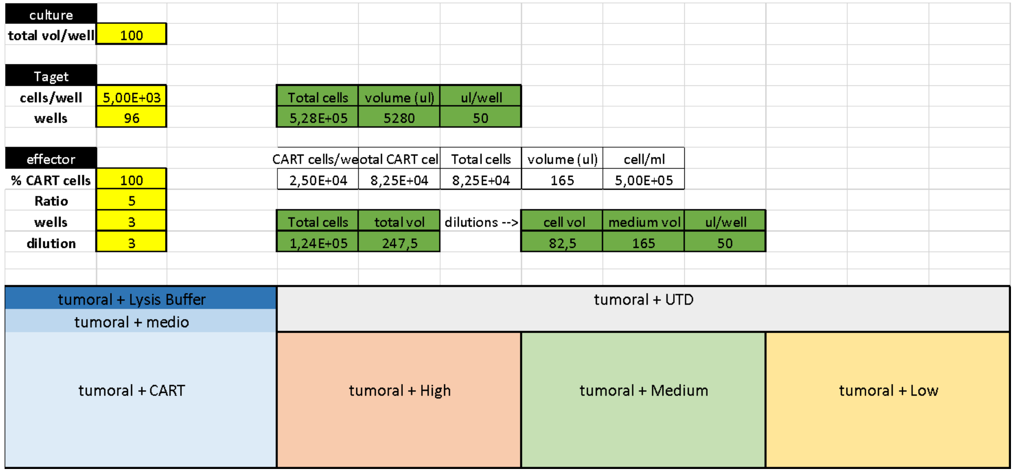
Fig. 1. Cytotoxicity example
Day 1. Bright-Glo Luciferase Assay (Cytotoxicity assay)
- Add 10 µl of Lysis Buffer to the maximum lysis wells (tumoral + lysis buffer). Incubate at 37°C, for 45 min.
- Equilibrate the medium RPMI 10% FBS to room temperature.
- Equilibrate the reagent to room temperature, which is near the temperature optimum of luciferase. Transfer the contents of one bottle of Bright-Glo™ Buffer (RT in the lab) to one bottle of Bright-Glo™ Substrate* (-20℃ in the lab). Mix by inversion until the substrate is thoroughly dissolved.
· Equilibration of the reagent prior to use is unnecessary when the buffer is stored at room temperature.
· If the reagent is stored frozen after reconstitution, the most convenient and effective method to thaw is to place it in a water bath at room temperature. Mix well after thawing. - Centrifuge multiwell plates for 5 min, 800 ×g. Put the supernatants in another p96w plate and store at -20°C (aprox 100 µl/well) for cytokine production measurement.
- Resuspend the cells with 100 µl of PBS (Wash step).
- Centrifuge 5 min, 800 ×g. Throw supernatant by plate inversion.
- Add 50 µl of medium RPMI 10% to each well, resuspend the cells and put them in a white 96w NUNC plate.
- Add 50 µl of reagent and mix. Wait 5 minutes to allow complete cell lysis, and measure in the luminometer.
- Store the remaining Bright-Glo™ Reagent at -80℃.
- To obtain the % of lysis, first, the average absorbance for the maximum lysis wells and for the spontaneous lysis wells (tumoral + media) is obtained from the 3 replicates. Then, the % of lysis of the experimental data is calculated for each well with the following formula:

*Approximate stability of Bright-Glo™ Reagent after reconstitution: 10% loss of luminescence per 5 hours at room temperature, 10% loss per 24 hours at 4°C, and <5% loss after one month at -70°C. The reagent may be subjected to up to seven freeze-thaw cycles with no effect on potency. Check the manufacture instructions for more details.
Cytokine production measurement (ELISA)
Materials and Reagents:
- F96 Maxisorp Nunc-Immuno plate (Thermo Scientific™). Ref. 442404
- Plate sealers (R&D systems). Catalog nº DY992
- BD OptEIA™ Reagent Set B (Cat. No. 550534)
- OptEIA™ Sets for human interleukin-2 (IL-2), tumor necrosis factor (TNF-α), interferon- gamma (IFN-γ), etc.
- Eppendorfs 1,5 ml
- Multichannel pipette + tips
- Reagent reservoirs
- Vortex
Experimental procedure:
Day 0. Coating
Coat microwells with 100 µl per well of Capture Antibody diluted in Coating Buffer. For recommended antibody coating dilution, see lot-specific Instruction/Analysis Certificate. Seal plate and incubate overnight at 4℃.
Day 1.
- Aspirate wells and wash 3 times with ≥ 300 µl /well Wash Buffer. After last wash, invert plate and blot on absorbent paper to remove any residual buffer.
- Block plates with ≥ 200 µl /well Assay Diluent. Incubate at RT for 1 hour.
- Prepare standard and sample dilutions in Assay Diluent. See “Standards Preparation and Handling” from technical data sheet.
- Aspirate/wash as in step 2.
- Pipette 100 µl of each standard, sample, and control into appropriate wells. Seal plate and incubate for 2 hours at RT.
- Aspirate/ wash as in step 2, but with 5 total washes.
- Add 100 µl of Working Detector (Detection Antibody + Streptavidin-HRP reagent) to each well. Seal plate and incubate for 1 hour at RT.
- Aspirate/ wash as in step 2, but with 7 total washes. NOTE: In this final wash step, soak wells in wash buffer for 30 seconds to 1 minute for each wash.
- Add 100 µl of Substrate Solution to each well. Incubate plate (without plate sealer) for 30 minutes at room temperature in the dark.
- Add 50 µl of Stop Solution to each well.
- Read absorbance at 450 nm within 30 minutes of stopping reaction. If wavelength correction is available, subtract absorbance at 570 nm from absorbance 450 nm.
Continuous repeated stimulation
CARHigh T and CARLow T cells were co-culture with irradiated tumor cells for 21 days.
Materials and Reagents:
- 12 well cell culture plates
- RPMI culture medium supplemented with 3% human serum (Sigma-Aldrich), 1% penicillin/streptomycin
Experimental procedure:
- Count sorted CARHigh T and CARLow T cells. Take 1.5×10⁶ cells of each population separately and centrifuge 5 min at 500 ×g. Discard supernatant and resuspend in 750 µl of complete RPMI culture medium.
- Take 3×10⁶ cells ARP1-LucGFP and irradiate at 54 cGy.
- Centrifuge ARP1-LucGFP cells 5 min at 500 ×g. Discard supernatant and resuspend in 1.5 ml of complete RPMI culture medium.
- Co-culture 1.5×10⁶ CARHigh T or CARLow T cells, at a 1:1 ratio, with 1.5×10⁶ cells in RPMI culture medium in a total volume of 1.5 ml in a 12 well plate.
- Incubate at 37°C, for 72 h.
- Count CAR T cells and add ARP1-LucGFP tumoral cells irradiated at 54 cGy at a 1:1 effector: target ratio.
- Repeat steps 5 and 6 every three days, until day 21 is reached.
- Hernaez, M, Rodriguez-Madoz, J and Prosper, F(2023). Cytotoxicity assay, cytokine production and continuos repeated stimulation. Bio-protocol Preprint. bio-protocol.org/prep2122.
- Rodriguez-Marquez, P., Calleja-Cervantes, M. E., Serrano, G., Oliver-Caldes, A., Palacios-Berraquero, M. L., Martin-Mallo, A., Calviño, C., Español-Rego, M., Ceballos, C., Lozano, T., San Martin-Uriz, P., Vilas-Zornoza, A., Rodriguez-Diaz, S., Martinez-Turrillas, R., Jauregui, P., Alignani, D., Viguria, M. C., Redondo, M., Pascal, M., Martin-Antonio, B., Juan, M., Urbano-Ispizua, A., Rodriguez-Otero, P., Alfonso-Pierola, A., Paiva, B., Lasarte, J. J., Inoges, S., Lopez-Diaz de Cerio, A., San-Miguel, J., Fernandez de Larrea, C., Hernaez, M., Rodriguez-Madoz, J. R. and Prosper, F.(2022). CAR density influences antitumoral efficacy of BCMA CAR T cells and correlates with clinical outcome. Science Advances 8(39). DOI: 10.1126/sciadv.abo0514
Do you have any questions about this protocol?
Post your question to gather feedback from the community. We will also invite the authors of this article to respond.