
- Home
- Protocols

-

Flow cytometric analysis to detect KIR+CD8+ T cells and KIR subtypes in human PBMCs
Last updated date: Dec 12, 2022 Views: 1069 Forks: 0
Antibodies and buffers
Antibodies: CD3-BUV805 (UCHT-1, BD), anti-CD56-PercP/Cy5.5 (HCD56, Biolegend), CD4-Alexa Fluor 700 (RPA-T4, Biolegend), anti-CD8-BV650 (RPA-T8, Biolegend), TCRαβ-BUV395 (IP26, BD), anti-KIR3DL1-PE (DX9, Biolegend), anti-KIR2DL2/3-PE (DX27, Biolegend), anti-KIR2DL5-PE (UP-R1, Biolegend), anti-KIR2DL1-PE (Clone 143211, R&D) and anti-KIR3DL2-PE (Clone 539304, R&D), anti-KIR3DL1-BV421 (DX9, Biolegend), anti-KIR2DL2/3-PE/Cy7 (DX27, Biolegend), anti-KIR2DL1-FITC (Clone 143211, R&D) and anti-KIR3DL2-APC (Clone 539304, R&D).
RPMI complete media: 500 ml of RPMI-1640 media supplemented with 50 ml of heat-inactivated human serum (10%), 5 ml of 100× L-Glutamax (1%) and 5 ml of penicillin/streptomycin solution (1%).
FACS buffer: 500 ml of PBS with 2 mM EDTA and 2% heat-inactivated FBS.
Staining of PBMCs
- Warm the RPMI complete media to 37oC.
- Aliquot 9 ml of pre-warmed RPMI complete media into 15 ml conical tubes and add 1 μl of Benzonase.
- Thaw the cryovials with frozen peripheral blood mononuclear cells (PBMCs) into a 37oC water bath. Remove the cryovial from the water bath before PBMCs are completely thawed. Do not allow the cells to warm completely to 37oC (see Note 2).
- Transfer 1 ml of cells from the cryovials into the 15 ml conical tubes with RPMI complete media containing Benzonase and centrifuge the cells at 600xg at 4oC for 5 min.
- Count the cells and determine cell viability with Trypan blue. Resuspend PBMCs at 107/ml in RPMI complete media. Plate the cells in a 24-well plate.
- Rest the PBMCs overnight (12-18h) in a 37oC, 5% CO2 incubator.
- After 12-18h, transfer the PBMCs to 1.5 ml Eppendorf tubes. Centrifuge the cells at 600xg at 4oC for 5 min. Discard the supernatants.
- Resuspend the PBMCs in FACS buffer at <107 PBMCs per 100 ul FACS buffer. Add 5 μl of Human TruStain FcX per 76.5 μl staining volume, mix and incubate at room temperature for 10 minutes.
- Make an antibody master mix of 1 μl of anti-CD3-BUV805, 1 μl of anti-CD56-PercP/Cy5.5, 1 μl of anti-CD4-Alexa Fluor 700, 1 μl of anti-CD8-BV650, 1 μl of anti-TCRαβ-BUV395, 1 μl of anti-KIR3DL1-PE, 1 μl of anti-KIR2DL2/3-PE, 1 μl of anti-KIR2DL5-PE, 2 μl of anti-KIR2DL1-PE, 2 μl of anti-KIR3DL2-PE, 1 μl of anti-KIR3DL1-BV421, 1 μl of anti-KIR2DL2/3-PE/Cy7, 2 μl of anti-KIR2DL1-FITC, 2 μl of anti-KIR3DL2-APC and 0.5 μl of LIVE/DEAD™ Fixable Near-IR Dead Cell Stain per 100 μl staining volume. Add 18.5 μl of the antibody master mix per 81.5 μl PBMCs. Incubate at 4oC for 30 min.
- Wash the cell twice with 1 ml of FACS buffer. Resuspend the PBMCs at 107 cells per 1 ml of FACS buffer.
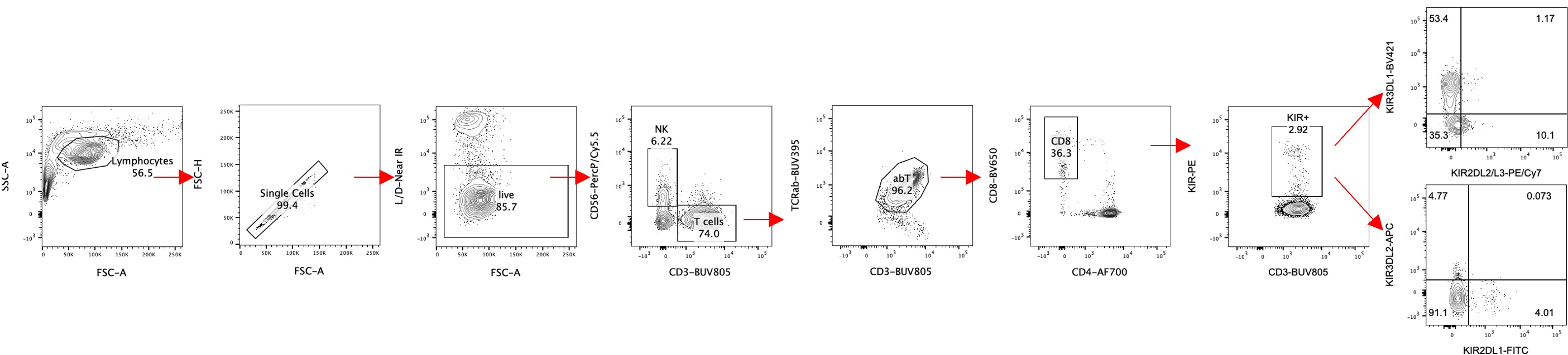
Flow cytometric analysis
Cells were acquired on an LSR II flow cytometer (BD), and data were analyzed using FlowJo X. Gating strategies are shown below.
- Li, J, Saligrama, N and Davis, M(2022). Flow cytometric analysis to detect KIR+CD8+ T cells and KIR subtypes in human PBMCs. Bio-protocol Preprint. bio-protocol.org/prep2078.
- Li, J., Zaslavsky, M., Su, Y., Guo, J., Sikora, M. J., van Unen, V., Christophersen, A., Chiou, S., Chen, L., Li, J., Ji, X., Wilhelmy, J., McSween, A. M., Palanski, B. A., Mallajosyula, V. V. A., Bracey, N. A., Dhondalay, G. K. R., Bhamidipati, K., Pai, J., Kipp, L. B., Dunn, J. E., Hauser, S. L., Oksenberg, J. R., Satpathy, A. T., Robinson, W. H., Dekker, C. L., Steinmetz, L. M., Khosla, C., Utz, P. J., Sollid, L. M., Chien, Y., Heath, J. R., Fernandez-Becker, N. Q., Nadeau, K. C., Saligrama, N. and Davis, M. M.(2022). KIR+CD8+ T cells suppress pathogenic T cells and are active in autoimmune diseases and COVID-19. Science. DOI: 10.1126/science.abi9591
Do you have any questions about this protocol?
Post your question to gather feedback from the community. We will also invite the authors of this article to respond.