
- Home
- Protocols

-

Cell suspension preparation for flow cytometry and flow cytometry analysis
Last updated date: Jul 5, 2022 Views: 589 Forks: 0
Geissmann Lab, Immunology Program, Memorial Sloan Kettering Cancer Center Standard Operating Procedure | |||
| Protocol No.: 027 | Title: Isolation of adipose tissue immune populations and particularly macrophages for analysis by flow cytometry
|
Page 1 of 3 | |
| Effective Date: 02/02/2016 | |||
Revision Number: Version 1 Date & Initials: 02/03/2016 LC | |||
| Originator: Crozet Lucile | Approved by: (sign and date) | ||
Isolation of adipose tissue macrophges for analysis by flow cytometry
Materials
- Reagents
- PBS 1X Without Ca2+ Mg2+
- PBS 1X BSA 0.5%, filtered with 0.22μm stericup
- CaCl2 1mol/L
- Facs buffer: PBS 1X + 0.5% BSA + 2mmol/L EDTA, filtered with 0.22μm stericup
- Collagenase II (sigma, ref: C6885-1g), prepared at 4 mg/mL in PBS + 0.5% BSA + CaCl2 5mmol/L. (For 50mL: 0.2g of collagenase II + 250μL CaCl2 1mol/L + 49.75mL PBS 1X + 0.5% BSA)
- Materials
- 6-well plate (Falcon, ref: 353046)
- 96-well plate (Thermo scientific, ref: 163320)
- 50mL Falcon tubes
- 100 μm strainers (Falcon, ref 352360)
- 70 μm strainers (Falcon, ref 352350)
- 10mL pipettes
- 37°C bug incubator, or any other incubator with shaking option
- Antibodies – on page 2 -
Procedure
Once the mouse is dead, collect the desired fat pads (for our experiments: mWAT, eWAT, iWAT, iBAT) see reference: Orr et al below – page 3 -
1. Adipose tissues are collected in 2mL of cold PBS, in a 6-well plate on ice. NO more than 1.2g of tissue per well
2. Mince the fat tissues into small pieces and transfer into a clean 50mL falcon tube
3. Wash the wells of the 6-well plate with 1mL PBS and then add to the 2mL already in the 50mL falcon.
4. Add 3mL of Collagenase II mix per tube (final concentration for the collagenase 2 mg/mL)
Geissmann Lab Standard Operating Procedure | |||
| Protocol No.: 027 | Title: Isolation of adipose tissue immune cells and macrophages for flow cytometry - analysis
|
Page 2 of 3 | |
| Effective Date: 02/02/2016 | |||
Revision Number: 1 Date & Initials: 02/03/2016 LC | |||
| Originator: Crozet Lucile | Approved by: (sign and date) | ||
5. Incubate for 20min on shaker under agitation at 37°C
6. After incubation, add 10mL of ice cold PBS to the falcon tube and pipette up and down several times with a 10mL pipette
7. Filter the cell suspension through a 100μm strainer and then transfer the sample into a new 50 ml falcon tube
8. Spin the sample for 10min at 500g in a swinging-bucket centrifuge
9. Remove supernatant and resuspend the pellet in 50 μL of FACS buffer + FcBlock
10. Transfer the cell suspension into a 96-well plate and add 50μL of antibody mix, incubate for 30min on ice.
11. Following the incubation period spin down the samples for 7min at 320g in a swinging-bucket centrifuge
12. Wash the cell pellet with 200μL of FACS buffer
13. Repeat 3.9 and 3.10 steps
14. You are now done!! Remember to run the samples through a 70μm strainer prior to flow cytometry analysis
Antibody mix proposition
| Antigen | fluorochrome | Clone | Final dilution |
| CD45.2 | APC-Cy7 | 104 | 1/100 |
| CD3 | BV711 | 145-2C11 | 1/200 |
| CD19 | BV711 | 1D3 | 1/200 |
| NKp46 | BV711 | 29A1.4 | 1/200 |
| SiglecF | PE | E50-2440 | 1/200 |
| Ly6G | PE | 1A8 | 1/200 |
| F4/80 | BV605 | BM8 | 1/200 |
| CD11b | PE-Cy7 | M1/70 | 1/400 |
| Tim4 | APC | RMT4-54 | 1/200 |
| CD11c | BV421 | HL3 | 1/100 |
| MHCII | AF700 | M5/114.15.2 | 1/200 |
Geissmann Lab Standard Operating Procedure | |||
| Protocol No.: 027 | Title: Isolation of adipose tissue immune cells and macrophages for flow cytometry - analysis
|
Page 3 of 3 | |
| Effective Date: 02/02/2016 | |||
Revision Number: 1 Date & Initials: 02/03/2016 LC | |||
| Originator: Crozet Lucile | Approved by: (sign and date) | ||
References
- Protocol adapted from Orr, Jeb S., Arion J. Kennedy, et Alyssa H. Hasty. 2013. « Isolation of Adipose Tissue Immune Cells », JOVE no 75 (mai): e50707. doi:10.3791/50707.
- For our experiments: mWAT, eWAT, iWAT, iBAT
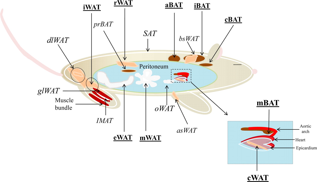
Schema from Waldén, Tomas B., Ida R. Hansen, James A. Timmons, Barbara Cannon, et Jan Nedergaard. 2012. « Recruited vs. Nonrecruited Molecular Signatures of Brown, “brite,” and White Adipose Tissues ». American Journal of Physiology - Endocrinology and Metabolism 302 (1): E19‑E31. doi:10.1152/ajpendo.00249.2011
- Cox, N and Geissmann, F(2022). Cell suspension preparation for flow cytometry and flow cytometry analysis. Bio-protocol Preprint. bio-protocol.org/prep1768.
- Cox, N., Crozet, L., Holtman, I. R., Loyher, P., Lazarov, T., White, J. B., Mass, E., Stanley, E. R., Elemento, O., Glass, C. K. and Geissmann, F.(2021). Diet-regulated production of PDGFcc by macrophages controls energy storage. Science 373(6550). DOI: 10.1126/science.abe9383
Do you have any questions about this protocol?
Post your question to gather feedback from the community. We will also invite the authors of this article to respond.