
- Home
- Protocols

-

Pressure myography
Last updated date: Oct 6, 2021 Views: 824 Forks: 0
Assessment of myogenic tone responses in mouse mesenteric arteries
Abstract: Myogenic vasoconstriction is important for the regulation of organ perfusion and peripheral resistance. In this protocol, we present the assessment of myogenic tone responses in mouse mesenteric arteries.
Background: An increase in intraluminal pressure in resistance arteries results in a contraction of the arteries [1]. This ability of vascular smooth muscle cells to respond to mechanical stretch has been observed in a variety of vascular beds. It has been suggested that myogenic vasoconstriction is involved in maintaining basal vascular tone to promote constant perfusion despite fluctuations in perfusion pressure [2-4]. Pressure myograph is a widely used technique to evaluate intrinsic myogenic contractions mediated by vascular smooth muscle cells. In pressure myography, an intact small segment of an artery is mounted onto two small glass cannulae and pressurized via a step-wise increase in transmural pressure. The changes in diameter with an increase in pressure steps are determined using a CCD camera. In addition, applying gradients of flow, flow-mediated dilation (FMD) responses are assessed at physiological pressure [5, 6]
Materials and methods: Tamoxifen, acetylcholine, phenylephrine, Krebs-Ringer bicarbonate-buffered salt solution (KREBS) contains (in mmol/l): 118.5 NaCl, 4.7 KCl, 2.5 CaCl2, 1.2 MgSO4, 1.2 KH2PO4, 25.0 NaHCO3 and 5.5. All chemicals can be purchased from
e.g. Sigma.
Equipment: Pressure myograph (Danish Myo Technology; 110P and 114P).
Software: Myoview 2 & 4 (Danish Myo Technology)
Procedure:
1. 10-14 days after the last tamoxifen injection, mice are euthanized with CO2.
2. First and second order mesenteric arteries are dissected from the mesentery and placed in cold KREBS solution. The buffer is pre-equilibrated with 95% O2 / 5% CO2 for 30 min.
3. Arterial segments of 3 mm length are mounted in a pressure myograph chamber (Danish Myo Technology; 110P and 114P; see below).
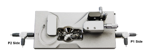
Pressure myograph chamber
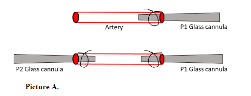
4. As a first step in the mounting process, P1 and P2 glass cannulae are gently perfused with KREBS buffer using a syringe connected to respective inlet valves at the P1 and P2 sides.
5. One end of the arterial segment is mounted onto the right side of the glass cannula (P1 side, see Picture A) and carefully tied with a nylon suture. With the help of a syringe, the arterial segment is flushed and filled with KREBS buffer via the P1 valve inlet.
6. The other end of the arterial segment is mounted onto the left side of the glass cannula (P2 side, see Picture A) and tied with a nylon suture. The chamber is then filled with KREBS buffer.
7. All arterial segments are pre-checked for any leakage by gently flushing buffer using a syringe connected to the P1 valve inlet.
Picture B.
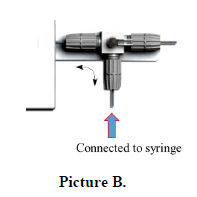
8. The Myo-interface panel on the pressure regulator of the pressure regulatory unit is set to ‘flow on’, then the KERBS buffer from the reservoir (see Picture C) is passed until all air/bubbles in the connecting tube have been removed.
Picture C.
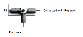
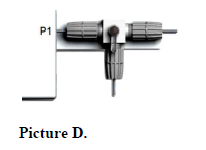
9. Once the buffer has been withdrawn, the 3-way valve is turned in the direction shown in Picture D to connect the P1 side of the chamber to the P1 reservoir.
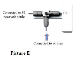
10. Next, the P2 valve inlet is connected to a syringe containing KREBS buffer (see Picture E). With the Myo-interface panel set to ‘flow on’, slowly perfuse the buffer manually. After all the bubbles have been removed, turn off the flow and close the inlet valve (syringe side) so that the P2 side of the chamber is connected to the P2 reservoir bottle.
11. The arterial segment is visualized with a camera attached to an inverted microscope, allowing to monitor vessel diameter and wall thickness using Myoview software.
12. The artery is initially pressurized with pressure steps 10, 70, and 140 mmHg to check for possible leakage.
13. Next, the arterial segment is incubated for 30 minutes under continuous aeration (95% O2 / 5% CO2) at 10 mmHg pressure at 37 ֯C.
14. At 60 mmHg pressure, endothelial integrity is assessed by pre-contracting with phenylephrine (10 µM) followed by acetylcholine (10 µM). Arterial segments that show less than 85% relaxation are not used for the experiment. The chamber is washed three times with KREBS buffer and then left for 15 min.
15. To assess myogenic tone responses, the intraluminal pressure of the arterial segment is gradually increased to the following pressure steps every 4 minutes: 20, 40, 60, 80, 100, 120, 140 mmHg.
16. Myogenic tone is expressed as the percentage of passive diameter ((passive diameter - active diameter) / passive diameter x 100).
Notes:
Siblings or age- and weight-matched animals are used in the experiments.
Competing interests: The authors declare no conflicts of interest.
Ethics: All animal care and experimental procedures in this study were approved by the local authorities (Regierungspräsidia Karlsruhe and Darmstadt, Germany). The approved animal proposal number for the experiments is B2-1166.
References:
1. Bayliss, W.M., On the local reactions of the arterial wall to changes of internal pressure. J Physiol, 1902. 28(3): p. 220-31.
2. Davis, M.J. and M.A. Hill, Signaling mechanisms underlying the vascular myogenic response.
Physiol Rev, 1999. 79(2): p. 387-423.
3. Loutzenhiser, R., A. Bidani, and L. Chilton, Renal myogenic response: kinetic attributes and physiological role. Circ Res, 2002. 90(12): p. 1316-24.
4. Chennupati, R., et al., Myogenic vasoconstriction requires G12/G13 and LARG to maintain local and systemic vascular resistance. Elife, 2019. 8.
5. Wang, S., et al., P2Y(2) and Gq/G(1)(1) control blood pressure by mediating endothelial mechanotransduction. J Clin Invest, 2015. 125(8): p. 3077-86.
6. Wang, S., et al., Endothelial cation channel PIEZO1 controls blood pressure by mediating flow-induced ATP release. J Clin Invest, 2016. 126(12): p. 4527-4536.
- Chennupati, R and Offermanns, S(2021). Pressure myography. Bio-protocol Preprint. bio-protocol.org/prep1393.
- Chennupati, R., Wirth, A., Favre, J., Li, R., Bonnavion, R., Jin, Y., Wietelmann, A., Schweda, F., Wettschureck, N., Henrion, D. and Offermanns, S.(2019). Myogenic vasoconstriction requires G12/G13 and LARG to maintain local and systemic vascular resistance. eLife. DOI: 10.7554/eLife.49374
Category
Do you have any questions about this protocol?
Post your question to gather feedback from the community. We will also invite the authors of this article to respond.