
- Home
- Protocols

-

Virus-like-particle production and viral entry assays.

Last updated date: Jul 23, 2021 Views: 893 Forks: 0
Ebola virus-like-particle production and viral entry assays
Abstract
Viruses from the Filoviridae family, such as the Ebola virus (EBOV) and Marburg virus (MARV), can cause sporadic outbreaks of highly lethal hemorrhagic fevers in humans. Due to the high virulence and fatality rates associated with filoviruses, the study of infectious filoviral particles must be conducted in biosafety level (BSL)-4 laboratories. However, co-expression of the EBOV glycoprotein (GP), the matrix protein (VP40), and nucleoprotein (NP) in cells leads to the production of Ebola viral-like-particles (VLPs) that exhibit the native characteristic filamentous morphology of filoviruses, representing valuable and convenient BSL-2 surrogate systems. In addition, the use of a VP40 construct fused to b-lactamase enables the rapid detection of viral entry in cells, the study of entry pathways and the testing of entry inhibitors. This approach and protocol can also be adapted to study viral entry of other filoviruses using heterologous GPs.
Keywords: Ebola virus, viral-like particles, viral entry, filoviruses
Materials and Reagents
Cell lines and cell culture reagents
- VLP producer cells: Human embryonic kidney cells (HEK293T) cells (ATCC, catalog number CRL- 3216), maintained in 10 cm dishes or T75 flasks in complete DMEM medium (see recipes)
- Susceptible cells: Vero E6 (ATCC, catalog number CRL-1586), maintained in 10 cm dishes or T75 flasks in complete MEM medium (see recipes)
- Dulbecco’s Modified Eagle Medium with 4.5 g/L Glucose and L-Glutamine (DMEM, Lonza catalog number CA12001-568)
- Minimum Essential Medium Eagle (MEM, VWR, catalog number M2279)
- Fetal bovine serum (FBS, Sigma catalog number F1051)
- Penicillin/Streptomycin/Glutamine 100X (VWR, catalog number CA10753-886)
- Phosphate buffered saline 10X solution (PBS, Fisher Scientific, catalog number BP399-1)
- Trypsin-EDTA (VWR, CA45001-082)
Transfection reagents
- jetPRIME transfection reagent and buffer (Polyplus transfection, catalog number 114-01)
- Plasmids: Empty vector (pCAGGs), plasmids encoding EBOV VP40-βlam, EBOV nucleoprotein (NP), and glycoprotein of choice
VLP purification materials and reagents
- Sucrose (VWR, catalog number CA71008-918)
- Sterile single use bottle top filters, 0.45µm (Thermo, catalog number 295-4545)
- Ultracentrifuge tubes (Beckman Coulter, catalog number 326823)
Viral entry assay reagents
- LiveBLAzer™ FRET-B/G Loading Kit with CCF2-AM (Invitrogen, catalog numberK1032)
- Probenecid (Sigma, catalog number P8761)
- Ammonium chloride (NH4Cl, Sigma, catalog number 12125-02-9)
- 0.45 µm syringe filter (Starstedt, catalog number 83.1826)
- (Optional) Poly-D-lysine (Sigma, catalog number P6407)
(Optional) Materials for using high throughput sampler
- Multichannel pipette and tips (optional) (Thermo, catalog number 14387970BT)
- 96-well flat bottom plates (VWR, catalog number CA62406-081)
Miscellaneous
- Tissue culture-treated 10 cm dishes (Sarstedt, catalog number 83.3902)
- 15 mL conical centrifuge tubes (Froggabio, catalog number, TR15-500)
- 50mL conical centrifuge tubes (Froggabio, catalog number TR50-500)
- 1.5mL microcentrifuge tubes (Sarstedt, catalog number 72.690.301)
- Tissue culture-treated 48-well plates (Sarstedt, catalog number 83.3923)
- 5mL round bottom polystyrene flow cytometry tubes (Fisher Scientific, catalog number 14-959- 2A)
- Racks for 15ml/50mL conical tubes and 1.5mL microtubes
- Sterile serological pipettes (5mL, 10mL, 50mL) and pipette dispenser
- Micropipettes and tips
- 70% ethanol or 0.5% accelerated hydrogen peroxide for decontamination
Equipment
- Incubator (5% CO2, 37°C) (VWR Symphony, catalog number 98000-372)
- Biosafety cabinet, Class II, Type A2 with vacuum line
- Water bath (VWR, catalog number 10805-322)
- Tabletop centrifuge with temperature control and adaptors for 50mL and 5mL conical tubes and multi-well plates (ex., Fisher Scientific ST40, catalog number 75-257-406, note this product has been discontinued)
- Microcentrifuge (Thermo, catalog number 75002432)
- Ultracentrifuge, rotor and buckets (Optima XPN-100, Sw32Ti rotor)
- Flow cytometer (BD Biosciences, FACSCelesta™ BVYG laser configuration, catalog number 660345),
BD FACSCelesta™ High Throughput Sampler (optional), catalog number 658946 - Vortex mixer (Fisher Scientific, catalog number 02-215-414)
- Analytical balance (Mettler Toledo, catalog number 30216623)
- Laboratory Balance (Sartorius, catalog number BCE1202I-1S)
- (optional) Mini shaker (VWR, catalog number 12620-938)
Software
Flowjo (version 1.7) or other flow cytometry analysis software
Procedure
Note: Cell culture, manipulation of DNA for transfection and handling of VLPs should be performed in a biosafety cabinet using aseptic technique.
VLP production
- Seed HEK293T cells in 10cm dishes to ensure approximately 90% confluency the next day. Each dish will be used to produce one type of VLP (ex., EBOV-GP VLPs, MARV-GP VLPs, Bald VLPs, etc).
- Twenty-four hours later, prepare transfection solutions and perform transfection according to the jetPRIME transfection protocol: dilute viral plasmids (plasmid ratios for one 10cm dish: 3µg NP, 3µg βlam-VP40, 4µg GP of choice) in 500uL of JetPrime buffer in a 1.5mL microtube. For bald VLP production, replace GP with 4µg of empty pCAGGs vector. See note A for optimization of transfection and recommended experimental controls.
- Vortex tubes on low speed for 5 sec to thoroughly mix DNA.
- Vortex jetPRIME reagent on high speed to mix thoroughly.
- Add 20uL of JetPrime reagent to each tube containing DNA. Vortex on high speed for 10 sec to mix thoroughly.
- Spin down tubes in a microcentrifuge (>10 x g) for 5 sec
- Incubate tubes at room temperature for 10 min.
- Change the media in the dish using 10mL of pre-warmed complete DMEM (see recipes). Add media slowly to prevent celldetachment.
- Add the transfection solution containing DNA, jetPRIME buffer, and jetPRIME reagent (~520µL total) dropwise to the 10cm dish using a micropipette. Shake plates gently to mix.
- Incubate cells (5% CO2, 37˚C) overnight.
- After 24 hrs, replace media with pre-warmed low FBS-containing DMEM (see recipes), 10mL per dish.
- Harvest the media supernatant containing VLPs at 48hrs, 72hrs, and 96hrs post-transfection (3 harvests, ~30mL total supernatant per 10cm dish). Gently aspirate the supernatant using a 10mL serological pipette and transfer to 50mL conical tubes. Supernatant from dishes producing the same type of VLPs can be combined into the same 50mL tube. Harvested VLPs should be stored at 4˚C and should be purified as soon as possible) (see note B on potential cytotoxicity of viral glycoproteins)
VLP Purification
- Prepare the ultracentrifuge rotor and buckets by spraying them thoroughly with 70% ethanol and allowing to dry. If possible, before preparing the samples, place the rotor (without the tube holders) into the ultracentrifuge, turn on vacuum and set to 4°C to equilibrate the rotor.
- Balance the 50mL tubes containing viral supernatants with sterile PBS if necessary.
- Centrifuge the harvested samples at (~525 x g, 5 min, 4˚C ) to pellet cells and large debris.
- Transfer the supernatant into ultracentrifuge tubes by gently removing the supernatant from the tubes using a 50mL serological pipette, taking care not to disturb the pellet. Add no more than 30mL of supernatant to each ultracentrifuge tube.
- Add 4.5mL of 20% sucrose in PBS (see recipes) to the bottom of the ultracentrifuge tubes using a 5mL serological pipette. Submerge the pipette to the very bottom of the ultracentrifuge tube and slowly add the sucrose solution to create a distinct sucrose cushion. Take care not to overly jostle the tubes after this point to keep the sucrose cushion intact.
- Fill the ultracentrifuge tubes to the top by slowly adding sterile PBS dropwise using a 5mL serological pipette. There should be no gap between the media and the top of the tubes.
- Gently place the ultracentrifuge tubes into the tube holders provide with the Sw32Ti rotor and seal with the tube holder lids. Using a laboratory balance, balance the tubes within 0.05g using sterile PBS.
- Load tubes into the Sw32Ti rotor, insert rotor into ultracentrifuge, and spin at 20,000 RPM for 1.5 hrs at 4°C.
- After ultracentrifugation is complete, release vacuum and remove samples. Remove the ultracentrifuge tubes carefully from the tube holders using forceps sprayed with 70% ethanol. Decant supernatant into a waste container, and leave tubes upside down on paper towel for 1–5 min to ensure all supernatant is removed.
- Reinvert tubes and gently add 300µL sterile PBS to the pellet. Do NOT re-suspend the pellet.
- Seal the top of the ultracentrifugation tubes using parafilm and leave them in a tube rack on an orbital shaker at 4°C for 2 hrs to overnight (100–200 RPM).
- Resuspend the VLPs by gently pipetting up and down with a micropipette. Ensure that there are no visible clumps. If clumps are still present following resuspension, transfer the samples to a 1.5mL microcentrifuge tube and spin the sample down (10 000 x g, 30 sec). Transfer the supernatant to a new 1.5mL microtube.
- Aliquot the VLP solution into 5µL–100µL samples in microtubes and store at -80°C until use. Multiple freeze-thaws are not recommended.
VLP entry assays
- The day before, seed target cells in 48-well plates. The number of cells is dependent on the size and growth rate of the chosen cell line, the general target is 80-90% confluence for viral entry assays. Retain two wells for staining controls (do not infect these wells). See note C for optimization of entry assays.
- Infection without spinoculation: Remove media and replace with 150µL/well of pre-warmed (37°C) media of choice. Add 50uL of VLPs diluted in media to cells. Infect cells with VLPs at a concentration to obtain entry in about 20-30% of cells. Place plate in incubator (37°C, 5% CO2) for 3 hrs to allow entry to proceed. A longer incubation period is acceptable, but it is not recommended to go beyond 6 hours. Proceed to step 4.
- Infection with spinoculation: Remove media and replace with 250µL/well of pre-chilled (4°C) media of choice. Add 50uL of VLPs diluted in media to cells. Infect cells with VLPs at a concentration to obtain entry in about 20-30% of cells. Place plate in centrifuge (pre-cooled to 4°C) equipped with plate adaptors and spin for 30 min (200 x g). Remove plate, aspirate media, and add 250µL/well of pre-warmed (37°C) media. Place plate in incubator (37˚C, 5% CO2) for 3 hrs to allow entry to proceed. A longer incubation period is acceptable, but it is not recommended to go beyond 6 hours. Proceed to step 4.
- Prepare CCF2-AM staining solution (see recipes). Optional: add probenecid (final concentration 2.5mM) to improve staining and NH4Cl (final concentration 15mM) to arrest EBOV entry. Prepare enough 1X staining solution for 100ul/well (48 well plate) as well as some extra to account for pipetting errors. See note D regarding the use of probenecid and NH4Cl and optimization of CCF2 staining.
- Wash cells carefully with ~150 µL room temperature serum-free media.
- Aspirate media and gently add 100µL of staining solution to each well via pipetting down the side of the wells. Keep one (uninfected) well in serum free media without stain (unstained control) and one stained, uninfected well.
- Incubate 1 to 2 hrs at room temperature, protected from light (staining solution is light sensitive).
- Aspirate staining solution and gently wash cells once with PBS.
- Add 100uL of trypsin-EDTA per well and allow cell detachment at 37°C.
- Resuspend cells in trypsin by pipetting up and down with a P1000, and transfer total volume to flow cytometry tubes pre-filled with 2mL of 2% FBS in PBS.
- Centrifuge tubes at 550 x g for 5 min (room temperature).
- Pour off supernatant into waste container in one fluid motion, taking care not to disturb pellet.
- Resuspend pellet in 100 µL 2% FBS in PBS.
- If analysing samples using a high throughput sampler, transfer samples to the wells of a 96 well plate (using a multichannel pipette if desired).
- Analyze cells by flow cytometry (see data analysis)
Data analysis
The recommended configurations for detection of CCF2-AM by using the BD FACSCelesta are as follows: krypton violet laser (405 nm), 525/50 bandpass filter (detection of uncleaved CCF2, “green”), 450/40 bandpass filter (detection of cleaved CCF2, “blue”). Similar configurations on other flow cytometers may be used.
For gating strategy, see figure 1. First gate for viable cells using forward scatter and side scatter on the unstained, uninfected sample. Next, gate for the stained, infected population using the stained, uninfected sample. Note that the magnitude of the shift in green fluorescence in the stained cells compared to the unstained cells will depend on cell type and efficiency of staining. It is normal to have a small amount of background (<1%) “infection” in the uninfected samples. You may choose to subtract this background percentage from the infected samples.
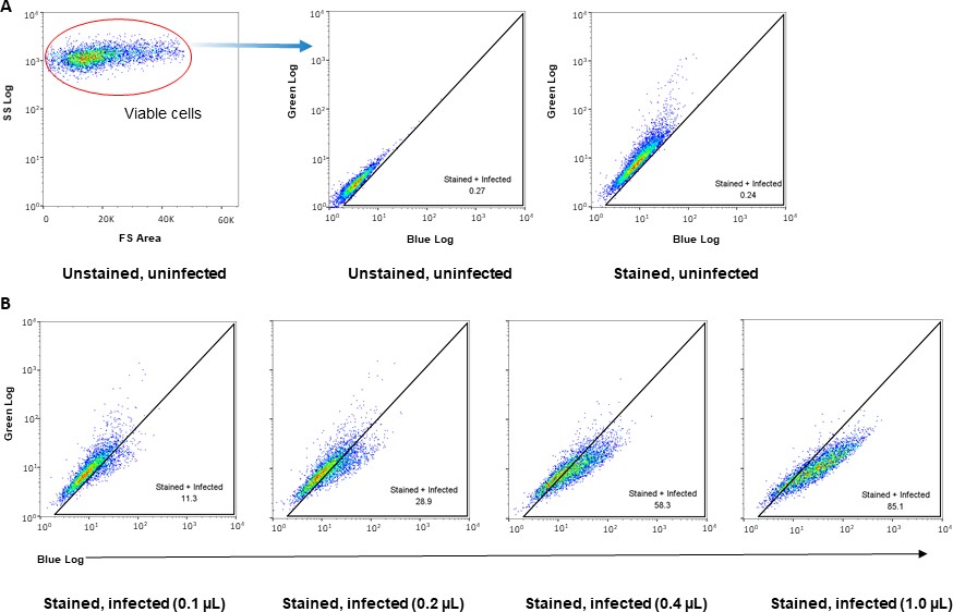
Figure 1. Gating strategy for detection of βlam-VLP entry in cells using a CCF2 FRET assay. Vero cells seeded in 48 well plates were incubated with increasing concentrations of EBOV-βlam VLPs for 3.5 hrs, followed by staining with CCF2-AM staining solution for 1 hr. Cells were analyzed on a BD FACSCelesta flow cytometer. (A) Control samples (unstained, uninfected and stained, uninfected) were used to gate for stained, infected cells. First viable cells were gated in the unstained, uninfected sample using forward scatter (FS) and side scatter (SS). Next, a gate for stained, infected cells was drawn using the stained, uninfected sample, and applied to all samples. (B) The stained, infected gate from (A) was used to gate for percentage of cells with cleaved CCF2 in cells infected with increasing concentrations of EBOV-βlam VLPs.
Notes
- HEK293T cells are frequently used for production of VLPs due to their high transfection efficiency. However, care should be taken to optimize transfection conditions to minimize cell death while maximising transfection efficiency. We have found the ideal confluency level of HEK293T for transfection to be around 90%. Decreasing the confluency of viral producer cells may enhance transfection efficiency, however, it can also increase cytotoxicity during transfection. The use of low FBS containing media for viral harvests is to minimize the presence of growth factors and other signalling molecules in the VLP preparations; this is particularly important if VLPs are to be used for cell signalling experiments. VLPs may also be harvested in serum-free DMEM to completely remove any signalling molecules that are present in the FBS, however note that this can increase cell death during transfection and harvests. If transfecting particularly toxic plasmids such as full-length EBOV GP, it may be necessary to increase the concentration of FBS up to 10% in the viral harvest media.
For cell signalling experiments using VLPs, we recommend also producing bald VLPs (VLPs lacking glycoprotein) as well as a mock transfected control (consisting of supernatant from cells transfected with pCAGGS alone, purified alongside VLP-containing cell supernatants). This is important as the mock transfected samples will contain any cell factors (such as microsomes) that may be copurified alongside VLPs during ultracentrifugation. - Depending on the toxicity of the glycoproteins used, there may be substantial cell detachment during harvests. Ensure very gentle aspiration and replenishment of media to reduce the number of cells detached. Some glycoproteins (ex., full length EBOV GP) induce significant cytotoxicity when expressed in cells, in which case only 1–2 harvests may be possible.
- If the target cells lift easily from plates (such as HEK293Ts), we recommend coating plates with poly- D-lysine. The plate format is flexible depending on the number of cells required for the assay; it is possible to scale up or down by increasing or decreasing the amount of staining solution used. The media of choice is dependent on cell line and the biological mechanisms being analyzed. For example, if signalling pathways are being analyzed, consider using low serum or serum-free media to minimize background activation of signalling pathways by growth factors present in serum.
Spinoculation is not required but may enhance VLP entry and can also enhance precision between technical replicates by synchronising VLP entry. - For optimal staining efficiency, use HBSS or serum-free media to prepare the staining solution. The presence of serum significantly decreases staining efficiency. Staining should always be done in the dark at room temperature, however, the staining time can be extended to up to 18 hrs (overnight). Note than increased staining incubations may enhance signal but can also increase background.
Staining solution components: Although it is not required to aliquot solution A (CCF2-AM in DMSO), we have found that aliquoting into 5-10 µL aliquots reduces degradation of CCF2-AM during freeze-thaws. We recommend using each aliquot only once. Solution B may form white precipitates if stored at lower temperatures. If this occurs, warm and mix solution B at 37°C until the precipitate dissolves. Probenecid is a non-specific inhibitor of anion transport that can enhance the amount of CCF2 retained in cells, however, note that it may cause cytotoxicity in some cell types. NH4Cl is a weak base that arrests entry of low-pH dependent viruses by neutralizing endosomal pH. It may be optionally added to reduce residual entry during staining.
Recipes
- Complete DMEM (10% FBS):
To 500 mL DMEM, add 50 mL FBS and 5.5 mL 100X Penicillin/Streptomycin/Glutamine. - Low FBS DMEM (2% FBS):
To 500 mL DMEM, add 10 mL FBS and 5.1 mL 100X Penicillin/Streptomycin/Glutamine. - Complete MEM:
To 500 mL MEM, add 50 mL FBS and 5.5 mL 100X Penicillin/Streptomycin/Glutamine - 1X PBS (500mL):
Dilute 50 mL 10X PBS stock in 450 mL milli-Q water. Autoclave to sterilize. - 20% (w/v) sucrose solution:
Dissolve 100 g sucrose in 400 mL 1X autoclave sterilized PBS. Top up with PBS to 500 mL. Filter using 0.45 µm bottle top filter. Store at 4°C. - 6X CCF2-AM staining solution:
Makes enough for 1 x 48-well plate (100 µl/well)
5 ul solution A (CCF2-AM in DMSO)
50 ul solution B
778 ul solution C
75 ul 1 M ammonium chloride
50 ul 250mM probenecid
Dilute to 1X in serum free cell media or HBSS. Solution is light sensitive, use within 2 hours of preparation. - Probenecid (250mM)
Dissolve 713.4 mg probenecid in 10 mL 500mM NaOH. Add mL 100 mM sodium phosphate buffer, pH 8.0
Adjust the pH of the resulting solution to pH 8.0 with 1 M HCl or 1 M NaOH. Divide into 0.5 – 1 mL aliquots and store at -20°C. - Ammonium chloride (1M)
Dissolve 534.91 mg ammonium chloride in 10 mL milli-Q water. Filter using a 0.45 µm syringe filter. Store at 4°C.
Acknowledgments
Adaptation of this protocol was funded by the Canadian Institutes of Health Research: grants ER1- 143489352509 to M.C. We would like thank Dr. Lijun Rong (University of Illinois at Chicago, Chicago) for sharing the EBOV NP and βlam-VP40 constructs, and Drs. James Cunningham (Brigham and Women’s Hospital, Boston) and Kartik Chandran (Albert Einstein College of Medicine, Bronx) for sharing the filovirus GP constructs. This protocol was modified from previous work by the Rong lab.
We declare that no conflicts of interest or competing interests exist.
References
Filoviruses Use the HOPS Complex and UVRAG To Traffic to Niemann-Pick C1 Compartments during Viral Entry. Bo Y, Qiu S, Mulloy RP, Côté M. J Virol. 2020 Jul 30;94(16):e01002-20. doi: 10.1128/JVI.01002-20.
Expression of Ebolavirus glycoprotein on the target cells enhances viral entry. Manicassamy B, Rong L. Virol J. 2009;6:75. doi:10.1186/1743-422X-6-75
- Qiu, S, Fu, K, Phan, A and Côté, M(2021). Virus-like-particle production and viral entry assays.. Bio-protocol Preprint. bio-protocol.org/prep1312.
- Bo, Y., Qiu, S., Mulloy, R. P. and Côté, M.(2020). Filoviruses Use the HOPS Complex and UVRAG To Traffic to Niemann-Pick C1 Compartments during Viral Entry. Journal of Virology 94(16). DOI: 10.1128/JVI.01002-20
Do you have any questions about this protocol?
Post your question to gather feedback from the community. We will also invite the authors of this article to respond.