
- Home
- Protocols

-

Liposome dye release assay
Last updated date: Apr 21, 2021 Views: 1124 Forks: 0
Indirect Viroporin Assay – Liposome Dye Release Assay
Reagents
- L-α-Phosphatidic acid (egg monosodium salt) (PA) (10 mgml-1 chloroform stock)
- L-α-Phosphatidylcholine (egg) (PC) (10 mgml-1 chloroform stock)
- L-α-Phosphatidylethanolamine with lissamine rhodamine B labelled head groups (PE) (10 mgml-1 chloroform stock)
- Triton x100
- Liposome Assay Buffer
- 10 mM HEPES pH 7.4
- 107 mM NaCl - (5)6-Carboxyfluorescien Buffer
- 50 mM (5)6-Carrboxyfluorescien
- 10 mM HEPES pH 7.4
- 10 mM NaCl
- 134 mM NaOH
Equipment
- Inert gas supply (Argon or Nitrogen)
- Desiccator
- Glass test tubes and vials
- Mini extruder and glass syringes (Avanti Polar Lipids)
- Nuclepore PC filters, 0.4 μm
- Support Filters: drain discs 10mm PE (Whatman)
- Ultracentrifuge (capable of 100,000 xg, capacity of 2 ml)
- Heat block
- Black 96 well microplates
- Fluorescent plate reader (capable of λex 492 nm/ λem 517)
Protocol
Liposome Preparation
Day 1
1. Measure out lipids under a stream of inert gas: 50 μl PA, 50 μl PC and 5 μl PE.
2. Dry down the lipid mixture using the stream of inert gas.
3. Place dried-down lipid mix in a vacuum for at least 2 hours.
4. Resuspend the dried-down lipids in 500 ml of (5)6-Carboxyfluorescien Buffer, incubate overnight at room temperature.
Day 2
1. Warm resuspended lipid mix, liposome assay buffer, extruder and syringes to 37 °C.
2. Assemble the extruder. Pass 1 ml of liposome assay buffer through to hydrate the filters.
3. Pass the resuspended lipid mix back-and-forth through the extruder at least 15 times. (You must make sure after the final pass that the lipid mix is on the opposite side to which it started.)
4. Purify the liposomes by centrifugation at 100,000 xg for 10 mins (or until liposomes have formed a red pellet), washing the pellet at least 3x in liposome assay buffer.
5. Liposomes are then resuspended in 300 ul of liposome assay buffer.
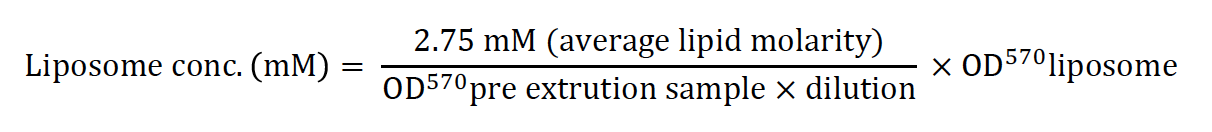
6. Concentration of liposomes is calculated by:
Liposome Dye Release Assay
1. Pipette peptide/protein of interest into a 96 well plate at desired concentration… if drug compounds are being added, add these now and incubate the plate for at least 15 minutes. Set up a control well containing 10 ul of 0.1% triton x100.
2. Set-up the plate reader (using filters in the range of 488/520) to take readings every minute over 30 minutes (or for a shorter length of time if saturation is reached before 30 minutes)
3. Place the plate in the plate reader, add 100 ul of 50 mM of liposomes to the triton x100 well. Use this to set the gain on the plate reader using the triton x100 control, so that the measurement is in the range of detection.
4. Then quickly add 100 ul of 50 mM of liposomes to each experimental well.
- Swinscoe, G E and Griffin, S(2021). Liposome dye release assay. Bio-protocol Preprint. bio-protocol.org/prep1028.
- Shaw, J., Gosain, R., Kalita, M. M., Foster, T. L., Kankanala, J., Mahato, D. R., Abas, S., King, B. J., Scott, C., Brown, E., Bentham, M. J., Wetherill, L., Bloy, A., Samson, A., Harris, M., Mankouri, J., Rowlands, D. J., Macdonald, A., Tarr, A. W., Fischer, W. B., Foster, R. and Griffin, S.(2020). Rationally derived inhibitors of hepatitis C virus (HCV) p7 channel activity reveal prospect for bimodal antiviral therapy. eLife. DOI: 10.7554/eLife.52555
Category
Do you have any questions about this protocol?
Post your question to gather feedback from the community. We will also invite the authors of this article to respond.