- Protocols

- Articles and Issues
- For Authors
- About
- Become a Reviewer

Polysome Profiling Analysis

Published: Vol 3, Iss 14, Jul 20, 2013 DOI: 10.21769/BioProtoc.833 Views: 54246
Reviewed by: Lin FangFanglian He
Original research article
The authors used this protocol in:
Dec 2012
Advertisement


Protocol Collections
Comprehensive collections of detailed, peer-reviewed protocols focusing on specific topics


Abstract
Polysome profiling is a method that allows monitoring of translation activity of mRNAs in cells and tissues. Once each polysome fractions are collected, the translation activity of each mRNA is analyzed using various molecular biology techniques such as Northern blotting, RT-PCR, microarray or deep-sequencing.
Keywords: TranslationMaterials and Reagents
- Sucrose
- 1 M HEPES-KOH (pH 7.6)
- 2 M KCl
- 1 M MgCl2
- PBS
- 1 M Tris-HCl (pH 7.5)
- 10 mg/ml cycloheximide (Sigma-Aldrich, catalog number: C7698 ) in MilliQ water
Note: Cycloheximide inhibits protein synthesis by blocking translation elongation. This molecule interferes with the translocation of tRNAs with the mRNA and the ribosome, resulting in fixed ribosomes on mRNAs. - Protease inhibitor cocktail (EDTA-free) (F. Hoffmann-La Roche, catalog number: 04 693 159 001 )
- RNase inhibitor (RNasin 40 units/μl) (Promega Corporation, catalog number: N2511 )
- 10% Triton X-100
- 10% Sodium deoxycholate (Sigma-Aldrich, catalog number: D6750 )
- RNaseZap (Life Technologies, Ambion®, catalog number: AM9780 )
- Trizol (Life Technologies, InvitrogenTM)
- Oligo dT primer and Super Script III (Life Technologies, InvitrogenTM, catalog number: 18418012 and 18080093 )
- 10x sucrose gradient buffer (see Recipes)
- 10-50% sucrose solutions (see Recipes)
- Hypotonic buffer (see Recipes)
Equipment
- Ultra centrifuge and rotor (Beckman, model: Optima L-80 ; SW40Ti rotor )
- UV detector and fraction collector (Teledyne ISCO)
- NanoDrop (Thermo Fisher Scientific)
- 0.22 µm filter
Software
- TracerDAQ software (MicroDAQ)
Procedure
I. Preparation of sucrose gradients
- Prepare 50 ml of 60% (w/v) sucrose solution in MilliQ water and 10 ml of 10x sucrose gradient buffer. Filter solutions with 0.22 μm filter.
- Prepare 10-50% sucrose solutions using 60% sucrose. Prepare gradients at least one day before cell lysis to allow gradient to diffuse overnight at 4 °C.
- Carefully add 1.2 ml of each sucrose solution (starting with the 50% sucrose solution) to Beckman tubes (12 ml total volume).To improve efficacy at this step, the gradients can be flash frozen in liquid nitrogen following each addition of sucrose solution. This helps in generating equally poured gradients and prevents too much diffusion.
- Cover tube with parafilm and incubate at 4 °C overnight. Alternatively, sucrose gradients can be stored at -80 °C indefinitely.
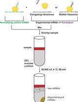
II. Isolation of polysomes
- Prepare cells at least one day before lysing. Confluency is very important. About 80-90% confluency gets the maximum polysomes. 1-5 15-cm dishes are needed to see polysome profile.
- Prior to lysing the cells, treat cells with 100 μg/ml cycloheximide at a final concentration for 5 min.
- Wash cells twice with 10 ml of ice-cold 1x PBS containing 100 μg/ml cycloheximide.
- Scrape cells with 5 ml of ice-cold 1x PBS containing 100 μg/ml cycloheximide, collect them in 15 ml tube and rinse with 5 ml of ice-cold 1x PBS containing 100 μg/ml cycloheximide.
- Centrifuge cells at 1,200 rpm (300 x g) for 5 min at 4 °C.
- Aspirate supernatant, resuspend cells in 425 μl of hypotonic buffer and transfer all to a new pre-chilled 1.5 ml tube.
- Then add:
5 μl of 10 mg/ml cycloheximide
1 μl of 1 M DTT
100 units of RNasin - Vortex for 5 sec.
- Then add:
25 μl of 10% Triton X-100
25 μl of 10% Sodium Deoxycholate - Vortex for 5 sec.
- Centrifuge at 15,000 rpm (21,000 x g, a maximal speed on a bench top centrifuge in 1.5 ml eppendorf tube) for 5 min at 4 °C.
- Transfer supernatant (about 500 μl) to a new pre-chilled 1.5 ml tube. Measure OD260nm for each sample using NanoDrop.

Figure 1. Polysome profiling from MEFs treated with DMSO or Ink1341 (250 nM) for 8 h from Dowling et al. (2010). 40 S, 60 S and 80 S denote the corresponding ribosomal subunits and monosome, respectively. - Load the same OD amount of lysate onto each gradient (10-30 OD260nm is loaded). Keep 10% of lysates as an input.
- Weight and balance each gradient.
- Centrifuge at 35,000 rpm for 2 h at 4 °C using SW40Ti rotor in a Beckman Coulter (Optima L-80 ultra centrifuge). No brake of brake at 1 (maximum) for deceleration. Acceleration does not matter much.
- While the samples are centrifuging, clean fraction collector with warm MilliQ water containing a bit of RNase ZAP (a few sprays).
- Carefully remove tubes from the rotor and place them at 4 °C until they are ready for running.
- Switch on computer, pump, UV detector and fraction collector. Set pump at 1.5 ml/min and the fraction collector by time. Place 2 ml tubes on fraction collector.
- Open TracerDAQ program.
- Run chasing solution (60% (w/v) sucrose with bromophenol blue) through the system until it reaches the needle. Make sure to see at least one drop coming out of the needle such that no bubbles are introduced into the gradient.
- Screw each tube onto the ISCO UV detector and then pierce the tube with the needle.
- Begin running the chasing solution through the gradient. Run solution with the pump at 1.5 ml/min. Click on acquire data button and press run on the fraction collector.
- Place fractions on dry ice or liquid nitrogen.
- Add 750 μl of Trizol for RNA isolation.
III. RNA isolation and RT-qPCRMQ
- Add 150 μl of chloroform to each fraction.
- Isolate RNA according to manufacturer’s Trizol protocol.
- Measure the RNA concentration of each fraction or input.
- Prepare the following solutions in 500 μl tubes or PCR tubes.
0.5 μg of RNA x μl
Oligo (dT) primer 1 μl
dNTP (10 mM each) 1 μl
Water x μl/total 13 μl - 65 °C for 5 min.
- On ice for at least 1 min.
- Add the following solutions (7 μl premix) on ice. Make premix before adding.
5x buffer 4 μl
1 M DTT 1 μl
RNasin 40 units/μl 1 μl
RTase 1 μl - Mix gently and spindown on ice.
- 50 °C for 60 min.
- 70 °C for 15 min.
- Keep at 4 °C until next step.
- Add 180 μl of MQ.
- qPCR according to manufacturer’s protocol.
2x SYBR 10 μl
5’ Primer (10 μM) 0.4 μl
3’ Primer (10 μM) 0.4 μl
RT sample 5 μl
MQ 4.2 μl/Total 20 μl
Recipes
- 10x sucrose gradient buffer (50 ml)
200 mM HEPES (pH 7.6)
1 M KCl
50 mM MgCl2
100 μg/ml cycloheximide
1x protease inhibitor cocktail (EDTA-free)
100 units/ml RNase inhibitor - 10-50% sucrose solutions
Final (%) 60% stock (ml) water (ml) 10x sucrose gradient buffer (ml) 50 8.3 0.7 1 45 7.5 1.5 1 40 6.7 2.3 1 35 5.8 3.2 1 30 5.0 4.0 1 25 4.2 4.8 1 20 3.3 5.7 1 15 2.5 6.5 1 10 1.7 7.3 1 - Hypotonic buffer5 mM Tris-HCl (pH 7.5)
2.5 mM MgCl2
1.5 mM KCl
1x protease inhibitor cocktail (EDTA-free)
References
- Alain, T., Morita, M., Fonseca, B. D., Yanagiya, A., Siddiqui, N., Bhat, M., Zammit, D., Marcus, V., Metrakos, P., Voyer, L. A., Gandin, V., Liu, Y., Topisirovic, I. and Sonenberg, N. (2012). eIF4E/4E-BP ratio predicts the efficacy of mTOR targeted therapies. Cancer Res 72(24): 6468-6476.
- Dowling, R. J., Topisirovic, I., Alain, T., Bidinosti, M., Fonseca, B. D., Petroulakis, E., Wang, X., Larsson, O., Selvaraj, A., Liu, Y., Kozma, S. C., Thomas, G. and Sonenberg, N. (2010). mTORC1-mediated cell proliferation, but not cell growth, controlled by the 4E-BPs. Science 328(5982): 1172-1176.
- Larsson, O., Morita, M., Topisirovic, I., Alain, T., Blouin, M. J., Pollak, M. and Sonenberg, N. (2012). Distinct perturbation of the translatome by the antidiabetic drug metformin. Proc Natl Acad Sci U S A 109(23): 8977-8982.
Article Information
Copyright
© 2013 The Authors; exclusive licensee Bio-protocol LLC.
How to cite
Morita, M., Alain, T., Topisirovic, I. and Sonenberg, N. (2013). Polysome Profiling Analysis. Bio-protocol 3(14): e833. DOI: 10.21769/BioProtoc.833.
Category
Molecular Biology > RNA > mRNA translation
Do you have any questions about this protocol?
Post your question to gather feedback from the community. We will also invite the authors of this article to respond.