


- Protocols

- Articles and Issues
- For Authors
- About
- Become a Reviewer


Cost-Effective Transient Expression of Viral Glycoproteins Using Mammalian Expi293F Cells

(§Technical contact: h.nijhuis1@amsterdamumc.nl) Published: Vol 16, Iss 23, Dec 5, 2026 DOI: 10.21769/BioProtoc.5899 Views: 38
Reviewed by: David PaulSowmya GunasekaranAnonymous reviewer(s)
 PDF
 Ask a question


Protocol Collections
Comprehensive collections of detailed, peer-reviewed protocols focusing on specific topics




Abstract
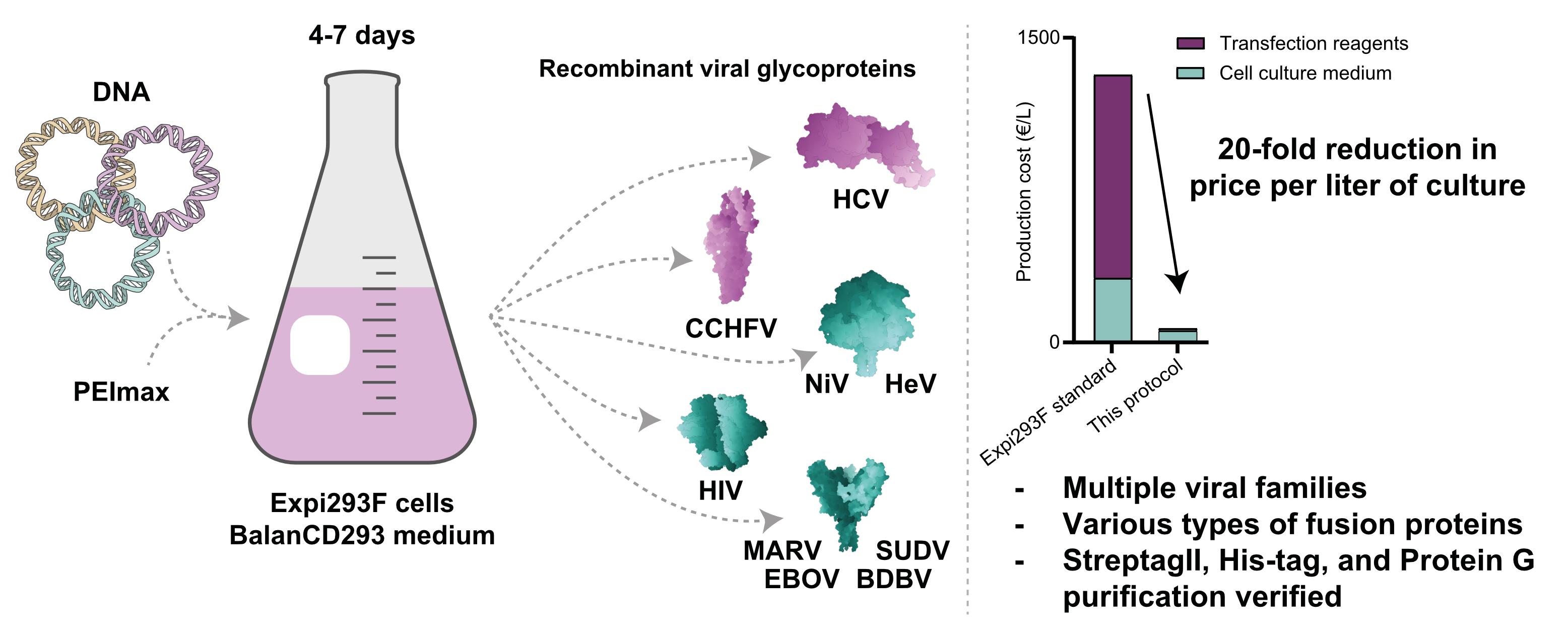
The efficacy of most recombinant viral vaccines relies on surface glycoproteins to induce immunity and protection from infection. However, many viral glycoproteins are notoriously difficult to manufacture both at lab and industrial scales for in vitro experiments and immunization studies. They usually comprise multiple subunits, the quaternary structure of which is often critical for the induction of relevant immune responses. Furthermore, they frequently contain complex, host-derived glycosylation patterns. Correct glycosylation, multimerization, and recapitulation of the native protein structure in a recombinant soluble form often depend on human-derived suspension cell culture. The associated high costs restrict the use of these systems to well-funded and/or specialized labs. Here, we describe the production of glycoproteins derived from diverse viruses (henipaviruses, filoviruses, human immunodeficiency virus, Andes virus, Crimean–Congo hemorrhagic fever virus, and hepatitis C virus) using human-derived Expi293F cells; we found these can be produced with existing infrastructure and commercially available components at less than 10% of the cost compared to conventional transfection protocols. This protocol combines general Expi293F cell culture principles with more economical and widely available reagents. First identified for the production of antibodies, it is now applied and expanded to produce a set of challenging recombinant viral proteins, not requiring any additional equipment beyond those necessary for basic suspension cell culture.
Key features
• Builds upon antibody production protocols and Expi293F transfection optimization previously examined by Zhou et al. [1] and Hederoth et al. [2].
• Uses BalanCD293 as medium, PEImax as transfection reagent, and glass flasks for culturing, which greatly reduce cost and plastic waste
• Minimal loss of expression for volumes between 5 and 600 mL.
• Provides additional information on cell supernatant storage and both StrepTagII and His-tag purification.
Keywords: Viral glycoproteins Expi293F culturing
Suspension cell culture
Transient protein expression
Tag-based purification
Graphical overview
Background
Several viral outbreaks in recent years have exemplified the importance of viral surface glycoproteins (VGPs) in vaccines and in immunological research. The Zaire ebolavirus outbreaks in West Africa in 2014–2015 and in the Democratic Republic of the Congo in 2018 are unlikely to recur because of two newly developed protein vaccines primarily based on VGPs [3]. SARS-CoV-2, the etiologic agent of COVID-19, was likewise eventually controlled by several vaccines that were based on VGPs [4–6]. In both cases, the relatively short time between outbreak and vaccine was reliant on decades of research on the form and function of similar viral proteins, not to mention their use in diagnostics and antibody discovery [5–8]. When recombinantly produced glycoproteins recapitulate their native form, they are instrumental in both fundamental research and clinical applications. Examples include, but are not limited to, structural biology, understanding viral entry mechanisms, determining tissue tropism, and (pre-)clinical applications, such as vaccine development, diagnostics, and antibody discovery.
VGPs are often flexible and multimeric in nature, which can make achieving their native form difficult. Their main function of receptor engagement and membrane fusion hinges on a metastable protein complex that can exist in different conformations, often pH- or host protease-dependent [6–10]. Three classes of viral fusion proteins have been described, each with its own fusion mechanism. Humoral immunity is mostly effective against the protein conformation present at the surface of a virion prior to infection, less so against the conformations that arise after receptor engagement, during refolding, or after membrane fusion [11]. This conformational diversity greatly complicates manufacturability of a stable, unchanging, native-like glycoprotein. A second aspect that complicates the manufacturability of VGPs is the glycans present on their surface. Glycans form immunological shields against the host immune system, are frequently involved in the early stages of receptor binding, and are therefore essential to be correctly displayed on recombinant proteins in research or clinical settings [12]. Not all host species glycosylate their proteins, nor do glycosylating species do so identically. This quickly limits the production cell lines available for manufacturing VGPs to a human origin [12]. Combining these characteristics, flexible, multimeric, and correctly glycosylated VGPs are difficult to produce. Mammalian suspension cell lines, such as Expi293F cells, are paramount to purifying enough protein to allow downstream analyses. However, the associated consumable costs make employing these cell lines untenable for many labs. Here, we describe a low-cost production of VGPs in Expi293F cells without expensive investments beyond basic suspension cell culture equipment (Figure 1).
This protocol builds upon the work described by Zhou et al. [1] and Hederoth et al. [2], in which the cost-effectiveness of antibody production in Expi293F cells was optimized by using PEImax as the transfection reagent and BalanCD293 as the growth medium. They compared the use of PEImax and BalanCD293 with the efficient but expensive ExpiFectamine293 and Expi293F expression media, which are sold specifically for Expi293F culture. We have expanded on their work by producing numerous VGPs from a variety of species and adopted these principles to develop 15 mL Expi293F cultures that allow for sufficient purified recombinant VGPs. Additionally, our protocol details other avenues of long-term cost reduction and reduced environmental impact, such as the use of reusable glass culturing flasks and reusable plastic buckets for centrifugation. Our protocol is limited by its scope, as production yield per liter of culture is only one of the first steps in characterization of a recombinant VGP. While having a yield at all is necessary to start, yield by itself is a poor indicator of protein quality. VGP yields are not primarily dictated by production conditions, but rather by inherent protein complexity. Protein-specific optimization could combat or reduce complexity but needs to be assessed on a protein-individual basis. Several of the protein stability optimizations used for constructs produced in this protocol are described, though not exhaustively, by Rutten et al. [13] for filovirus GP stabilization, Loomis et al. [14] for henipavirus F stabilization, Sanders et al. [15] for HIV Env stabilization, and Capella-Pujol et al. [16] for HCV E1E2 stabilization. Like Zhou et al. [1] and Hederoth et al. [2], we have successfully used this protocol to produce monoclonal antibodies (mAbs) and Fabs at comparable yields. The diversity of VGPs we produce here suggests other proteins requiring mammalian expression could also be produced using our protocol.
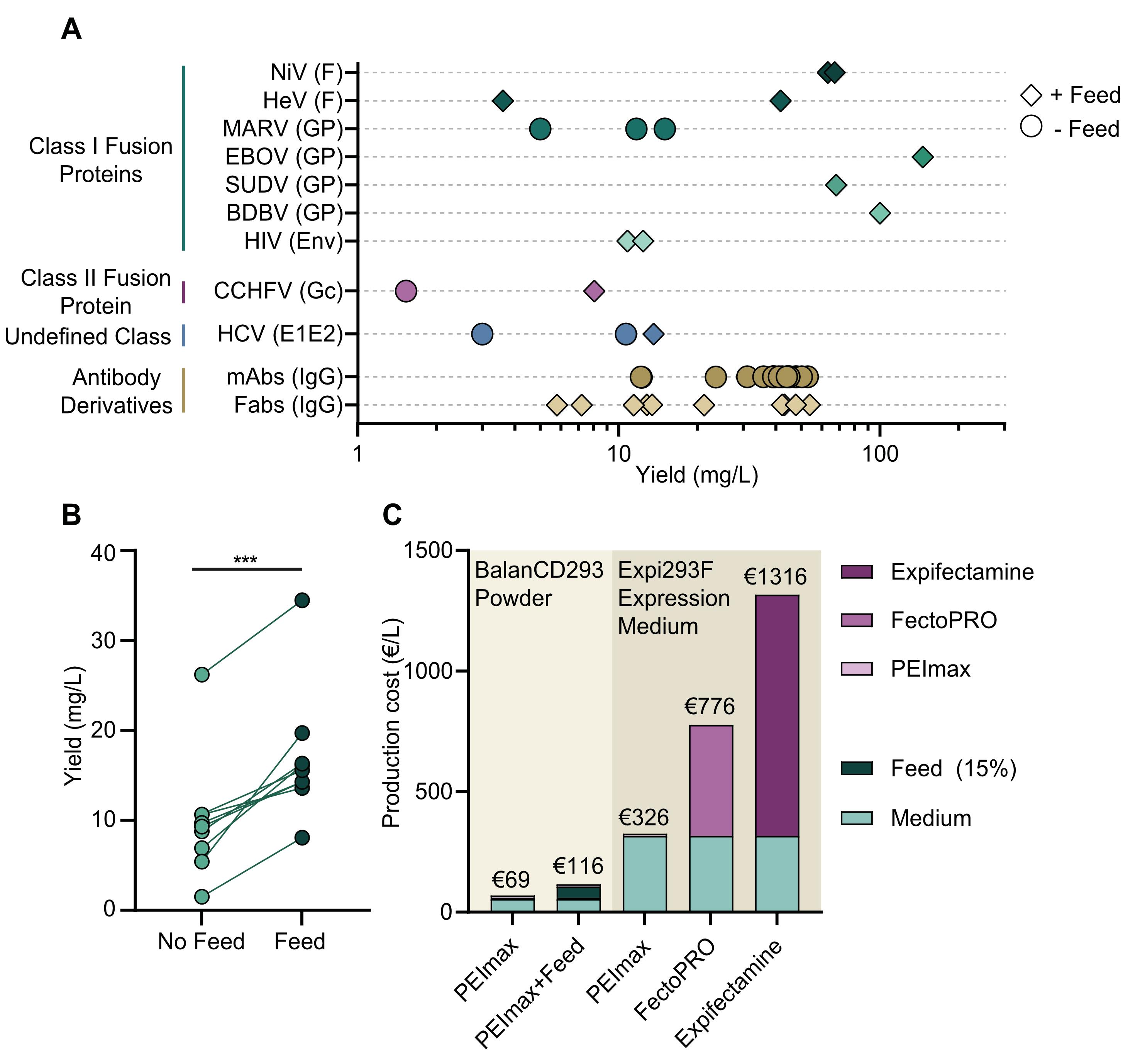
Figure 1. Viral surface glycoproteins from several families can be produced cost-effectively. (A) Production yield in milligrams of protein per liter of culture of glycoproteins derived from several viral families, with (diamond) or without feed (circle), and scaled from various smaller cultures. All data points shown are single biological replicates. Nipah virus (NiV), Hendra virus (HeV), Marburg virus (MARV), Zaire ebolavirus (EBOV), Sudan ebolavirus (SUDV), Bundibugyo ebolavirus (BDBV), human immunodeficiency virus (HIV), Crimean–Congo hemorrhagic fever virus (CCHFV), hepatitis C virus (HCV), monoclonal antibodies (mAbs), Fab-arm only antibodies (Fab), fusion protein (F), glycoprotein (G), envelope (Env), glycoprotein C/N-terminal (Gc/n), envelope protein 1 and 2 (E1E2). See General note 5 for elaboration on viral species. (B) Increased production yields by using BalanCD293 Feed of several unspecified VGPs and mAbs. ***p < 0.005 by paired t-test. (C) Cost of transfection in EUR per liter of Expi293F culture for various commercial options as of August 2026 at our location (the Netherlands).
Materials and reagents
Biological materials
1. Expi293F cells (Thermo Fisher, Gibco, catalog number: A14527)
Reagents
1. BalanCD 293 powder (Fuji Film, catalog number: 94137), storage: 4 °C
2. BalanCD feed (Fuji Film, catalog number: 91166), storage: 4 °C
3. L-glutamine (200 mM) (Fuji Film, catalog number: 9317), storage: -20 °C
4. Sodium bicarbonate (NaHCO3) (Sigma-Aldrich, catalog number: S5761)
5. PEImax hydrochloride (Kyfora Bio, catalog number: 24765-1)
6. OptiMEM (Gibco, catalog number: 31985-054), storage: 4 °C
7. Trypan Blue (Sigma-Aldrich, catalog number: T8154)
8. Hydrogen chloride (HCl) (Millipore, catalog number: 1.00317.1011)
Caution: This chemical is corrosive.
9. 70% ethanol (Added Pharma, catalog number: ROL1642610)
Caution: This chemical is flammable.
10. Ultrapure water (Elga, Purelab flex)
Optional reagents
11. Salmon semen DNA (Invitrogen, catalog number: 15632011)
12. Sodium azide (Sigma-Aldrich, catalog number: S2002)
Caution: This chemical is toxic.
13. Valproic acid (Sigma-Aldrich, catalog number: P4543)
Caution: This chemical is corrosive.
14. Peptone (Sigma-Aldrich, catalog number: P4963)
15. Glucose (Sigma-Aldrich, catalog number: G6152)
16. StraptactinXT beads (iba, catalog number: 2-5010-010)
17. Protein G beads (Thermo Fisher, catalog number: 20397)
18. Ni Sepharose excel resin (Cytiva, catalog number: 17371201)
Solutions
1. BalanCD Culture medium (see Recipes)
2. PEImax transfection reagent (see Recipes)
Recipes
1. BalanCD culture medium
| Reagent | Final concentration | Quantity or volume |
|---|---|---|
| BalanCD powder | 21.32 g/L | 213.2 g |
| NaHCO3 | 2.2 g/L | 22 g |
| L-Glutamine (200 mM stock) | 4 mM | 200 mL |
| Ultrapure water | n/a | 9.8 L |
| Total | n/a | 10 L |
Note: BalanCD293 is also available as a liquid, but the powdered form is more cost-effective. Preparation of the medium is also described by the manufacturer and can be done at room temperature [17].
a. Add 21.32 g/L BalanCD 293 powder medium to ultrapure water in an appropriately sized container.
Critical: Do not use demiwater.
b. Mix the solution at 300–350 rpm for 10 min or until the powder is fully dissolved.
Note: The solution may still appear cloudy at this stage.
c. Add 2.20 g/L NaHCO3 to the solution and mix at 300 rpm.
d. Adjust pH to 6.7–7.4 with HCl if needed.
e. Supplement with 20 mL/L of 200 mM L-Glutamine.
Note: L-Glutamine 200 mM should be kept at -20 °C prior to use and should be used within two weeks when stored at 4 °C.
f. Filter-sterilize through a 0.22 μm Steritop filter into a sterile bottle.
Critical: Filter inside a laminar flow hood or add/remove Steritop inside a laminar flow hood only.
Critical: Use one Steritop filter per 5 L of medium maximum, as more volume can compromise the membrane.
Note: The solution can be stored in the fridge (4 °C) for up to 1 year.
2. PEImax transfection reagent
| Reagent | Final concentration | Quantity or volume |
|---|---|---|
| PEImax hydrochloride (MW 40,000) | 1 g/L | 100 mg |
| HCl (37%) | n/a | Until pH 7.0 |
| Ultrapure water | n/a | 100 mL |
| Total | 100 mL |
a. Weigh 0.1 g of PEImax hydrochloride (MW 40,000).
b. Dissolve in 100 mL of ultrapure water.
c. Set pH to 7.0 using 37% HCl.
Note: pH affects transfection efficiency.
Caution: This chemical is corrosive.
d. Filter-sterilize using 0.22 μm filters and aliquot in the flow hood using sterile tubes.
Note: Aliquots of 1–5 mL are recommended.
e. Store at -20 °C for up to 2 years.
f. Can be stored at 4 °C for a week.
Note: Refreezing PEImax solution is not recommended.
Laboratory supplies
1. Tubes (sterile: to hold reagents during transfection; non-sterile: to store small volumes of clarified supernatant): 1.2 mL cryovials (Sarstedt, catalog number: 72.377), 1.5 mL Eppendorf (Sarstedt, catalog number: 72.069.001), 15 mL tubes (Sarstedt, catalog number: 62.554.502), 5 mL tubes (Eppendorf, catalog number: 0030119401), and 50 mL tubes (Sarstedt, catalog number: 62.547.254)
2. Pipette tips (sterile and non-sterile: to transfer small volumes): 10 μL (Greiner, catalog number: 771354), 20 μL (Greiner, catalog number: 775355), and 1,000 μL (Greiner, catalog number: 778354)
3. Serological pipettes (sterile and non-sterile: to transfer larger volumes): 5 mL (Corning, catalog number: 4051), 10 mL (Corning, catalog number: 4101), 25 mL (Corning, catalog number: 4251), and 50 mL (Corning, catalog number: 4501)
4. Centrifugation vessels (non-sterile: to centrifuge supernatant during harvesting): 250 mL plastic (Nalgene, catalog number: 3141-0250), 1 L plastic (Hettich, catalog number: 4239), and 2 L plastic (Hettich, catalog number: 550)
5. Syringes (sterile or non-sterile: to filter-sterilize transfection mix; sterile recommended): 5 mL (BD, catalog number: 309649), 10 mL (BD, catalog number: 300912), 20 mL (BD, catalog number: 300629), 30 mL (BD, catalog number: 301229), and 50 mL (BD, catalog number: 300865)
6. Filters (sterile: to filter-sterilize prepared BalanCD 293, transfection mix, and supernatant during harvesting): syringe filter 0.2 μm (Cytiva, catalog number: 10462200), Steritop 0.22 μm (Millipore, catalog number: S2GPT05RE), AcroPrep 24-wells 0.22 μm (Cytiva, catalog number: 97026), and Spin-X 0.22 μm (Costar, catalog number: 8160)
7. Culturing vessels (sterile: to culture Expi293F cells): 50 mL mini-bioreactors (Greiner, catalog number: 227245), 125 mL Plastic (Corning, catalog number: 431405), 250 mL plastic (Corning, catalog number: 431407), 500 mL plastic (Corning, catalog number: 431401), 1,000 mL plastic (Corning, catalog number: 431147), 2,000 mL plastic (Corning, catalog number: 431256), 250 mL glass (DWK Life Sciences, catalog number: 212833655), 500 mL glass (DWK Life Sciences, catalog number: 212834454), and 1,000 mL glass (DWK Life Sciences, catalog number: 212835459)
8. Bottles (sterile or non-sterile: to store supernatant after filtering/prior to purification; sterile recommended): 150 mL plastic (Corning, catalog number: 431175), 250 mL plastic (Corning, catalog number: 430281), 500 mL plastic (Corning, catalog number: 430282), 1,000 mL plastic (Corning, catalog number: 430518), 250 mL glass (VWR, catalog number: 215-1593), 500 mL glass (VWR, catalog number: 215-1594), and 1,000 mL glass (VWR, catalog number: 215-1595)
9. Counting slides (Bio-Rad, catalog number: 1450011) (non-sterile: to count cells during subculturing or prior to transfection)
10. pH strips (Millipore, catalog number: 1.00543.0001) (non-sterile: to measure BalanCD 293 pH)
11. 10 L buckets (Bel Art, catalog number: 15590986) (non-sterile: vessel to prepare one bottle of BalanCD 293 powder)
12. Magnetic stir bar (any brand) (non-sterile: to dissolve BalanCD 293 powder in ultrapure water)
13. 96-well plate U bottom (Sarstedt, catalog number: 53.3926) (non-sterile: to prepare Trypan Blue mixture prior to cell counting)
14. Tube rack (any brand) (non-sterile: used to hold tubes within or outside the flow hood)
Equipment
1. Incubator (Eppendorf, Newbrunswick S41i) (for culturing Expi293F cells) with mini-bioreactor rack insert (Eppendorf, model: M1289-0060) and sticky pads for flask culture (Eppendorf, model: M1250-9700)
2. Laminar flow hood (BioVanguard, Greenline) (for all handling of Expi293F cells, see General note 1)
3. Cell counter (Bio-Rad, model: TC20) (to count cells prior to subculturing or transfection)
4. Microscope (any brand) (to visually inspect cell health)
5. Water bath (Lauda, model: LD0726) (to prewarm culturing medium)
6. Fridge 2–8 °C (any brand) (to store culturing medium)
7. -20 °C freezer (any brand) (to store prepared PEImax solution)
8. -150 °C freezer (PHCBI, model: MDF-1156-PE) (to store Expi293F aliquots)
9. Pipettes (to transfer liquids within or outside the laminar flow hood): 2–20 μL (Gilson Pipetman), 20–200 μL (Gilson Pipetman), 100–1,000 μL (Gilson Pipetman), and Pipetboy (Hirschmann, Pipetus)
10. Stir plate (Heidolph, model: MR3002) (to dissolve BalanCD 293 powder in ultrapure water)
11. Vacuum pump (KNF Laboport, model: N840) (to filter-sterilize BalanCD 293 medium or cell supernatant)
12. Autoclave (HMC, model: HV85L) (to decontaminate biological waste)
13. Ultrapure water machine (Elga, Purelab flex) (to use for dissolving PEImax and BalanCD 293 powder)
14. Centrifuge (to centrifuge culture volumes during harvest): ≤250 mL buckets (Hettich, model: Rotanta 460R) and ≥250 mL buckets (Hettich, model: Roto Silenta 930 RS)
15. Centrifuge rotors (several sizes to accommodate several vessels): 15 or 50 mL tubes (Hettich, model: Rotanta 460R, Rotor 5699-R), 250 mL buckets (Hettich, model: Rotanta 460R, Rotor 5646), and 1 and 2 L buckets (Hettich, model: Roto Silenta 930 RS, Rotor 4176)
16. Scale(s) to measure 1 mg to 500 g (any brand) (to weigh PEImax and BalanCD 293 powder)
17. Mr. Frosty (Nalgene, model: 5100-0001) (to freeze Expi293F when storing aliquots)
18. NanoDrop One (Thermo Fisher, model: ND-ONE-W)
Procedure
A. Thaw Expi293F cells
Note: Expi293F establishment is also described in detail in the manufacturer’s instructions, besides the use of BalanCD 293 medium instead of Expi293 Expression medium to reduce costs [18].
1. Prewarm BalanCD medium to 37 °C.
2. Add 30 mL of prewarmed BalanCD 293 to a 125 mL flask.
3. Remove a vial of Expi293F cells from liquid nitrogen and swirl at 37 °C until a small clump of ice remains.
Critical: Do not submerge the vial in the water bath; keep swirling to ensure homogeneous thawing of the cells.
4. In one swift motion, pour the contents of the vial into the prepared flask.
Note: The cells can also be pipetted if desired.
5. Place cells in a shaking incubator with >80% relative humidity, 8% CO2, 37 °C, and set to 125 rpm.
Critical: Shaking speed recommendations differ based on shaking diameter and culture volume. Consult the manufacturer’s instructions for more details [18].
6. Between 3 and 7 days post-thaw, the cells should have reached a density of 3 × 10⁶/mL, with a viability of >90%.
7. Once cells reach 3 × 10⁶/mL, continue with subculturing the cells.
Critical: Cells can only be used for transfection after at least three passages post-thaw.
B. Subculture Expi293F cells
Note: Expi293F subculturing is also described in detail in the manufacturer’s instructions, besides the use of BalanCD 293 medium instead of Expi293 Expression medium to reduce costs [18].
1. Expi293F cells should be subcultured when they reach a density of 3 × 10⁶ to 5 × 10⁶/mL and diluted to a density of 0.3 × 10⁶/mL for 4-day intervals, or 0.5 × 10⁶/mL for 3-day intervals.
Example: Dilute cells to 0.5 × 10⁶/mL on Monday and 0.3 × 10⁶/mL on Thursday to be able to transfect cells on Tuesday and Friday and to avoid needing to handle the cells on weekends.
Critical: Never exceed one-third of the total flask volume with culture volume, e.g., <333 mL in a flask of 1 L.
Critical: Always use prewarmed medium to make dilutions.
Critical: Be extra careful to use sterile technique when handling the cells.
Notes:
1. We recommend against using glass flasks to subculture cells; see General note 6.
2. Flasks used for subculture can be reused for at least five passages and for subsequent transfections to save on flask usage.
2. Place cells in a shaking incubator with >80% relative humidity, 8% CO2, 37 °C, and set to 125 rpm.
Critical: Shaking speed recommendations differ based on shaking diameter and culture volume. Consult the manufacturer’s instructions for more details [18].
3. Cells can be reliably used up to passage number 30.
C. Store Expi293F cells
Note: Expi293F storage is also described in detail in the manufacturer’s instructions, besides the use of BalanCD 293 medium instead of Expi293 Expression medium to reduce costs [18].
1. Grow a volume of cells with a viable cell density of 3 × 10⁶ to 5 × 10⁶/mL to store 1 × 10⁷ cells per 1 mL aliquot.
Note: We advise storing multiple vials at as low a passage as possible to ensure long-term availability of cells.
2. Prepare freeze medium by making a solution of BalanCD 293 + 10% DMSO and cool to 4 °C prior to use.
3. Make a cell pellet by centrifuging the culture volume at 300× g for 5 min at RT.
4. Discard the medium and resuspend the cells in ice-cold freeze medium to a final density of 1 × 10⁷/mL.
Critical: Limit the amount of time between centrifugation and freezing as much as possible.
5. Freeze the cells using a Mr. Frosty or similar controlled-freezing device.
Note: Temperature decrease should be approximately 1 °C/min.
6. Store the cells in liquid nitrogen.
D. PEImax transfection (flasks)
Notes:
1. OptiMEM is often used as a complexation medium because of stable pH, stable osmolality, no serum, and its basis as a growth medium. All these factors benefit transfection efficiency and cell health directly after transfection.
2. PEImax, like other transfection reagents, is able to efficiently form complexes with DNA, enter the cells, and escape into the cytoplasm. It differs from other transfection reagents by being relatively cheap and less toxic to the cells, which allows the omission of changing the medium commonly seen with other transfection reagents.
1. Thaw PEI, DNA, and OptiMEM prior to transfection and warm to room temperature.
Note: According to the manufacturer, BalanCD293 culture medium also works as a complexation medium. According to Zhou et al. [1], PBS pH 7.2 should work as well.
2. Prewarm BalanCD293 culture medium to 37 °C.
3. One day prior to transfection, the cells should be in early log-phase growth (3 × 10⁶ to 5 × 10⁶/mL), healthy (viability >95%), and split down to 3 × 10⁶/mL.
Note: See General note 6 regarding the use of reusable glass flasks to save costs.
4. On the day of transfection, the cells should have grown to approximately 1.5× (4.5 × 106/mL) and should be diluted to 3 × 10⁶/mL.
5. Prepare three tubes per transfection:
a. One to mix DNA with OptiMEM (DNA tube).
b. One to mix PEImax with OptiMEM (PEImax tube).
c. One to mix filter-sterilized PEImax + OptiMEM and filter-sterilized DNA + OptiMEM (complexation tube).
6. The transfection ratios are as follows:
a. 1.0 μg DNA/mL of transfection volume.
Notes:
1. When using multiple plasmids, the total amount should still be 1 μg DNA/mL.
2. According to Hederoth et al. [2], up to 80% of input DNA can be replaced with salmon semen DNA to reduce cost.
b. 8.0 μL of PEImax/mL of transfection volume.
c. Total complexation volume should be 10% of transfection volume.
Example:
• 100 μg of DNA in 5 mL of OptiMEM for a 100 mL culture.
• 800 μL of PEImax in 5 mL of OptiMEM for a 100 mL culture.
7. Add and mix DNA plasmid(s) to OptiMEM in the DNA tube.
8. Add PEImax to OptiMEM and immediately filter-sterilize into the complexation tube.
Critical: PEImax will start complexation without DNA and will get caught in the 0.22 μm filters if not immediately filter-sterilized.
Note: The PEImax solution was already filter-sterilized during its preparation, so it could be added without filtering. To prevent precipitated PEImax from entering the culture and ensure sterility, we recommend filter-sterilizing.
9. Immediately filter-sterilize the prepared DNA + OptiMEM mix from step D7 into the complexation tube.
Critical: PEImax will start complexation without DNA and will not efficiently complex with the DNA if it is not immediately added.
10. Mix gently by inversion and incubate the complexation mix for approximately 15 min at room temperature.
11. Add the complexation mix to the cells, swirl the cell flask gently to incorporate the complexation mix with the culture, and put it back in the incubator immediately.
Notes:
1. Medium does not have to be changed after transfection.
2. The use of a positive control transfection is recommended.
12. Cells should be harvested before viability drops below 70%, typically between 4 and 7 days post-transfection.
E. PEImax transfection (mini bioreactors)
Note: The use of bioreactors can greatly increase the production throughput but is limited to approximately 20 mL of total volume and requires a dedicated incubator to facilitate a higher rpm necessary for proper cell health.
1. PEImax transfection of mini bioreactors follows the same general principle and ratios as flask transfection, using the following laboratory supplies at designated steps below to expedite the process:
a. When filtering <24 samples, use Spin-X filters to easily sterilize small volumes. Centrifuge DNA + OptiMEM or PEImax + OptiMEM at 1,000× g for 2 min or until the volume has passed through.
b. When filtering ≥24 samples, use AcroPrep filters to easily sterilize 24 samples at once. Centrifuge DNA + OptiMEM or PEImax + OptiMEM at 1,000× g for 5 min or until the volume has passed through.
F. Adding feed (optional)
Note: Adding BalanCD HEK293 feed will significantly increase your overall yield by extending the culture time to at least 7 days, but will increase the overall cost of transfection by approximately 70%/L. For more background regarding BalanCD HEK293 feed, see the manufacturer’s manual [19]. Background is not necessary for the procedure below.
1. Prewarm BalanCD HEK293 feed to 37 °C.
2. Add a total of 15% of your transfected volume as feed, spread over as many days as feasible, starting 24 h after transfection.
Notes:
1. Transfection mix added during transfection does not increase the volume of cells transfected; e.g., transfecting 100 mL of cells with 10 mL of transfection mix results in a transfected volume of 100 mL.
2. Up to 20% of your transfected volume can be supplemented as feed; e.g., 20 mL of feed added to 100 mL of transfected cell volume.
Example 1: Add 5 mL of feed 1, 2, and 3 days post-transfection to a volume of 100 mL.
Example 2: Add 15 mL of feed 1, 2, or 3 days post-transfection to a volume of 100 mL.
G. Harvesting cell supernatant
1. Harvest cell supernatant before cell viability drops below 70%, typically around day 5 (without feed) or day 7 (with feed).
2. Transfer the cells to a centrifugation container of choice and centrifuge them at 4,000× g for 30 min at room temperature.
Notes:
1. See General note 6 regarding usage of reusable plastic buckets to save costs.
2. Mini bioreactors can be spun down immediately without transfer to a new tube.
3. Filter the supernatant using Steritop filters and a vacuum pump for volumes >50 mL or syringes and syringe filters for volumes <50 mL.
Note: Multiple Steritop filters are usually necessary to filter the supernatant when volumes are >500 mL.
4. At this point, one can purify the clarified supernatant as they see fit. The recommendations below are purification method–specific optimizations that we have used. Other methods of purification might require similar optimization to maximize yield. See also General note 4.
a. His-tag purification:
i. Use Ni Sepharose Excel resin for purification.
Note: Other resins might not be compatible with BalanCD293 medium.
ii. Add 5 mL of 1 M Tris, pH 8, per 100 mL of supernatant.
iii. Add 7 mL of 5 M NaCl per 100 mL of supernatant.
b. StrepTagII purification: Use 1.8 mL of BioLock per 100 mL of supernatant and wait 15 min to capture the excess biotin prior to adding Streptavidin/StrepTactinXT coated beads.
5. Supernatant can be stored in multiple ways, depending on your specific downstream use. Below, we list some recommendations for short- and long-term storage:
a. Filter the supernatant inside a laminar flow hood to keep the supernatant sterile for longer periods (>1 week, 4 °C).
b. Add 1 mL of PBS + 0.05% sodium azide per 100 mL of supernatant to keep contaminants out for intermediate periods (2–7 days, 4 °C or room temperature).
Caution: High concentrations of sodium azide are toxic and should be handled according to your local chemical safety regulations.
c. Nothing needs to be added when protein is purified the day after (4 °C).
Data analysis
VGP and antibody concentrations shown in Figure 1 were measured using a NanoDrop One. Specific extinction coefficients and molecular weights of VGPs and Fabs were determined by ProtParam and used in the E & MW preset of the protein A280 function [20]. Monoclonal antibodies were measured using the IgG preset of the protein A280 function.
Validation of protocol
This protocol has not been used and validated in published research articles but has been used in various manuscripts that are currently in preparation.
General notes and troubleshooting
General notes
1. All steps in this protocol, until harvesting, should be conducted in a sterile environment (e.g., a laminar flow hood), and general sterile technique should be applied when handling Expi293F cells or any reagents added to those cells.
2. General Expi293F cell maintenance is done exactly according to the manufacturer’s instructions, except for the use of BalanCD193 medium.
3. When discarding excess and/or contaminated Expi293F cells, do so according to your local biosafety regulations.
4. Using purification methods besides His-tag or StreptagII might warrant optimization when using BalanCD293 culture medium. Of note, Protein A and G purification of antibodies is not affected by BalanCD293 culture medium.
5. VGPs are extremely diverse with regard to yield, purity, and folding, and as such, this protocol cannot guarantee high yields with every construct transfected. While most of the purified VGPs displayed the expected antigenic profile, we have noticed that some HIV envelope protein productions failed to meet our antigenic standards, likely because of small differences in glycosylation compared to HEK293F cells or cell culture media composition. Optimization of poorly producing or folding constructs is beyond the scope of this protocol.
6. Other cost-saving steps can be undertaken besides the usage of BalanCD293 culture medium and PEImax transfection reagent:
a. The use of autoclavable glass flasks with vented caps can greatly reduce the cost of culturing in the long term. Glass flasks are approximately twice as expensive as plastic ones and can be reused for years. The glass flasks appear slightly more prone to infection than individually wrapped plastic flasks and should thus only be used for transfections, not for subculturing, and should be placed under decontaminating UV light for 5 min prior to use. Glass flasks are cleaned as follows:
i. Remove cap and store separately.
ii. Clean flasks in the dishwasher.
Critical: Do not wash the caps, as this will compromise the membrane.
iii. Screw on the cap and autoclave the flask.
b. Instead of using disposable plastic to centrifuge cells during harvest, using plastic buckets in varying sizes can greatly reduce cost over time
i. Volumes <250 mL: Centrifuge Hettich Rotanta 460R, rotor 5699-R or 5646.
ii. Volumes >250 mL: Centrifuge Hettich Roto-Silenta 930 RS, rotor: 4176.
Critical: 2-L buckets should only be filled with 2 L, not less, or they will collapse under centrifugal forces.
7. Many other techniques and reagents can be used to possibly further improve yields or reduce costs. Below, we list approaches we have explored but ultimately did not include because of lack of efficacy in our limited experience:
a. Valproic acid to halt cell division
b. 45% glucose as feed
c. Peptone as feed
d. NaHCO3 to balance pH
8. Our protocol is limited by its scope, as production yield per liter of culture is only the first step in characterizing recombinant VGPs. This is by no means primarily dictated by production conditions but rather inherent protein stability. This could be remedied by protein-specific optimization, which is impossible to generalize. Several of the protein stability optimizations used for constructs produced in this protocol are described, though not exhaustively, by Rutten et al. [13] for filovirus GP stabilization, Loomis et al. [14] for henipavirus F stabilization, Sanders et al. [15] for HIV Env stabilization, and Capella-Pujol et al. [16] for HCV E1E2 stabilization.
Troubleshooting
Problem 1: Cells are not growing at the proper rate.
Possible cause: Improper dilution, improper medium preparation, underlying infection, high passage number, or incorrect flask size.
Solution: Take extra care when splitting the cells to not go below 0.3 × 10⁶/mL and use a new flask of medium. If after two passages cells do not recover, disregard this line and thaw a new vial. If the problem persists after thawing a new vial, prepare a new batch of medium, check the incubator for errors, and/or properly disinfect the entire flow cabinet. Do not exceed 30% fill volume of the culturing flask. Cell health prior to transfection is paramount to good protein yields.
Problem 2: Cells are dying within 5 days post-transfection with no signs of infection.
Possible cause: Transfected protein affects cells’ health.
Solution: Optimize harvest time.
Problem 3: Protein yields reduce over time despite good cell health.
Possible cause: High passage number (>25).
Solution: Start a new culture.
Problem 4: No protein yields despite good cell health and low passage number.
Possible cause: Unsuccessful transfection.
Solution: Retry with new cells and adhere to the order of filtering time and order of mixing to prevent PEImax complexation without DNA. Plasmid quality could also affect complexation and thus transfection efficiency.
Acknowledgments
This research was supported in part by Bill and Melinda Gates Foundation grants (INV-063951, INV-0106616) to R.W.S., a European Defense Fund grant (101103023) to M.J.G, and a Coefficient Giving grant to K.S. The authors thank Laura Radic for recommending this work to be published. The authors acknowledge the work of Zhou et al. [1] and Hederoth et al. [2], which greatly inspired this protocol.
Author contributions
Conceptualization, H.N., S.O., J.S., K.S.; Investigation, H.N., S.O., J.S., F.M., W.O., S.P., J.C.P., C.M.S., T.C.; Writing—Original Draft, H.N.; Writing—Review & Editing, H.N., S.O., J.S., F.M., W.O., S.P., J.C.P., C.M.S., I.B., T.C., M.G., R.S.; Funding acquisition, K.S., M.G., R.S.
Competing interests
The authors declare no conflicts of interest.
References
- Zhou, J., Yan, G., Cluckey, D., Meade, C., Ruth, M., Sorm, R., Tam, A., Lim, S., Petridis, C., Lin, L., et al. (2023). Exploring Parametric and Mechanistic Differences between Expi293FTM and ExpiCHO-STM Cells for Transient Antibody Production Optimization. Antibodies. 12(3): 53. https://doi.org/10.3390/antib12030053
- Hederoth, G., de la Rosa, A., Godec, A., Aguilar, X., Napoleone, A., Petrovic, A., Metzendorf, N. G. and Hultqvist, G. (2025). Updated transient gene expression protocol in Expi293F cells using PEI. Front Bioeng Biotechnol. 13: e1661193. https://doi.org/10.3389/fbioe.2025.1661193
- Saphire, E. O. (2020). A Vaccine against Ebola Virus. Cell. 181(1): 6. https://doi.org/10.1016/j.cell.2020.03.011
- Baden, L. R., El Sahly, H. M., Essink, B., Kotloff, K., Frey, S., Novak, R., Diemert, D., Spector, S. A., Rouphael, N., Creech, C. B., et al. (2021). Efficacy and Safety of the mRNA-1273 SARS-CoV-2 Vaccine. N Engl J Med. 384(5): 403–416. https://doi.org/10.1056/nejmoa2035389
- Voysey, M., Clemens, S. A. C., Madhi, S. A., Weckx, L. Y., Folegatti, P. M., Aley, P. K., Angus, B., Baillie, V. L., Barnabas, S. L., Bhorat, Q. E., et al. (2021). Safety and efficacy of the ChAdOx1 nCoV-19 vaccine (AZD1222) against SARS-CoV-2: an interim analysis of four randomised controlled trials in Brazil, South Africa, and the UK. Lancet. 397(10269): 99–111. https://doi.org/10.1016/s0140-6736(20)32661-1
- Polack, F. P., Thomas, S. J., Kitchin, N., Absalon, J., Gurtman, A., Lockhart, S., Perez, J. L., Pérez Marc, G., Moreira, E. D., Zerbini, C., et al. (2020). Safety and Efficacy of the BNT162b2 mRNA Covid-19 Vaccine. N Engl J Med. 383(27): 2603–2615. https://doi.org/10.1056/nejmoa2034577
- Harrison S. C. (2008). Viral membrane fusion. Nat Struct Mol Biol. 15(7): 690–698. https://doi.org/10.1038/nsmb.1456
- White, J. M., Delos, S. E., Brecher, M. and Schornberg, K. (2008). Structures and Mechanisms of Viral Membrane Fusion Proteins: Multiple Variations on a Common Theme. Crit Rev Biochem Mol Biol. 43(3): 189–219. https://doi.org/10.1080/10409230802058320
- Podbilewicz, B. (2014). Virus and Cell Fusion Mechanisms. Annu Rev Cell Dev Biol. 30(1): 111–139. https://doi.org/10.1146/annurev-cellbio-101512-122422
- Harrison, S. C. (2015). Viral membrane fusion. Virology. 479–480: 498–507. https://doi.org/10.1016/j.virol.2015.03.043
- Murin, C. D., Wilson, I. A. and Ward, A. B. (2019). Antibody responses to viral infections: a structural perspective across three different enveloped viruses. Nat Microbiol. 4(5): 734–747. https://doi.org/10.1038/s41564-019-0392-y
- Bagdonaite, I. and Wandall, H. H. (2018). Global aspects of viral glycosylation. Glycobiology. 28(7): 443–467. https://doi.org/10.1093/glycob/cwy021
- Rutten, L., Gilman, M. S., Blokland, S., Juraszek, J., McLellan, J. S. and Langedijk, J. P. (2020). Structure-Based Design of Prefusion-Stabilized Filovirus Glycoprotein Trimers. Cell Rep. 30(13): 4540–4550.e3. https://doi.org/10.1016/j.celrep.2020.03.025
- Loomis, R. J., Stewart-Jones, G. B. E., Tsybovsky, Y., Caringal, R. T., Morabito, K. M., McLellan, J. S., Chamberlain, A. L., Nugent, S. T., Hutchinson, G. B., Kueltzo, L. A., et al. (2020). Structure-Based Design of Nipah Virus Vaccines: A Generalizable Approach to Paramyxovirus Immunogen Development. Front Immunol. 11: e00842. https://doi.org/10.3389/fimmu.2020.00842
- Sanders, R. W., Derking, R., Cupo, A., Julien, J. P., Yasmeen, A., de Val, N., Kim, H. J., Blattner, C., de la Peña, A. T., Korzun, J., et al. (2013). A Next-Generation Cleaved, Soluble HIV-1 Env Trimer, BG505 SOSIP.664 gp140, Expresses Multiple Epitopes for Broadly Neutralizing but Not Non-Neutralizing Antibodies. PLoS Pathog. 9(9): e1003618. https://doi.org/10.1371/journal.ppat.1003618
- Capella-Pujol, J., Mulder, F., Cannac, F., Peters, S., Newby, M. L., Poniman, M., Zon, I., Olijhoek, W., Beaumont, T., Crispin, M., et al. (2026). Structure-based design of stable recombinant hepatitis C virus E1E2 heterodimers. Nat Commun. 17(1): e1038/s41467–026–75744–9. https://doi.org/10.1038/s41467-026-75744-9
- BalanCD HEK293 – Powder. Accessed on August 19th, 2026, https://fujifilmbiosciences.fujifilm.com/us/balancd-hek293-powder.html
- Expi293 Expression system protocol guide. Accessed on August 19th, 2026, https://www.thermofisher.com/nl/en/home/life-science/protein-biology/protein-expression/mammalian-protein-expression/transient-mammalian-protein-expression/expi293-expression-system/expi293-expression-system-protocol-guide.html
- BalanCD HEK293 – Feed. Accessed on August 20th, 2026, https://fujifilmbiosciences.fujifilm.com/us/balancd-hek293-feed.html
- Gasteiger, E., Hoogland, C., Gattiker, A., Duvaud, S., Wilkins, M. R., Appel, R. D. and Bairoch, A. (2005). Protein Identification and Analysis Tools on the ExPASy Server. In John M. Walker. (Ed). The Proteomics Protocols Handbook. pp: 571–607. https://doi.org/10.1385/1-59259-890-0:571
Article Information
Publication history
Received: Aug 26, 2026
Accepted: Oct 26, 2026
Available online: Nov 18, 2026
Published: Dec 5, 2026
Copyright
© 2026 The Author(s); This is an open access article under the CC BY license (https://creativecommons.org/licenses/by/4.0/).
How to cite
Nijhuis, H., Oeverdieck, S., Snitselaar, J. L., Mulder, F., Olijhoek, W., Peters, S., Capella-Pujol, J., Silva, C. M., Bontjer, I., Caniels, T. G., van Gils, M. J., Sanders, R. W. and Sliepen, K. (2026). Cost-Effective Transient Expression of Viral Glycoproteins Using Mammalian Expi293F Cells. Bio-protocol 16(23): e5899. DOI: 10.21769/BioProtoc.5899.

 Download Citation in RIS Format
Do you have any questions about this protocol?
Post your question to gather feedback from the community. We will also invite the authors of this article to respond.
 Post a Question


