


- Protocols

- Articles and Issues
- For Authors
- About
- Become a Reviewer


Capturing Proteins Co-Sorted into Budding Vesicles via an Integrated Proximity Labeling and In Vitro Vesicle Reconstitution Assay


Published: Vol 16, Iss 23, Dec 5, 2026 DOI: 10.21769/BioProtoc.5897 Views: 34
Reviewed by: Anik TuladharDevika AndhareAnonymous reviewer(s)
 PDF
 Ask a question

Original research article
The authors used this protocol in:
Jun 2026

Protocol Collections
Comprehensive collections of detailed, peer-reviewed protocols focusing on specific topics




Abstract
Protein trafficking relies on the selective packaging of cargo into transport vesicles, yet the molecular machinery governing specific cargo sorting remains incompletely understood. Co-immunoprecipitation, although widely used, often fails to capture weak or transient interactions within dynamic vesicle populations. Conventional proximity labeling methods also fail to distinguish bona fide vesicle-associated interactors from proteins merely near a cargo. Here, we describe a stepwise protocol that integrates proximity biotinylation with an in vitro vesicle reconstitution assay to selectively capture proteins that are co-packaged with a cargo of interest into budding vesicles. The cargo is fused to ascorbate peroxidase 2 (APEX2), and the resulting cargo–APEX2 fusion mediates proximity biotinylation of nearby proteins in live cells. The cells are then subjected to a cell-free vesicle formation reaction, followed by affinity capture of biotinylated proteins that are specifically incorporated into vesicles. This strategy reduces background contributions. Validated here with ShhN-APEX2 in HEK293T cells, this assay is expected to be adaptable to other secretory cargoes and provides a platform for systematic identification of trafficking regulators in development and disease.
Key features
• This protocol integrates proximity biotinylation with an in vitro vesicle formation assay to capture proteins co-sorted into budding vesicles with a specific cargo protein.
• This method minimizes background noise by isolating only biotinylated proteins that are specifically co-incorporated into reconstituted transport vesicles.
• This method was validated using Sonic hedgehog (Shh), applicable to diverse secretory cargoes.
Keywords: Proximity biotinylation APEX2
Vesicle reconstitution
Cargo sorting
Protein trafficking
Graphical overview
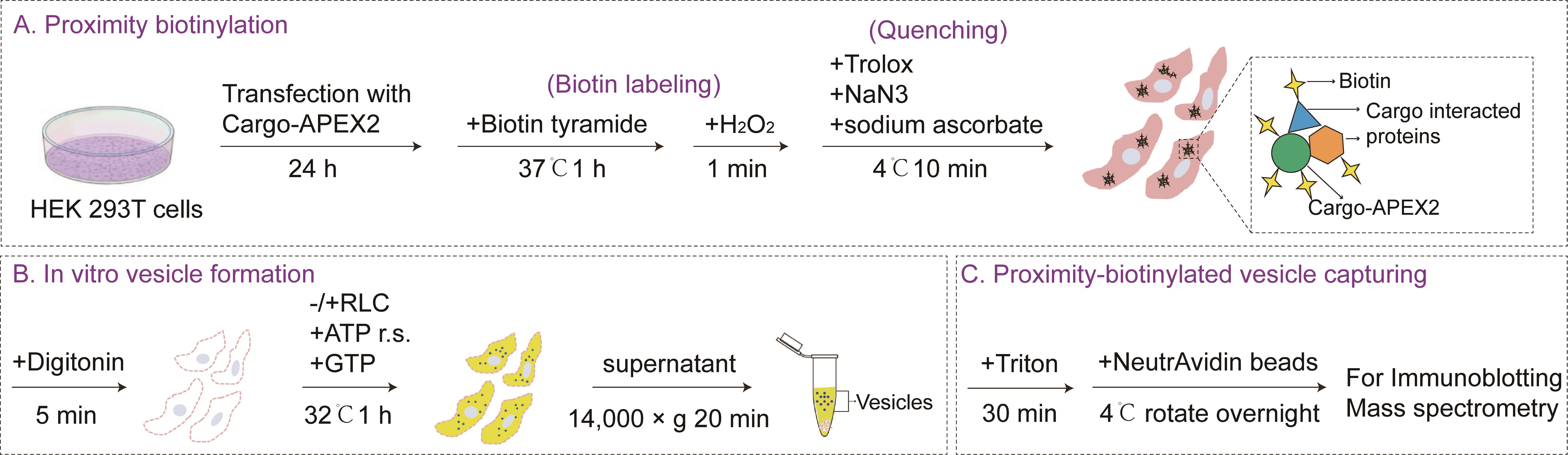
Schematic overview of the combined proximity biotinylation and in vitro vesicle reconstitution assay. Methods for proximity biotinylation, in vitro vesicle formation, and proximity-biotinylated vesicle capture are described in sections A, B, and C, respectively, of the Procedure. Efficiency of packaging of cargos into vesicles and the precipitation of biotinylated proteins were measured by immunoblotting, as described in steps C5–6 of the Procedure. RLC, rat liver cytosol. This figure is adapted from Song et al. [1].
Background
Intracellular protein trafficking relies on the selective packaging of cargo proteins into transport vesicles, a process fundamental to cellular organization, signaling, and development. However, the molecular mechanisms underlying cargo-specific vesicle sorting remain largely unclear. A critical challenge is to identify the protein mediators that govern cargo recognition, vesicle budding, and cargo delivery [2]. Conventional approaches for identifying trafficking regulators include co-immunoprecipitation (Co-IP) and proximity biotinylation [3,4]. Co-IP often fails to capture weak or transient interactions within dynamic vesicle populations. Proximity biotinylation labels proteins in the spatial vicinity of a bait but cannot discriminate between bona fide vesicle-associated interactors and abundant residents of donor compartments, leading to high background and frequent false positives. Both methods provide information about protein proximity but cannot directly distinguish vesicle-associated candidates from background proteins.
To overcome these limitations, we developed a protocol that integrates proximity biotinylation and an in vitro vesicle reconstitution assay. In this approach, the cargo of interest is fused to the engineered ascorbate peroxidase 2 (APEX2) and biotinylated in live cells. The cells are then permeabilized and subjected to a cell-free vesicle budding reaction [5,6]. Briefly, digitonin permeabilizes the plasma membrane, allowing cytosolic proteins to be removed by washing while intracellular membranes remain intact. The addition of rat liver cytosol (RLC), the ATP regeneration system (ATP r.s.), and GTP then supports vesicle budding, and the released vesicles are separated from the donor membranes by centrifugation. Biotinylated proteins that are co-packaged with the cargo into the released vesicles are subsequently enriched by affinity capture using NeutrAvidin agarose, effectively reducing background proteins from static organelles or non-vesicular compartments. Notably, proteins recovered by this approach represent candidates that are co-sorted or co-packaged with the cargo into vesicle-containing fractions. This enrichment does not necessarily indicate a direct physical interaction with the cargo, and such interactions require further validation.
This protocol has the potential to be adapted to map vesicle-associated interactomes of a wide range of secretory cargoes, including morphogens, growth factors, and disease-relevant cargoes. It may also be modified to study trafficking in different cell types or at distinct steps of the secretory pathway. Furthermore, the platform could be combined with CRISPR-based screening for high-throughput screening [7]. Overall, this protocol will provide a flexible tool for discovering trafficking regulators across diverse biological contexts.
Materials and reagents
Biological materials
1. HEK 293T cells; kindly provided by the University of California–Berkeley Cell Culture Facility, confirmed by short tandem repeat profiling and tested negative for Mycoplasma contamination
2. ShhN-HA-APEX2 plasmid; the ShhN-HA-APEX2 construct encodes the N-terminal fragment of mouse Shh (ShhN, aa 25–198) fused to a C-terminal 3×HA tag and APEX2; generated by BGI (Beijing Genomics Institute)
Note: The position of APEX2 should be considered during construct design, as it determines the subcellular scope of proximity labeling. For soluble cargoes such as ShhN, APEX2 labeling is restricted to the lumen side and is unlikely to reach cytosolic proteins involved in vesicle budding across an intact membrane. For transmembrane cargoes, the accessible protein populations depend on the topology of APEX2. If APEX2 is fused to a luminal domain, only luminal proteins are labeled. If it is fused to a cytosolic domain, cytosolic proteins can be labeled prior to permeabilization. Furthermore, when APEX2 is positioned on the cytosolic side, the order of the assay can be adjusted according to the experimental purpose. The vesicle budding reaction can be performed first, followed by biotin labeling, to capture cytosolic factors supplied by the RLC.
Reagents
1. Dulbecco's modified Eagle medium (DMEM) (Thermo Fisher Scientific, catalog number: 12800082)
2. Opti-MEM (Gibco, catalog number: 31985070)
3. Fetal bovine serum (FBS) (Thermo Fisher Scientific, catalog number: 10270106)
4. Penicillin-streptomycin (Thermo Fisher Scientific, catalog number: 15140122)
5. Polyethylenimine (Polysciences, catalog number: 23966-1)
6. 0.25% trypsin-EDTA (Thermo Fisher Scientific, catalog number: 25200056)
7. Sodium chloride (NaCl) (Sigma-Aldrich, catalog number: S5886)
8. Potassium chloride (KCl) (VWR Chemicals BDH®, catalog number: 2576)
9. Di-sodium hydrogen phosphate dihydrate (Na2HPO4·2H2O) (Scharlab, catalog number: SO03390500)
10. Potassium dihydrogen phosphate (KH2PO4) (Thermo Fisher Scientific, catalog number: A12142.36)
11. Biotin tyramide (Sigma-Aldrich, catalog number: SML2135)
12. Dimethyl sulfoxide (DMSO) (Sigma-Aldrich, catalog number: D8418)
13. Hydrogen peroxide solution (Millipore, catalog number: 1.07298)
14. Sodium ascorbate (Supelco, catalog number: PHR1279)
15. Trolox (MedChemExpress, catalog number: HY-101445)
16. Sodium azide (NaN3) (Sigma-Aldrich, catalog number: S8032)
17. HEPES (Acros Organics, catalog number: 172571000)
18. Acetic acid, potassium salt (KOAc) (Fisher Scientific, catalog number: 127082)
19. Magnesium acetate tetrahydrate [Mg(OAc)2] (Sigma-Aldrich, catalog number: M0631)
20. Digitonin (Sigma-Aldrich, catalog number: D141)
21. Creatine phosphate (Roche, catalog number: 10621722001)
22. Creatine kinase (Roche, catalog number: 10736988001)
23. Adenosine 5'-triphosphate (ATP) (Sigma-Aldrich, catalog number: 2383)
24. Guanosine 5'-triphosphate disodium salt (GTP) (FUJIFILM Wako Pure, catalog number: 073-03113)
25. Triton® X-100 (Sigma-Aldrich, catalog number: X100)
26. Protease inhibitor cocktail tablets (Roche, catalog number: 05056489001)
27. DL-dithiothreitol (DTT) (Sigma-Aldrich, catalog number: D9779)
28. PierceTM NeutrAvidinTM agarose (Thermo Fisher Scientific, catalog number: 29201)
29. Sodium dodecyl sulfate (SDS) (Sigma-Aldrich, catalog number: 151213)
30. Bromophenol blue (Sigma-Aldrich, catalog number: 114391)
31. Tris (Affymetrix, catalog number: 75825)
32. Glycerol (Sigma-Aldrich, catalog number: G7757)
33. 2-Mercaptoethanol (β-ME) (Sigma-Aldrich, catalog number: M6250)
34. Antibodies
a. Rabbit anti-HA (Cell Signaling Technology, catalog number: 3724), 1:2,000
b. Rabbit anti-calnexin (Abcam, catalog number: ab22595), 1:4,000
c. Mouse anti-SURF4 antibodies; kindly provided by Prof. Xiaowei Chen (Peking University, China), 1:2,000; generated by the Proteintech Group (www.ptglab.com) against the C-terminal epitope of SURF4 (N-glllvvalpgpgvsmedkekkw-C) [8]
d. Horseradish peroxidase (HRP)-coupled goat antibodies against rabbit IgG (Beyotime, catalog number: A0208), 1:4,000
e. HRP-coupled goat antibodies against mouse IgG (Beyotime, catalog number: A0216), 1:4,000
Solutions
A. Buffer solutions (see Recipes)
1. 1× PBS
2. 1× KHM buffer
3. Lysis buffer
4. Labeling medium
5. Quenching solution
6. High-salt KHM buffer
B. Stock solutions (see Recipes)
1. 10× PBS, pH 7.4
2. 1 M KOAc
3. 1 M HEPES-KOH, pH 7.2
4. 1 M Mg(OAc)2
5. 50 mM biotin tyramide
6. 1 M H2O2
7. 0.5 M Trolox
8. 3% NaN3
9. 0.25 M sodium ascorbate
10. 40 mg/mL digitonin
11. ATP regeneration system (ATP r.s.)
12. 10 mM GTP
13. 20 mg/mL rat liver cytosol (RLC)
14. 10% Triton® X-100
15. 100× protease inhibitors
16. 500 mM DTT
17. 1 mg/mL polyethylenimine
18. 1 M Tris-HCl, pH 6.8
19. 5× SDS-PAGE protein loading buffer
Recipes
A. Buffer solutions
1. 1× PBS (1 L)
Add 100 mL of 10× PBS to 900 mL of ddH2O.
2. 1× KHM buffer (1 L)
a. Add 5 mL of 1 M Mg(OAc)2, 20 mL of 1 M HEPES-KOH, pH 7.2, and 110 mL of 1 M KOAc to 0.5 L of ddH2O.
b. Adjust volume to 1 L.
c. Filter the buffer.
d. Store at 4 °C.
3. Lysis buffer (1 mL)
Add 0.05 mL of 10% Triton® X-100, 10 μL of 100× protease inhibitors, and 2 μL of 500 mM DTT in 938 μL of KHM buffer.
4. Labeling medium (10 mL)
Add 100 μL of 50 mM biotin tyramide to 10 mL of cell culture (DMEM containing 10% FBS, 10 milliunits/mL of penicillin, and 0.1 mg/mL of streptomycin).
5. Quenching solution (10 mL)
Add 0.4 mL of 0.25 M sodium ascorbate, 0.1 mL of 0.5 M Trolox, and 0.217 mL of 3% NaN3 in 10 mL of 1× PBS.
6. High-salt KHM buffer (1 L)
a. Dissolve 98.14 g of potassium acetate in 0.5 L of ddH2O.
b. Add 5 mL of 1 M Mg(OAc)2 and 20 mL of 1 M HEPES-KOH, pH 7.2.
b. Adjust volume to 1 L.
c. Filter the buffer.
d. Store at 4 °C.
B. Stock solutions
1. 10× PBS, pH 7.4 (2 L)
a. Dissolve 160 g of NaCl, 4 g of KCl, 28.8 g of Na2HPO4, and 4.8 g of KH2PO4 in 1.9 L of ddH2O.
b. Adjust pH to 7.4 with NaOH.
c. Add ddH2O to 2 L.
d. Sterilize by autoclaving and store at room temperature (RT).
2. 1 M KOAc (1 L)
a. Dissolve 98.14 g of potassium acetate in 1 L of ddH2O.
b. Sterilize by autoclaving and store at RT.
3. 1 M HEPES-KOH, pH 7.2 (1 L)
a. Dissolve 238.3 g of HEPES in 0.5 L of ddH2O.
b. Adjust pH to 7.2 with KOH
c. Make up to 1 L with ddH2O.
d. Store at RT.
4. 1 M Mg(OAc)2 (1 L)
a. Dissolve 155.41 g of Mg(OAc)2 in 1 L of ddH2O.
b. Sterilize by autoclaving and store at RT.
5. 50 mM biotin tyramide (1 mL)
a. Dissolve 18.17 mg of biotin tyramide in 1 mL of DMSO.
b. Store at -20 °C.
6. 1 M H2O2 (1 mL)
a. Add 102.1 μL of 30% H2O2 to 897.9 μL of ddH2O.
b. Prepare fresh and protect from light.
7. 0.5 M Trolox (1 mL)
a. Dissolve 0.125 g of Trolox in 1 mL of DMSO.
b. Store at -20 °C.
8. 3% NaN3 (50 mL)
a. Dissolve 1.5 g of NaN3 in 50 mL of ddH2O.
b. Store at RT.
9. 0.25 M sodium ascorbate (1 mL)
a. Dissolve 49.5 mg of sodium ascorbate in 1 mL of ddH2O.
b. Store at 4 °C.
10. 40 mg/mL digitonin (10 mL)
a. Dissolve 400 mg of digitonin in 10 mL of ddH2O.
b. Aliquot in volumes of 30 μL and store at -20 °C.
11. ATP regeneration system (20 mL)
a. Dissolve 2.04 g of creatine phosphate, 40 mg of creatine kinase, and 101.44 mg of ATP in 20 mL of 1× KHM.
b. Aliquot in small volumes and store at -80 °C.
12. 10 mM GTP (20 mL)
a. Dissolve 113.43 mg of GTP in 20 mL of 1× KHM.
b. Aliquot in small volumes and store at -80 °C.
13. 20 mg/mL RLC
RLC was prepared as described previously [9]. Briefly, livers from Sprague–Dawley rats were homogenized and subjected to sequential centrifugation at 1,000× g for 10 min, 18,459× g for 20 min, and 125,171× g for 1 h at 4 °C. The deep-red cytosolic fraction was collected, and the high-speed spin was repeated 2–3 times. The protein concentration was determined by the Bradford assay, and aliquots were snap-frozen in liquid nitrogen and stored at -80 °C. Avoid repeated freeze/thaw cycles.
14. 10% Triton® X-100 (50 mL)
a. Dissolve 5 mL of Triton® X-100 in 45 mL of ddH2O.
b. Store at 4 °C.
15. 100× protease inhibitors (1 mL)
a. Dissolve two protease inhibitor cocktail tablets in 1 mL of ddH2O.
b. Aliquot into volumes of 30 μL and store at -20 °C.
16. 500 mM DTT (1 mL)
a. Dissolve 0.077 g of DL-dithiothreitol in 1 mL of ddH2O.
b. Aliquot in volumes of 30 μL and store at -20 °C.
17. 1 mg/mL polyethylenimine (100 mL)
a. Dissolve 0.1 g of polyethylenimine in 100 mL of ddH2O.
b. Filter using a 0.22 μm filter.
c. Aliquot in small volumes and store at -80 °C.
18. 1 M Tris-HCl, pH 6.8 (1 L)
a. Dissolve 121.1 g of Tris in 0.8 L of ddH2O.
b. Adjust pH to 6.8 with HCl.
c. Add ddH2O to 1 L.
d. Sterilize by autoclaving and store at RT.
19. 5× SDS-PAGE protein loading buffer
a. Dissolve 3.75 g of SDS and 93.75 mg of bromophenol blue in a small beaker with 12.5 mL of 1 M Tris, pH 6.8, with constant stirring.
b. Mix in 25 mL of glycerol.
c. Adjust volume to 37.5 mL with ddH2O.
d. Aliquot to 750 μL/tube and store at -20 °C.
e. Add 250 μL of β-ME to an aliquot fresh before use.
Laboratory supplies
1. 10 cm tissue culture dishes (Biofil, catalog number: TCD-010100)
2. 15 mL centrifuge tubes (Biofil, catalog number: CFT-011150)
3. Falcon® 50 mL centrifuge tubes (Corning, catalog number: 352070)
4. 1,000 μL tips (USA Scientific, catalog number: 11112021)
5. 200 μL tips (Axygen, catalog number: T200-Y)
6. 10 μL tips (Axygen, catalog number: T300)
7. 1.5 mL microtubes (Axygen, catalog number: 20220415)
Equipment
1. 100 mL, 500 mL, 1 L, and 5 L beakers
2. 4 °C, -20 °C, and -80 °C freezers
3. Water bath
4. Eppendorf® 5418R centrifuge, refrigerated, with rotor FA-45-18-11 and rotor lid (Eppendorf, model: 5418R)
5. Light microscope with a 10× or 20× objective (any simple or compound light microscope is fine)
6. Barnstead Thermolyne (Thermo Fisher, model: DB16520-26)
7. Elite dry bath incubator (Major Science, model: EL-02)
8. Benchmark RotoMini rotator with tube holders (Benchmark, model: BMSR2020)
9. Thermo Scientific FormaTM Series 3 Water Jacketed CO2 Incubator (Thermo Fisher, model: 4120)
Software and datasets
1. ImageLab software v6.1
2. Adobe Photoshop
3. Adobe Illustrator
4. Proteome Discoverer
5. Perseus software (v1.6.6.0)
6. R packages: Vennerable (v3.1.0.9), clusterProfiler (v4.12.6), and ggplot2 (v3.5.12)
7. Alternatively, other image analysis programs and graphics software can be used, such as Fiji.
Procedure
Note: All procedures are performed on ice, and all centrifugations are performed at 4 °C unless otherwise stated.
A. Proximity biotinylation
1. Culture 10 cm plates of HEK 293T cells in DMEM containing 10% FBS, 10 milliunits/mL of penicillin, and 0.1 mg/mL of streptomycin at 37 °C in a cell culture CO2 incubator providing 5% CO2 and around 95% relative humidity. The cells will be grown to ~60%–70% confluence on the day of transfection.
Note: One 10-cm plate of HEK293T cells provides enough donor membranes for one 1.3 mL budding reaction. Therefore, the number of 10 cm plates required equals the number of budding reaction conditions tested.
2. Transfect the ShhN-HA-APEX2 plasmid 24 h before the experiment. For each 10 cm plate, dilute 15 μg of the ShhN-HA-APEX2 plasmid and 30 μL of 1 mg/mL polyethylenimine (DNA: polyethylenimine ratio = 1: 2) separately in 1 mL of Opti-MEM and incubate at room temperature for 5 min. Mix the two solutions and incubate for another 15 min. Change the culture medium to Opti-MEM and add the DNA–polyethylenimine mixture dropwise to the cells. After incubation at 37 °C for 6 h, replace the medium with complete medium. Cells will reach 80%–100% confluence on the day of the experiment.
3. Aspirate media from the plates of HEK 293T cells 24 h after transfection.
4. Wash the cells twice with 5 mL of PBS (Recipe A1).
5. Add 10 mL of labeling medium (Recipe A4) into the plate and incubate the cells at 37 °C for 1 h.
6. Add 1 M H2O2 to the plate to a final concentration of 1 mM and incubate the cells for 1 min with gentle shaking.Note: For peroxide-sensitive systems (e.g., peroxisomes), lower concentrations (e.g., 0.5 mM) and shorter durations (e.g., 30 s) may be required.
Caution: H2O2 is corrosive and a strong oxidant. Handle it with appropriate personal protective equipment and avoid contact with skin and eyes.
7. Remove the labeling medium immediately and add 10 mL of quenching solution (Recipe A5).
Caution: The quenching solution contains sodium azide, which is highly toxic. Handle it with appropriate personal protective equipment and avoid inhalation or skin contact. Dispose of sodium azide–containing waste according to institutional safety regulations. Do not mix sodium azide with acidic solutions or heavy metals to avoid generating toxic gases.
8. Incubate the cells at 4 °C in the dark for 10 min with gentle shaking.
9. Remove the quenching solution and wash the cells twice with 5 mL of PBS.
B. In vitro vesicle formation
1. Collect the cells using a cell scraper in 8 mL of ice-cold PBS and transfer the cells to a 15 mL Falcon tube.
2. Centrifuge at 300× g for 3 min at 4 °C. Aspirate the supernatant and resuspend the cells in 4 mL of 1× KHM buffer (Recipe A2) in each tube.
3. Add digitonin to the resuspended cells at a final concentration of 40 μg/mL. Mix well and incubate on ice for 5 min.
Note: Digitonin permeabilization is a highly sensitive step. In preliminary experiments, conditions were optimized by assessing membrane integrity using a Trypan Blue exclusion assay. An acceptable preparation was defined as over 90% of cells taking up Trypan Blue while retaining their overall cellular outline without extensive fragmentation. Based on this, we established the optimal condition for HEK 293T cells as incubation with 40 μg/mL digitonin on ice for exactly 5 min. If applying this protocol to other cell types, re-optimize the conditions.
4. Add 8 mL of 1× KHM buffer to the cells, invert the tube, and immediately pellet the cells at 300× g for 3 min at 4 °C.
5. Aspirate the supernatant, resuspend the cell pellet in 1 mL of 1× KHM buffer, and transfer to a 1.5 mL microcentrifuge tube.
6. Incubate the cells on ice for 5 min, pellet the cells at 10,000× g for 5 s at 4 °C, and aspirate the supernatant. Resuspend the pellet in 130 μL of 1× KHM buffer. This suspension serves as the donor membrane (DM) for subsequent vesicle formation assays.
Note: To ensure equivalent amounts of donor membranes across experimental groups, the DM suspension is thoroughly mixed by gentle pipetting before aliquoting equal volumes into each reaction tube. For scaling up, the number of 10 cm plates is increased proportionally to the number of reactions, and the harvested DM are pooled into a single master mix before being equally divided among all reaction tubes.
7. Assemble vesicle budding reactions by adding the ingredients listed in Table 1.
Table 1. Vesicle formation assay
| Ingredient | -RLC | +RLC |
|---|---|---|
| 1× KHM buffer | 1,014 μL | 884 μL |
| ATP regeneration system | 130 μL | 130 μL |
| GTP | 26 μL | 26 μL |
| DM (donor membrane) | 130 μL | 130 μL |
| RLC (final concentration 2 mg/mL) | 0 μL | 130 μL |
Note: Each column represents a single experimental group. Each row represents a component of the reaction and the volume added to the reaction in μL. The total volume of the reaction is 1.3 mL. One 10 cm dish of cells at ~80%–100% confluence is sufficient to generate one donor membrane preparation, which supports one 1.3 mL budding reaction. The experimental group without RLC serves as a negative control to monitor background. RLC was prepared as described previously [9].
8. Incubate the reaction mixture at 32 °C for 1 h.
9. Centrifuge at 14,000× g for 20 min at 4 °C in an Eppendorf® 5418R refrigerated centrifuge using the FA-45-18-11 rotor to remove cell debris and large membranes.
10. Transfer the supernatant to a 1.5 mL microcentrifuge tube, which represents the total vesicle fraction.
C. Proximity-biotinylated vesicle capturing
1. Add Triton® X-100, protease inhibitors, and DTT to the vesicle fraction to final concentrations of 0.5%, 1×, and 1 mM, respectively, and incubate on ice for 30 min.
2. Centrifuge at 14,000× g for 5 min at 4 °C and collect the supernatant.
3. Wash 30 μL of NeutrAvidin agarose twice with 1 mL of ice-cold lysis buffer (Recipe A3). Incubate the agarose with the supernatant obtained above and rotate overnight at 4 °C.
4. Wash the agarose five times with 1 mL of ice-cold lysis buffer (Recipe A3). The proteins co-sorted into budding vesicles remain bound to the agarose.
5. Prepare the following samples for analysis:
a. Biotinylated proteins: Add 2× SDS-PAGE protein loading buffer to the agarose (from step C4).
b. DM fraction: Add 2% DM from step B6 to 20 μL of lysis buffer (Recipe A3). Incubate on ice for 30 min and centrifuge at 14,000× g for 5 min at 4 °C. Collect the supernatant and add 5× SDS-PAGE loading buffer (Recipe B19) to the supernatant to a final concentration of 1×.
c. Vesicle fraction: Add 5× SDS-PAGE loading buffer to the vesicle lysate (from step C2) to a final concentration of 1×.
6. Incubate the samples at 55 °C for 30 min and proceed with immunoblotting or mass spectrometry analysis. Immunoblotting was performed as described previously [9]. As shown in Figure 1 (adapted from [1]), samples prepared through the above procedures were subjected to immunoblotting.
Data analysis
1. Export immunoblot images from ImageLab software v6.1 as .tif files.
2. Use Adobe Photoshop and Adobe Illustrator to process images.
3. For mass spectrometry data analysis, protein identification and label-free quantification were performed using Proteome Discoverer software. Protein intensities were normalized against the total ion current (TIC) of each sample. Differentially expressed proteins between the +RLC and -RLC groups were identified using Perseus software, with significance assessed by Student's t-test and corrected via permutation-based false discovery rate (FDR) adjustment (q-value). The Venn diagram and Gene Ontology (GO) cellular component enrichment analysis were generated using the Vennerable and clusterProfiler R packages, respectively.
Validation of protocol
This protocol has been used and validated in the following research article:
• Song et al. (2026). An integrated proximity labeling and vesicle reconstitution assay identifies novel regulators of Sonic hedgehog secretion. Molecular Biology of the Cell [1] (Figure 1 and Supplemental figure 1).
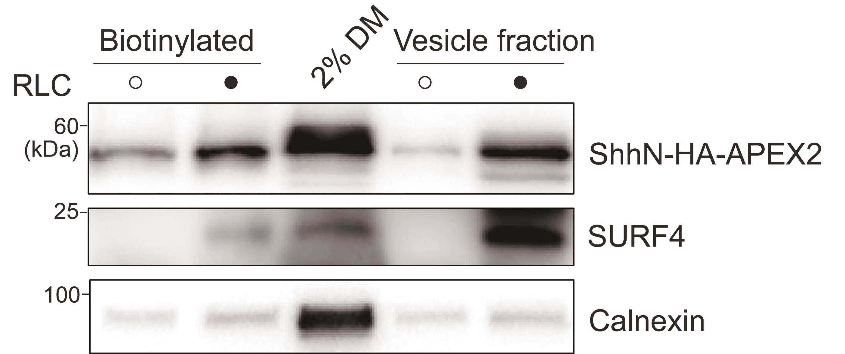
Figure 1. Proximity biotinylation and vesicle formation assay. HEK293T cells were transfected with a plasmid encoding ShhN-HA-APEX2. Twenty-four hours after transfection, proximity biotinylation and vesicle formation assays were performed in the absence or presence of rat liver cytosol (RLC). The biotinylated proteins in the vesicles were precipitated by NeutrAvidin agarose and analyzed by immunoblotting using anti-HA, anti-SURF4, or anti-calnexin antibodies. Data are representative of at least three independent experiments (Republished from Song et al. [1]).
General notes and troubleshooting
General notes
1. This protocol was developed and validated using ShhN fused to APEX2 in HEK 293T cells. It can be adapted to other secretory cargoes by generating the corresponding APEX2 fusion constructs. For cell types that are difficult to transfect, lentiviral transduction or stable cell line generation is recommended prior to the proximity labeling step.
2. In this protocol, the labeling condition uses 1 mM H2O2 for 1 min. For peroxide-sensitive systems (e.g., peroxisomes), lower concentrations (e.g., 0.5 mM) and shorter durations (e.g., 30 s) may be required.
3. In this protocol, the -RLC condition serves as a negative control to monitor nonspecific biotinylated proteins that remain in the vesicle fraction. For more comprehensive background filtering, we recommend including an APEX2-only labeling control.
Troubleshooting
Problem 1: High background in the -RLC control group.
Possible cause: Incomplete removal of cytosol during donor membrane preparation, leading to residual cytosolic proteins that persist in the -RLC condition.
Solutions: Increase the number and volume of washes after digitonin permeabilization; include a 5-min wash with 1 mL of high-salt buffer (Recipe A6) to remove residual cytosolic proteins.
Problem 2: Low vesicle budding efficiency.
Possible causes: Inactive RLC, ATP r.s., or GTP; damaged semi-intact cells due to excessive digitonin permeabilization or harsh handling.
Solutions: Verify activity of the reagents by performing a parallel budding reaction with a well-characterized cargo, such as Sec22B; ensure that the reagents are freshly thawed and stored in single-use aliquots to minimize repeated freeze/thaw cycles; check cell morphology after permeabilization to exclude over-permeabilization.
Problem 3: Weak or absent APEX2 biotinylation.
Possible causes: Degraded H2O2; insufficient labeling time; low APEX2 expression.
Solutions: Prepare fresh H2O2 and protect it from light; verify APEX2 expression and localization by immunoblotting or fluorescence before labeling; optimize labeling time.
Problem 4: Excessive APEX2 background.
Possible causes: Too high H2O2 concentration or too long labeling time; incomplete quenching; nonspecific biotinylation.
Solutions: Titrate H2O2 concentration and shorten labeling time; ensure fresh quenching solution is used and cells are washed thoroughly; compare +RLC and -RLC groups to identify nonspecific background.
Problem 5: Over- or under-permeabilization with digitonin.
Possible causes: Inappropriate digitonin concentration or incubation time; batch-to-batch variation in digitonin activity.
Solutions: Re-optimize digitonin concentration and incubation time using Trypan Blue staining; aliquot the digitonin stock to avoid repeated freeze/thaw cycles.
Problem 6: High calnexin or donor-membrane contamination in the vesicle fraction.
Possible causes: Incomplete removal of donor membranes; excessive membrane fragmentation; inappropriate centrifugation conditions.
Solutions: Handle semi-intact cells gently; optimize centrifugation speed and time to separate released vesicles from donor membranes; verify the vesicle fraction by immunoblotting for vesicle markers (e.g., Sec22B) and donor membrane markers (e.g., Calnexin).
Problem 7: Poor recovery from NeutrAvidin agarose.
Possible cause: Protein loss during washes.
Solution: Carefully aspirate the wash buffer to avoid losing or disturbing the agarose beads during washes.
Problem 8: Variability between RLC preparations.
Possible cause: Batch-to-batch differences in rat liver cytosol activity.
Solutions: Prepare RLC in large batches, aliquot into single-use tubes, and store at -80 °C; avoid repeated freeze/thaw cycles; test each new batch in a standard budding reaction with a well-characterized cargo such as Sec22B.
Problem 9: Loss of vesicle budding following repeated freeze/thaw cycles.
Possible cause: Freeze-thaw damage to RLC, ATP r.s., GTP, or other reaction components.
Solution: Aliquot RLC and other reagents into single-use aliquots; avoid repeated freeze/thaw cycles.
Problem 10: Poor expression or mislocalization of a new cargo-APEX2 fusion.
Possible causes: The APEX2 tag interferes with cargo folding or trafficking; low transfection efficiency; incorrect tag position.
Solutions: Optimize transfection conditions; test different tag positions (N- or C-terminal); verify expression and localization by immunofluorescence and immunoblotting; include a well-characterized positive control such as ShhN-HA-APEX2.
Acknowledgments
This protocol was modified from previous work [1,5,9]. We thank Dr. Xiaowei Chen (Peking University, China) for providing antibodies against SURF4. This work was supported by grants from the National Key R&D Program of China (2025YFA0923000 to Y.G.), Shenzhen Medical Research Fund (SMRF) (B2501003 to Y.G.), National Natural Science Foundation of China (NSFC32300589 to X.T.) and Anhui Province Higher Education Research Project (2022AH050174 to X.T.). This work was also supported by the Outstanding Innovative Research Team for Molecular Enzymology and Detection in Anhui Provincial Universities (2022AH010012), Biotechnology “Double-Teachers and Double-Abilities” Type Teacher Team in Anhui Provincial Universities (2025), the Key Laboratory of Biomedicine in Gene Diseases and Health of Anhui Higher Education Institutes, and Anhui Provincial Engineering Research Centre for Molecular Detection and Diagnostics. This work was also supported by Hong Kong Research Grants Council Grants (16104423, 16103624, AoE/M-402/25-N to Y.G.), and the Sinovac Fellowship Program to Y.G.
Author contributions
Conceptualization, X.T. and Y.G.; Investigation, Z.S., X.Z., and X.T.; Writing—Original Draft, X.Z. and X.T.; Writing—Review & Editing, X.T. and Y.G.; Funding acquisition, X.T. and Y.G.; Supervision, X.T. and Y.G.
Competing interests
The authors declare no conflicts of interest.
References
- Song, Z., Chen, Z., Tian, Y., Wang, Q., Zhao, X., Guo, Y. and Tang, X. (2026). An integrated proximity labeling and vesicle reconstitution assay identifies novel regulators of Sonic hedgehog secretion. Mol Biol Cell. 37(6): ee25–12–0627. https://doi.org/10.1091/mbc.e25-12-0627
- Barlowe, C. and Helenius, A. (2016). Cargo Capture and Bulk Flow in the Early Secretory Pathway. Annu Rev Cell Dev Biol. 32(1): 197–222. https://doi.org/10.1146/annurev-cellbio-111315-125016
- Huttlin, E. L., Bruckner, R. J., Paulo, J. A., Cannon, J. R., Ting, L., Baltier, K., Colby, G., Gebreab, F., Gygi, M. P., Parzen, H., et al. (2017). Architecture of the human interactome defines protein communities and disease networks. Nature. 545(7655): 505–509. https://doi.org/10.1038/nature22366
- Hung, V., Udeshi, N. D., Lam, S. S., Loh, K. H., Cox, K. J., Pedram, K., Carr, S. A. and Ting, A. Y. (2016). Spatially resolved proteomic mapping in living cells with the engineered peroxidase APEX2. Nat Protoc. 11(3): 456–475. https://doi.org/10.1038/nprot.2016.018
- Tang, X., Chen, R., Mesias, V. S. D., Wang, T., Wang, Y., Poljak, K., Fan, X., Miao, H., Hu, J., Zhang, L., et al. (2022). A SURF4-to-proteoglycan relay mechanism that mediates the sorting and secretion of a tagged variant of sonic hedgehog. Proc Natl Acad Sci USA. 119(11): e2113991119. https://doi.org/10.1073/pnas.2113991119
- Tang, X., Tian, Y., Wang, Q., Song, Z., Zhao, X. and Guo, Y. (2026). GRIA1 regulates TGN export and secretion of Sonic hedgehog. J Biol Chem. 302(2): 111084. https://doi.org/10.1016/j.jbc.2025.111084
- Christopher, J. A., Stadler, C., Martin, C. E., Morgenstern, M., Pan, Y., Betsinger, C. N., Rattray, D. G., Mahdessian, D., Gingras, A. C., Warscheid, B. et al. (2021). Subcellular proteomics. Nat Rev Methods Primers. 1: 32. https://pmc.ncbi.nlm.nih.gov/articles/PMC8451152/
- Wang, X., Wang, H., Xu, B., Huang, D., Nie, C., Pu, L., Zajac, G. J., Yan, H., Zhao, J., Shi, F., et al. (2021). Receptor-Mediated ER Export of Lipoproteins Controls Lipid Homeostasis in Mice and Humans. Cell Metab. 33(2): 350–366.e7. https://doi.org/10.1016/j.cmet.2020.10.020
- Tang, X., Yang, F. and Guo, Y. (2020). Cell-free Reconstitution of the Packaging of Cargo Proteins into Vesicles at the trans Golgi Network. Bio Protoc. 10(5): e3537. https://doi.org/10.21769/bioprotoc.3537
Article Information
Publication history
Received: Aug 24, 2026
Accepted: Oct 28, 2026
Available online: Nov 17, 2026
Published: Dec 5, 2026
Copyright
© 2026 The Author(s); This is an open access article under the CC BY-NC license (https://creativecommons.org/licenses/by-nc/4.0/).
How to cite
Readers should cite both the Bio-protocol article and the original research article where this protocol was used:
- Zhao, X., Song, Z., Guo, Y. and Tang, X. (2026). Capturing Proteins Co-Sorted into Budding Vesicles via an Integrated Proximity Labeling and In Vitro Vesicle Reconstitution Assay. Bio-protocol 16(23): e5897. DOI: 10.21769/BioProtoc.5897.
- Song, Z., Chen, Z., Tian, Y., Wang, Q., Zhao, X., Guo, Y. and Tang, X. (2026). An integrated proximity labeling and vesicle reconstitution assay identifies novel regulators of Sonic hedgehog secretion. Mol Biol Cell. 37(6): ee25–12–0627. https://doi.org/10.1091/mbc.e25-12-0627

 Download Citation in RIS Format
Do you have any questions about this protocol?
Post your question to gather feedback from the community. We will also invite the authors of this article to respond.
 Post a Question


