


- Protocols

- Articles and Issues
- For Authors
- About
- Become a Reviewer


Rapid Purification of Ion-Transporter Proteins in Arabidopsis thaliana

(*Contributed equally to this work, §Technical contact: 22482385@life.hkbu.edu.hk) Published: Vol 16, Iss 23, Dec 5, 2026 DOI: 10.21769/BioProtoc.5891 Views: 26
Reviewed by: Yuko KuritaAbdul ShirajAnonymous reviewer(s)
 PDF
 Ask a question


Protocol Collections
Comprehensive collections of detailed, peer-reviewed protocols focusing on specific topics




Related protocols
Abstract
Plant ion transporters are membrane-localized proteins that play critical roles in nutrient uptake and distribution, ion homeostasis, and stomatal movement. To date, the majority of research on ion transporters has focused on regulation of their activity, post-translational modifications, and endomembrane trafficking. In such work, it is often necessary to purify the full-length proteins. However, these transporters typically contain multiple transmembrane (TM) domains and exhibit low solubility, making conventional purification methods inefficient. Thus, there is an urgent need to develop simple, rapid, and cost-effective approaches for extracting and purifying these proteins. Here, we present a detailed protocol for the extraction and purification of full-length ion transporters and demonstrate its utility by isolating the Na+/H+ antiporter SOS1 and the vacuolar metal transporter NRAMP3.
Key features
• Requires no density gradient and ultracentrifugation.
• Can be completed in 50 min.
• Extracted proteins are directly compatible with western blotting.
Keywords: Arabidopsis thaliana Protein purification
Ion transporter
Plasma membrane protein
Vacuolar membrane protein
Tonoplast protein
SOS1
NRAMP3
Graphical overview
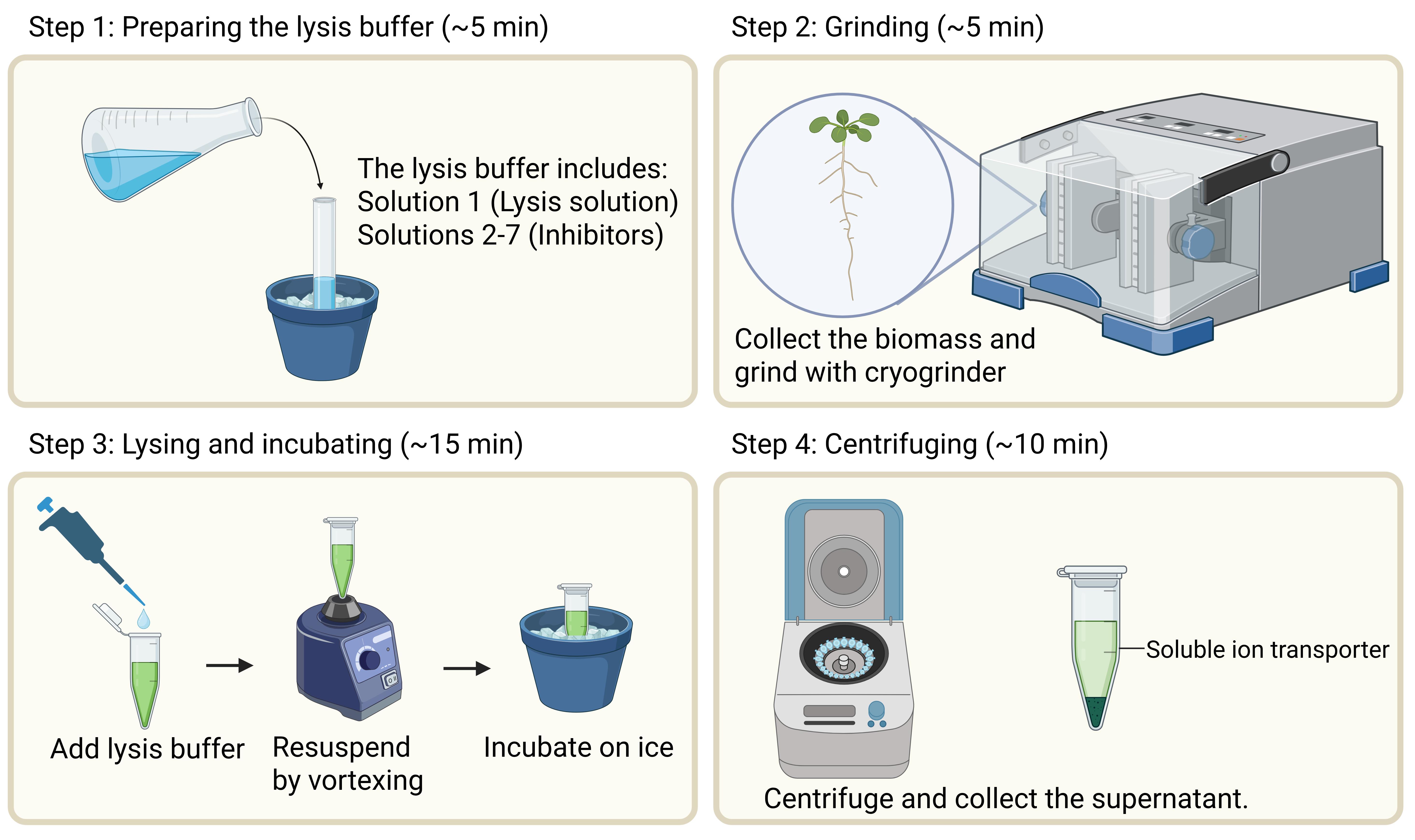
Overview of the workflow of purifying ion transporters from Arabidopsis. This protocol consists of four steps: 1) Mixing seven solutions to prepare the lysis buffer (~5 min), 2) grinding biomass (~5 min), 3) lysing and incubating (~15 min) the samples, and 4) centrifuging the mixtures (~10 min). The whole workflow can be completed within 1 h.
Background
Plant ion transporters are a class of membrane-localized proteins that play critical roles in diverse physiological processes [1]. In ion transporter studies, it is often required to purify the full-length transporter proteins [2,3]. However, ion transporters usually contain multiple transmembrane (TM) domains and exhibit low solubility, and purifying full-length plant ion transporters remains challenging. Therefore, there is an urgent need to develop a simple, rapid, and cost-effective protein extraction method.
Currently, the methods for purifying these ion transporters can be broadly categorized into two categories. The first is the two-phase partitioning method. For instance, the purification of the Na+/H+ antiporter SOS1 starts with low-speed centrifugation to remove cellular debris, followed by dextran-polyethylene glycol (PEG) ultracentrifugation [4]. While the SOS1 protein purified through this method is highly pure, the procedure involves multiple steps, is time-consuming, and is not suitable for routine experiments, such as co-immunoprecipitation (Co-IP) or phosphorylation assays. The second category is to purify the soluble fragment without transmembrane domains. For instance, when studying the phosphorylation of H+-ATPase 1 (AHA1), a common strategy is to divide full-length AHA1 into several fragments, including the soluble intracellular domains [5]. However, this strategy may miss other potential phosphorylation events that occur within the loop or TM domain. It also fails to preserve the native structure, leading to artificial folding or the unnatural exposure of cryptic residues and the generation of artifacts in biochemical assays.
Here, we synthesize and simplify several ion transporter purification protocols reported in published articles [6–8], eliminating the need for density gradients and ultracentrifugation. In contrast to the published protocols, our method provides a simpler and faster workflow with lower equipment requirements. Nevertheless, this method does not distinguish transporters from different organelles. Therefore, it should be used with caution when downstream experiments require organelle-specific ion transporters or highly purified protein preparations.
Materials and reagents
Biological materials
1. SOS1::SOS1:GFP in sos1: Transgenic Arabidopsis plants in the sos1 mutant background expressing SOS1 fused with GFP under the control of the SOS1 promoter (T2 homozygous plants were previously generated by Prof. FU Haiqi [2])
2. 35S::NRAMP3:mRFP: Transgenic Arabidopsis plants in the Col-0 background expressing NRAMP3 fused with mRFP under the control of the 35S promoter (T2 homozygous plants were generated by our laboratory)
Reagents
1. HEPES (Sigma-Aldrich, catalog number: H4034-100G)
2. NaOH (VWR, catalog number: 28244.295)
3. MgCl2 (Sigma-Aldrich, catalog number: M8266-1KG)
4. Glycerol (Sangon Biotech, catalog number: A100854-0500)
5. Triton X-100 (USBiological, catalog number: 9002-93-1)
6. NaCl (Sigma-Aldrich, catalog number: S3014-1KG)
7. NaF (Aladdin, catalog number: S111590-100G)
8. Na2MoO4 (Sigma-Aldrich, catalog number: 243655-100G)
9. Na2C4H4O6 (Sigma-Aldrich, catalog number: 6106-24-7)
10. Na3VO4 (Sigma-Aldrich, catalog number: S6508-50G)
11. DTT (Sigma-Aldrich, catalog number: 3483-12-3)
12. Protease inhibitor cocktail, mini-tablet (EDTA-free) (MCE, catalog number: HY-K0011)
13. Radioimmunoprecipitation assay buffer (Beyotime, catalog number: P0013B)
Solutions
1. HEPES-NaOH lysis solution (see Recipes)
2. 100× NaF stock (see Recipes)
3. 100× Na2MoO4 stock (see Recipes)
4. 100× Na2C4H4O6 stock (see Recipes)
5. 100× Na3VO4 stock (see Recipes)
6. 200× DTT stock (see Recipes)
7. 10× cocktail stock (see Recipes)
Recipes
1. HEPES-NaOH lysis solution (pH 7.2)
| Reagent | Final concentration | Quantity or volume |
|---|---|---|
| HEPES | 50 mM | 5.96 g |
| NaOH | 50 mM | 1.01 g |
| MgCl2 | 1.5 mM | 0.071 g |
| Glycerol | 10% (v/v) | 50 mL |
| Triton X-100 | 1% (v/v) | 5 mL |
| NaCl | 150 mM | 4.38 g |
| ddH2O | n/a | Fill up to 500 mL |
| Total | n/a | 500 mL |
Role of solution: Membrane lysis and solubilization matrix.
Vial specifics: Sterile glass bottle (500 mL).
Shelf life: 3–6 months at 4 °C.
2. 100× NaF stock (100 mM)
| Reagent | Final concentration | Quantity or volume |
|---|---|---|
| NaF | 100 mM | 0.21 g |
| ddH2O | n/a | Fill up to 50 mL |
| Total | n/a | 50 mL |
Role of solution: Acid phosphatase inhibitor.
Vial specifics: Sterile polypropylene conical tube (50 mL).
Shelf life: 3–6 months at 4 °C. Check for any precipitates before use. If present, vortex it until the precipitates are completely dissolved.
3. 100× Na2MoO4 stock (200 mM)
| Reagent | Final concentration | Quantity or volume |
|---|---|---|
| Na2MoO4 | 200 mM | 2.06 g |
| ddH2O | n/a | Fill up to 50 mL |
| Total | n/a | 50 mL |
Role of solution: Acid phosphatase inhibitor.
Vial specifics: Sterile polypropylene conical tube (50 mL).
Shelf life: 3–6 months at 4 °C. Check for any precipitates before use. If present, vortex it until the precipitates are completely dissolved.
4. 100× Na2C4H4O6 stock (400 mM)
| Reagent | Final concentration | Quantity or volume |
|---|---|---|
| Na2C4H4O6 | 400 mM | 4.60 g |
| ddH2O | n/a | Fill up to 50 mL |
| Total | n/a | 50 mL |
Role of solution: Acid phosphatase inhibitor.
Vial specifics: Sterile polypropylene conical tube (50 mL).
Shelf life: 3–6 months at 4 °C. Check for any precipitates before use. If present, vortex it until the precipitates are completely dissolved.
5. 100× Na3VO4 stock (200 mM) (optional)
| Reagent | Final concentration | Quantity or volume |
|---|---|---|
| Na3VO4 | 200 mM | 9.20 g |
| ddH2O | n/a | Fill up to 50 mL |
| Total | n/a | 50 mL |
Role of solution: Tyrosine phosphatase and alkaline phosphatase inhibitor.
Vial specifics: Sterile polypropylene conical tube (50 mL).
Shelf life: 3–6 months at 4 °C. Check for any yellow precipitates before use. If present, discard it and prepare a fresh solution.
6. 200× DTT stock (1 M)
| Reagent | Final concentration | Quantity or volume |
|---|---|---|
| DTT | 1 M | 0.15 g |
| ddH2O | n/a | Fill up to 1 mL |
| Total | n/a | 1 mL |
Role of solution: Reducing agent.
Vial specifics: Sterile polypropylene conical tube (1.5 mL)
Shelf life: 1 month at -20 °C. Aliquot into 100 μL to avoid repeated freeze/thaw cycles.
7. 10× Cocktail stock
| Reagent | Final concentration | Quantity or volume |
|---|---|---|
| Protease inhibitor cocktail tablet | n/a | 1 pellet |
| ddH2O | n/a | Fill up to 1 mL |
| Total | n/a | 1 mL |
Role of solution: Protease inhibitor.
Vial specifics: Sterile polypropylene conical tube (1.5 mL).
Shelf life: 1 month at -20 °C, protected from light. Aliquot into 100 μL to avoid repeated freeze/thaw cycles.
Laboratory supplies
1. 3 mm stainless steel grinding beads
2. 1.5 mL polypropylene conical tubes (Eppendorf, catalog number: HS4325)
3. 50 mL polypropylene conical tubes (Falcon, catalog number: 352070)
Equipment
1. Eppendorf tabletop centrifuge 5418R (Sigma-Aldrich, catalog number: EP5401000137)
2. Vortex-Genie mixer (Sigma-Aldrich, catalog number: Z258415)
3. Cryogenic grinder (Jingxin, catalog number: JXFSTPRP-24)
Procedure
A. Preparing the lysis buffer
Note: All solutions must be prepared in advance and stored at 4 °C (Recipes 1–5) or at -20 °C (Recipes 6 and 7) before the experiment starts. To prepare the working solution for each experiment, retrieve the pre-chilled Recipe 1 and stock solutions (Recipes 2–7), and mix them according to Table 1.
Table 1. Lysis buffer recipe (1 mL)
| Composition | Volume |
|---|---|
| 100× NaF stock (Recipe 2) | 10 μL |
| 100× Na2MoO4 stock (Recipe 3) | 10 μL |
| 100× Na2C4H4O6 stock (Recipe 4) | 10 μL |
| 100× Na3VO4 stock (Recipe 5) | 10 μL |
| 200× DTT stock (Recipe 6) | 5 μL |
| 10× Cocktail stock (Recipe 7) | 100 μL |
| HEPES-NaOH lysis solution (Recipe 1) | 855 μL |
Notes:
1. The lysis buffer must be prepared freshly before use and maintained strictly on ice throughout the procedure.
2. Before starting the experiment, estimate the required volume of lysis buffer based on the amount of ground biomass.
3. Omit Na3VO4 (Recipe 5) if the experiment is phosphorylation detection.
B. Grinding
Note: This protocol is designed for the purification of ion transporters from Arabidopsis thaliana. Biomass can be collected either from MS agar medium–grown young seedlings or from soil-grown plant tissues.
1. Prepare a 1.5 mL Eppendorf tube containing 3-mm stainless steel beads and transfer the required sample into the tube.
Note: For fresh materials, use Kimtech paper to wipe off any water droplets on the surface.
2. Freeze the samples in liquid nitrogen. Set the cryogenic grinder to 90 Hz and grind for 3 min until a fine powder is obtained.
Note: Alternatively, if the cryogenic grinder is not available, samples can be ground using a mortar and pestle.
3. Repeat the grinding cycle once until no unbroken tissue debris remains.
Pause point: If the ground powder is not used immediately, it can be stored in a liquid nitrogen tank or kept in a -80 °C freezer.
C. Lysis and incubation
1. Pipette the prepared lysis buffer into the ground powder at a 1:1 ratio (v/v) on ice.
Note: Typically, the lysis buffer and ground biomass are mixed at 1:1 (v/v), but the ratio can be increased or decreased depending on the experiment.
Critical: This step must be conducted quickly to prevent the ground powder from thawing. If there is a large number of samples, it is recommended to process them in batches.
2. Immediately vortex the tube vigorously until the powder is completely dissolved.
3. Incubate the mixture on ice for 10 min.
D. Centrifuging
1. Centrifuge the mixture at 12,000× g at 4 °C for 10 min.
2. Transfer the supernatant to a new 1.5 mL Eppendorf tube and discard the pellet and steel beads.
3. Repeat step D1 once; the supernatant is ready to use.
Pause point: If the purified proteins are not used immediately, they can be stored on ice for short-term storage. Following our protocol, SOS1 remains stable on ice for up to three days. However, it remains uncertain whether ion-transporter proteins can be stored at -80 °C for longer periods of time. To achieve optimal results, it is highly recommended to use the protein immediately after purification.
Validation of protocol
To validate the efficacy of this protocol, we selected the Arabidopsis Na+/H+ antiporter SOS1, which contains 12 TM domains, and compared its purification efficiency using this lysis buffer and RIPA buffer (Radio ImmunoPrecipitation Assay buffer). The result demonstrated that SOS1-GFP was successfully detected using the lysis buffer, whereas the RIPA buffer failed to purify SOS1 (Figure 1A). Furthermore, we evaluated three distinct denaturation temperatures and found that SOS1-GFP could be robustly detected under all these denaturation conditions. To confirm the versatility of this protocol, we also tested another 12-TMD Arabidopsis metal transporter, NRAMP3 (Figure 1B), which was also successfully purified and detected. Taken together, these data indicate that our modified lysis buffer can significantly enhance the solubility of multi-pass transmembrane proteins.
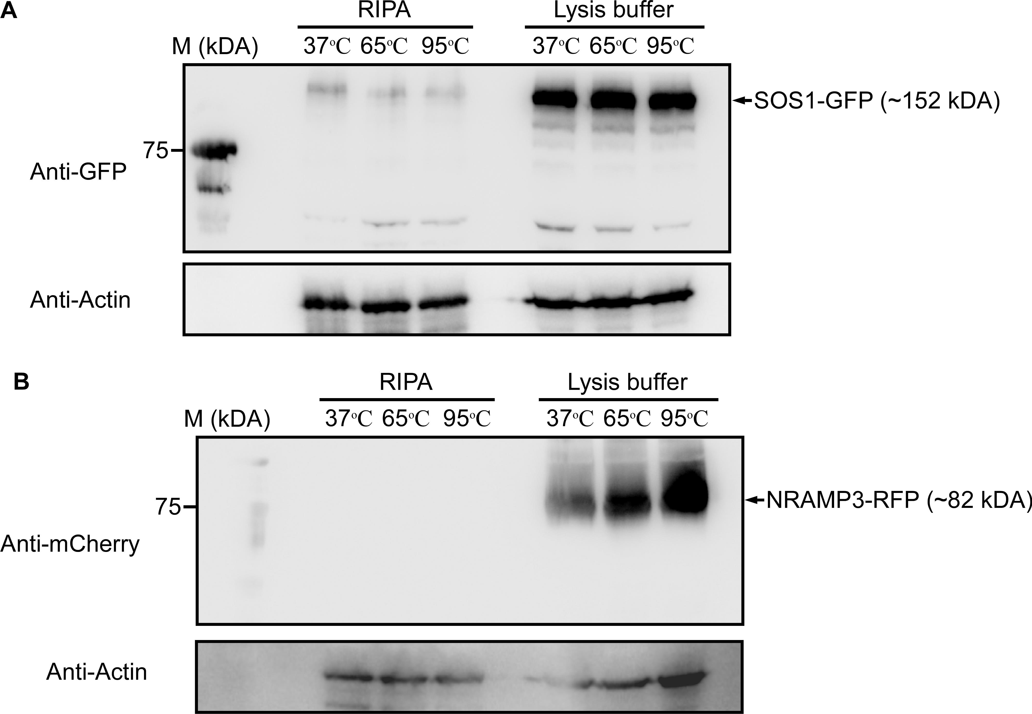
Figure 1. Immunoblot assay of ion transporter purification efficiency. (A) SOS1-GFP was purified using either the RIPA lysis buffer method (left) or the current protocol (right). The assays were performed at three denaturation temperatures (37, 65, and 95 °C) for 3 min. ACTIN was used as a loading control. Each lane contains 20 μg of Arabidopsis total protein. (B) NRAMP3-RFP was purified using either the RIPA lysis buffer method (left) or the current protocol (right). The assays were performed at three denaturation temperatures (37, 65, and 95 °C) for 3 min. ACTIN was used as a loading control. Each lane contains 20 μg of Arabidopsis total protein.
General notes and troubleshooting
General notes
For an initial trial, we recommend setting a gradient of denaturation temperatures and assessing the purification efficiency via immunoblot assay.
Troubleshooting
Problem 1: No target bands detected following this protocol.
Possible cause: Protein aggregation due to the high denaturing temperature (~95 °C or higher).
Solution: Lower the denaturing temperature to 37 or 65 °C for 10–15 min; if you still fail to detect target bands, try loading the samples at room temperature or omit the denaturation step before the immunoblot assay.
Problem 2: For phosphorylation-related experiments, the target bands are smeared or blurry.
Possible cause: The phosphate groups (PO43-) compete with the vanadate ions (VO43-) in the lysis buffer.
Solution: No need to add Na3VO4 (Solution 5) if the downstream experiment is on protein phosphorylation.
Problem 3: How to store the purified ion transporter proteins and how long can they be stored?
They can be kept on ice short-term (no more than 3 days). For the ion transporters we purified in this protocol (SOS1, NRAMP3), they remain stable on ice for approximately 3 days, but we have not tested longer storage times.
Acknowledgments
This work was supported by the Research Grants Council (RGC) of the Hong Kong Special Administrative Region, China General Research Fund #12104723 (to L.X. and Y.X.)
The following figures were created using BioRender: Graphical overview, BioRender.com/o18hhoq.
Author contributions
Protocol Development, Z.Z.; Protocol Optimization or Validation, Z.Z.; Writing—Original Draft, Z.Z.; Writing—Review & Editing, Z.Z. and L.X.; Funding acquisition, J.L., Y.X. and L.X.; Supervision, J.L., Y.X., and L.X.
Competing interests
We declare that we have no competing interests.
Ethical considerations
There are no ethical considerations.
References
- Pantoja, O. (2021). Recent Advances in the Physiology of Ion Channels in Plants. Annu Rev Plant Biol. 72(1): 463–495. https://doi.org/10.1146/annurev-arplant-081519-035925
- Fu, H., Yu, X., Jiang, Y., Wang, Y., Yang, Y., Chen, S., Chen, Q. and Guo, Y. (2022). SALT OVERLY SENSITIVE 1 is inhibited by clade D Protein phosphatase 2C D6 and D7 in Arabidopsis thaliana. Plant Cell. 35(1): 279–297. https://doi.org/10.1093/plcell/koac283
- Liu, G., Yu, X., Zeng, Y., Li, B., Wang, R., Wang, X., Zhao, X., Jiang, L. and Guo, Y. (2025). SOS2‐FREE1 regulates SOS1 tonoplast sorting to promote Na+ compartmentalization in vacuole during salt stress response. J Integr Plant Biol. 67(10): 2545–2560. https://doi.org/10.1111/jipb.13970
- Qiu, Q. S., Guo, Y., Dietrich, M. A., Schumaker, K. S. and Zhu, J. K. (2002). Regulation of SOS1, a plasma membrane Na+ /H+ exchanger in Arabidopsis thaliana, by SOS2 and SOS3. Proc Natl Acad Sci USA. 99(12): 8436–8441. https://doi.org/10.1073/pnas.122224699
- Liu, X., Zhang, D., Yang, Y. and Ma, L. (2026). The CPK8-AHA1 phosphorylation module dampens plasma membrane H+-ATPase activity to confer drought tolerance. Plant Physiol. 200(4): e1093/plphys/kiag128. https://doi.org/10.1093/plphys/kiag128
- Thor, K., Jiang, S., Michard, E., George, J., Scherzer, S., Huang, S., Dindas, J., Derbyshire, P., Leitão, N., DeFalco, T. A., et al. (2020). The calcium-permeable channel OSCA1.3 regulates plant stomatal immunity. Nature. 585(7826): 569–573. https://doi.org/10.1038/s41586-020-2702-1
- Lanquar, V., Ramos, M. S., Lelièvre, F., Barbier-Brygoo, H., Krieger-Liszkay, A., Krämer, U. and Thomine, S. (2010). Export of Vacuolar Manganese by AtNRAMP3 and AtNRAMP4 Is Required for Optimal Photosynthesis and Growth under Manganese Deficiency. Plant Physiol. 152(4): 1986–1999. https://doi.org/10.1104/pp.109.150946
- Xue, Y., Yang, Y., Yang, Z., Wang, X. and Guo, Y. (2018). VAMP711 Is Required for Abscisic Acid-Mediated Inhibition of Plasma Membrane H+-ATPase Activity. Plant Physiol. 178(3): 1332–1343. https://doi.org/10.1104/pp.18.00499
Article Information
Publication history
Received: Jul 14, 2026
Accepted: Oct 25, 2026
Available online: Nov 15, 2026
Published: Dec 5, 2026
Copyright
© 2026 The Author(s); This is an open access article under the CC BY-NC license (https://creativecommons.org/licenses/by-nc/4.0/).
How to cite
Zhang, Z., Li, J., Xia, Y. and Xiong, L. (2026). Rapid Purification of Ion-Transporter Proteins in Arabidopsis thaliana. Bio-protocol 16(23): e5891. DOI: 10.21769/BioProtoc.5891.

 Download Citation in RIS Format
Category
Plant Science > Plant biochemistry > Protein > Isolation and purification
Biochemistry > Protein > Isolation and purification
Do you have any questions about this protocol?
Post your question to gather feedback from the community. We will also invite the authors of this article to respond.
 Post a Question


