


- Protocols

- Articles and Issues
- For Authors
- About
- Become a Reviewer


Establishment and Proliferation of Primary Nilgai Fibroblast Cultures from Adult Tissue

Published: Vol 16, Iss 22, Nov 20, 2026 DOI: 10.21769/BioProtoc.5888 Views: 37
Reviewed by: Samantha HallerXuesong SuiThirupugal Govindarajan
 PDF
 Ask a question


Protocol Collections
Comprehensive collections of detailed, peer-reviewed protocols focusing on specific topics




Abstract
Primary fibroblast cultures provide a renewable source of living cells that support a wide range of molecular, genomic, and conservation applications. For understudied species such as the nilgai (Boselaphus tragocamelus), establishing primary fibroblast cultures creates a valuable biological resource for long-term biobanking and comparative genomic research. Here, we describe a reproducible protocol for the isolation, establishment, proliferation, and cryopreservation of primary fibroblast cultures from nilgai ear tissue. The protocol includes tissue processing, cell culture establishment, routine maintenance, passaging, and long-term storage of viable cells. The resulting cultures provide a sustainable source of DNA, RNA, chromatin, and protein, enabling downstream applications such as whole-genome sequencing, transcriptomics, epigenetic profiling, and other sequencing-based analyses. As a member of the Bovidae family, the nilgai offers opportunities for comparative genomic studies with domestic cattle and other bovids, providing insights into genome evolution and conserved regulatory mechanisms. This protocol establishes a reliable resource for wildlife biobanking and facilitates future research in comparative genomics, conservation biology, and livestock-related genomic studies.
Key features
• Adapted from previously described methods in domestic livestock species [1].
• Describes a reproducible workflow for the establishment, proliferation, and preservation of nilgai primary fibroblast cell cultures.
• Uses tissue explant culture for gentle tissue processing and minimizes cellular damage.
Keywords: Fibroblast Cell culture
Nilgai
Explant culture
Ruminant
Graphical overview
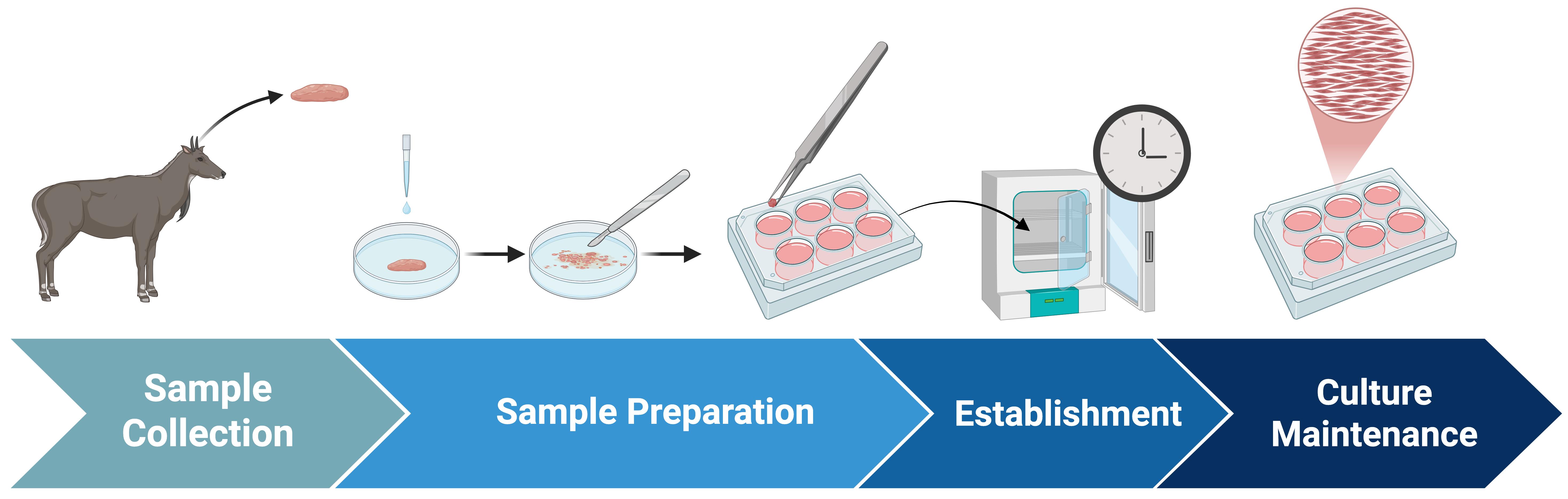
Experimental overview of nilgai primary fibroblast cell culture establishment, proliferation, and maintenance
Background
The nilgai (Boselaphus tragocamelus), the largest Asian antelope, is a member of the Bovidae family and shares an evolutionary relationship with domestic cattle (Bos taurus) [2]. Despite this relationship, genomic and cellular resources for nilgai remain limited compared with those available for domestic livestock species. The establishment of primary fibroblast cultures provides a sustainable source of biological material for whole-genome sequencing, comparative genomics, epigenetic profiling, and other functional applications [3]. Because fibroblasts can be expanded over multiple passages and cryopreserved for long-term storage, they represent an important component of wildlife biobanking and genetic resource conservation [4].
Comparative studies between nilgai and domestic cattle may also provide insight into the evolution of bovine genomes, chromosomal organization, species-specific adaptations, and disease susceptibility. As both species belong to Bovidae, comparative analyses may improve our understanding of conserved regulatory elements, genomic variation, and mechanisms underlying traits relevant to livestock biology [2]. Furthermore, properly preserved fibroblast cultures establish a renewable resource that can support future technologies, including genome assembly, gene editing, and reproductive biotechnology [3,5,6].
The protocol presented here describes a reproducible method for establishing, expanding, and preserving primary fibroblast cultures from nilgai ear tissue. The resulting cultures provide a valuable cellular resource for conservation biology, comparative genomics, and future applications in wildlife and livestock research.
Materials and reagents
Biological materials
1. Nilgai tissue samples (ear or skin biopsy)
Reagents
1. Dulbecco’s phosphate-buffered saline without calcium and magnesium (DPBS) (Gibco, catalog number: 14190-144)
2. Dulbecco’s modified Eagle medium (DMEM) (Gibco, catalog number: 11885-084)
3. Fetal bovine serum (FBS) (Gibco, catalog number: 12483-020)
4. Antibiotic-antimycotic 100× (anti-anti 100×) (Gibco, catalog number: 15240-062)
5. 0.05% Trypsin-EDTA (Sciencell Research Laboratories, catalog number: NC1672820)
6. Dimethyl sulfoxide Hybri-Max (DMSO) (Millipore Sigma, catalog number: D2650-5X5ML)
7. Skin prep solution Betadine 4 oz. bottle 10% strength (Fisher Scientific, catalog number: 503043366)
8. 75% Ethanol
9. Commercial antibacterial soap such as Dial Antibacterial or Hibiclens
Alternative: Diluted commercial detergent, such as 10%–20% Dawn detergent, may also be used; however, it can be less effective at reducing microbial contamination than antibacterial alternatives.
Solutions
1. Ruminant fibroblast medium, 20% FBS (RFM-20%) (see Recipes)
2. DPBS wash (see Recipes)
3. Freezing media (see Recipes)
4. Collection media (optional) (see Recipes)
Recipes
Before you begin: Aliquot frozen reagents (i.e., FBS, trypsin-EDTA 0.05%, dimethyl sulfoxide, anti-anti 100×) into appropriate volumes before storage at -20 °C to minimize multiple freeze/thaw cycles. When possible, use freshly thawed aliquots for each recipe and store mixed solutions in small volumes (indicated on individual recipe instructions) once mixed. Doing so will preserve the integrity of solutions and allow for longer storage and more consistent results.
1. RFM-20%
| Reagent | Final concentration | Volume | Volume |
|---|---|---|---|
| DMEM | 79% v/v | 39.5 mL | 197.5 mL |
| FBS | 20% v/v | 10 mL | 50 mL |
| Anti-anti 100× | 1% v/v (1× final concentration) | 500 μL | 2.5 mL |
| Total | n/a | 50 mL | 250 mL |
Mix the reagents to the desired volume of RFM-20% under a sterile flow hood, adding anti-anti 100× last. Ensure thorough mixing of each reagent before adding. Filter through a 0.2 μm vacuum filter within 20 min of mixing. Store in 20–50 mL aliquots at 4 °C for up to 30 days. Media should be orange-red in color. Do not use if media becomes yellow, turbid, or putrid.
2. DPBS wash
| Reagent | Final concentration | Volume | Volume |
|---|---|---|---|
| DPBS | 99% v/v | 49.5 mL | 247.5 mL |
| Anti-anti 100× | 1% v/v (1× final concentration) | 500 μL | 2.5 mL |
| Total | n/a | 50 mL | 250 mL |
Mix the reagents to the desired volume of DPBS wash under a sterile flow hood, adding anti-anti 100× last. Ensure thorough mixing of reagents before adding. Optional: Filter through a 0.2 μm vacuum filter if desired. Store in 20–50 mL aliquots at 4 °C for up to 30 days. Do not use if wash becomes colored or turbid.
3. Freezing media
| Reagent | Final concentration | Volume |
|---|---|---|
| DMEM* | 80% v/v | 1.2 mL |
| FBS | 10% v/v | 150 μL |
| DMSO | 10% v/v | 150 μL |
| Total | n/a | 1.5 mL |
*You may use FBS, PBS, or DPBS in place of DMEM, but ideal results are obtained with DMEM or FBS.
Make fresh for each use. Mix cold reagents under a sterile flow hood to make 1.5 mL of freezing medium for each cryo vial. Store at 4 °C until immediately before use. Do not use older than 12 h.
4. Collection media (optional)
| Reagent | Final concentration | Volume |
|---|---|---|
| DMEM | 99% v/v | 9.9 mL |
| Anti-anti 100× | 1% v/v | 100 μL |
| Total | n/a | 10 mL |
Mix reagents to the desired volume of collection medium under a sterile flow hood, adding anti-anti 100× last. Ensure thorough mixing of reagents before adding. Store at 4 °C for up to two weeks. For ideal results, use within three days of mixing.
Laboratory supplies
1. 15 mL conical centrifuge tube (Thermofisher Scientific, catalog number: 12565269)
2. 50 mL conical centrifuge tube (Thermofisher Scientific, catalog number: 12565271)
3. Falcon 6-well flat-bottom plates with lids (Corning, catalog number: 21100-002)
4. SureOne aerosol barrier pipette tips, 1,000 μL, filtered (Fisher Scientific, catalog number: 02707404)
5. T25 EasYFlask, TC surface, filter cap, 25 cm2 (Thermofisher Scientific, catalog number: 2565351)
6. Cryo vial, int thread, 2 mL (Corning, catalog number: 1232E26)
7. 60 mm × 15 mm Petri dish (Bellco Glass Corporate, catalog number: 23A00L845)
8. Carbon steel scalpel blades #10 (VWR International, catalog number: 76457-448)
9. Scalpel knife handle No.3 (VWR International, catalog number: 76457-416)
10. Metal tweezers (United Scientific Supplies Inc., catalog number: S111016), autoclave-sterilized
11. Sterile polystyrene serological pipettes, 10 mL (Fisher Scientific, catalog number: 1367610J)
12. 250 mL vacuum filter/storage bottle system, 0.2 μm, nylon (Corning, catalog number: 097612)
13. Scissors, sterilized
Equipment
1. HeracellTM 150i CO2 incubator 150 L (ThermoFisher Scientific, catalog number: 50116047), set to 37–38 °C and 5% CO2
2. Laminar flow hood, e.g., AirClean 600 PCR Workstation Flow Hood (AirClean Systems, model number: AC632LFUVC)
3. Water bath, e.g., PrecisionTM General Purpose Water Bath (Fisher Scientific, catalog number: TSFP10PMO, supplier number: TSGP10DUCK), set to 37–38 °C
4. Inverted microscope, e.g., Motic AE31 Elite 30W Trinocular Inverted Microscope (Motic Microscopy, SKU: 1100100200841)
Procedure
A. Sample collection of nilgai ear tissue
Critical: Ensure that all animal materials are collected in accordance with local guidelines and institutional regulations.
1. After the animal has been appropriately dispatched, thoroughly spray the area with 75% ethanol and rub to remove excess dirt and contaminants. Collect the ear by cutting at its base using sterile scissors.
2. Store the ear in a sealed bag or an airtight container at 4 °C or on ice until processing. Wash ear thoroughly with commercial antibacterial soap and warm water (see General note 1). Rinse the sample thoroughly with water to remove residual soap.
Alternative: After properly restraining the animal, thoroughly clean the sample area with 75% ethanol and rub to remove excess dirt and contaminants. Using sterile scissors, ear notch, or biopsy punch, collect 1–2 cm2 skin or ear tissue and store in collection media at 4 °C until processing.
B. Sample processing of nilgai ear tissue
Note: Before you begin, warm RFM-20% and DPBS wash in a water bath for at least 30 min.
1. Using sterile scissors, cut a 1–2 cm2 section of the ear from the conchal region of the pinna. If possible, avoid the helix and peripheral margins of the external ear (Figure 1).
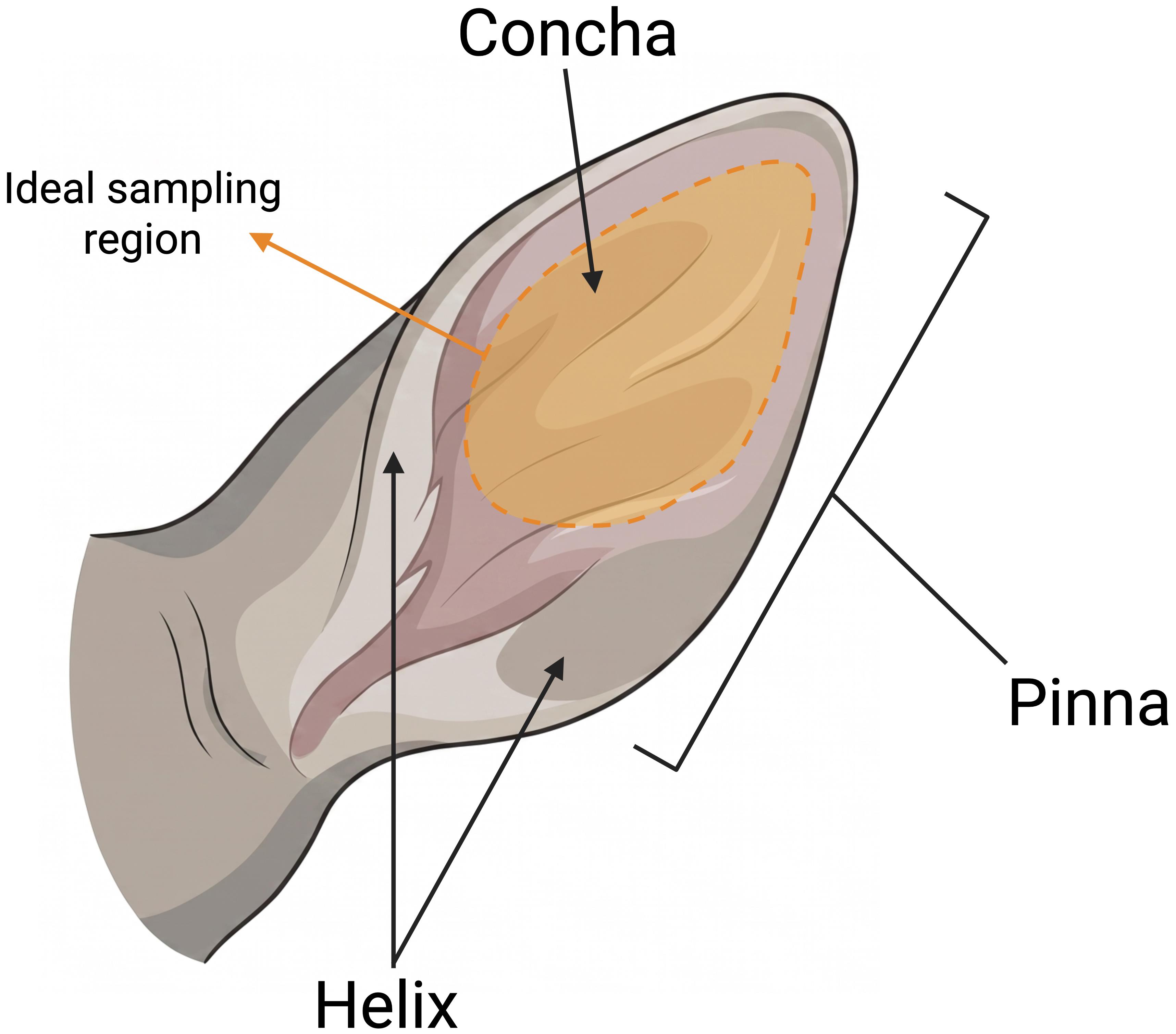
Figure 1. Illustration of nilgai ear with ideal sample region highlighted in orange
2. Wash the sample once with 75% ethanol and three times with warm water and commercial antibacterial soap. Be sure to rinse the sample well between each wash. Rinse sample thoroughly with water to remove residual soap. See General Note 2. (Optional) Pat sample dry with a paper towel to remove excess water.
3. Transport the cleaned sample to a clean laminar flow hood in a sterile 60 mm Petri dish.
Critical: The following steps should be conducted under a laminar flow hood using aseptic techniques and sterile instruments. A 10-min UV sterilization of the hood and supplies may also be done to reduce contamination risk.
(Optional) Add 1–2 mL of DPBS wash to the Petri dish to rinse and moisten the sample. Performing step B5 in the presence of DPBS wash can prevent hair sticking to the blade and enable brief rinsing as needed, but is not required.
4. Using a sterile scalpel blade and handle, shave any hair from the sample. Do not remove any layers of skin. Use tweezers to orient and stabilize the sample.
5. Once all hair is removed, rinse the tissue in a new Petri dish with 3–4 mL of DPBS wash. Repeat with fresh DPBS wash three times or until hair remnants are removed.
6. Rinse tissue quickly in 75% ethanol for no more than 30 s. Ensure that both sides of the tissue are rinsed.
7. Rinse tissue in DPBS wash one to three times. Ensure that both sides of the tissue are rinsed.
8. Coat the tissue with a few drops of betadine. Allow the tissue to rest for 30–60 s. Tap off excess and rinse in DPBS wash twice.
9. Place the cleaned sample on a sterilized cutting surface or fresh Petri dish. Using a new sterile blade, finely mince tissue into 1 mm2 tissue seeds.
10. Add 2 mL of prewarmed RFM-20% to each well of a 6-well plate. Using sterile tweezers, transfer 5–7 seeds to each well (Figure 2).
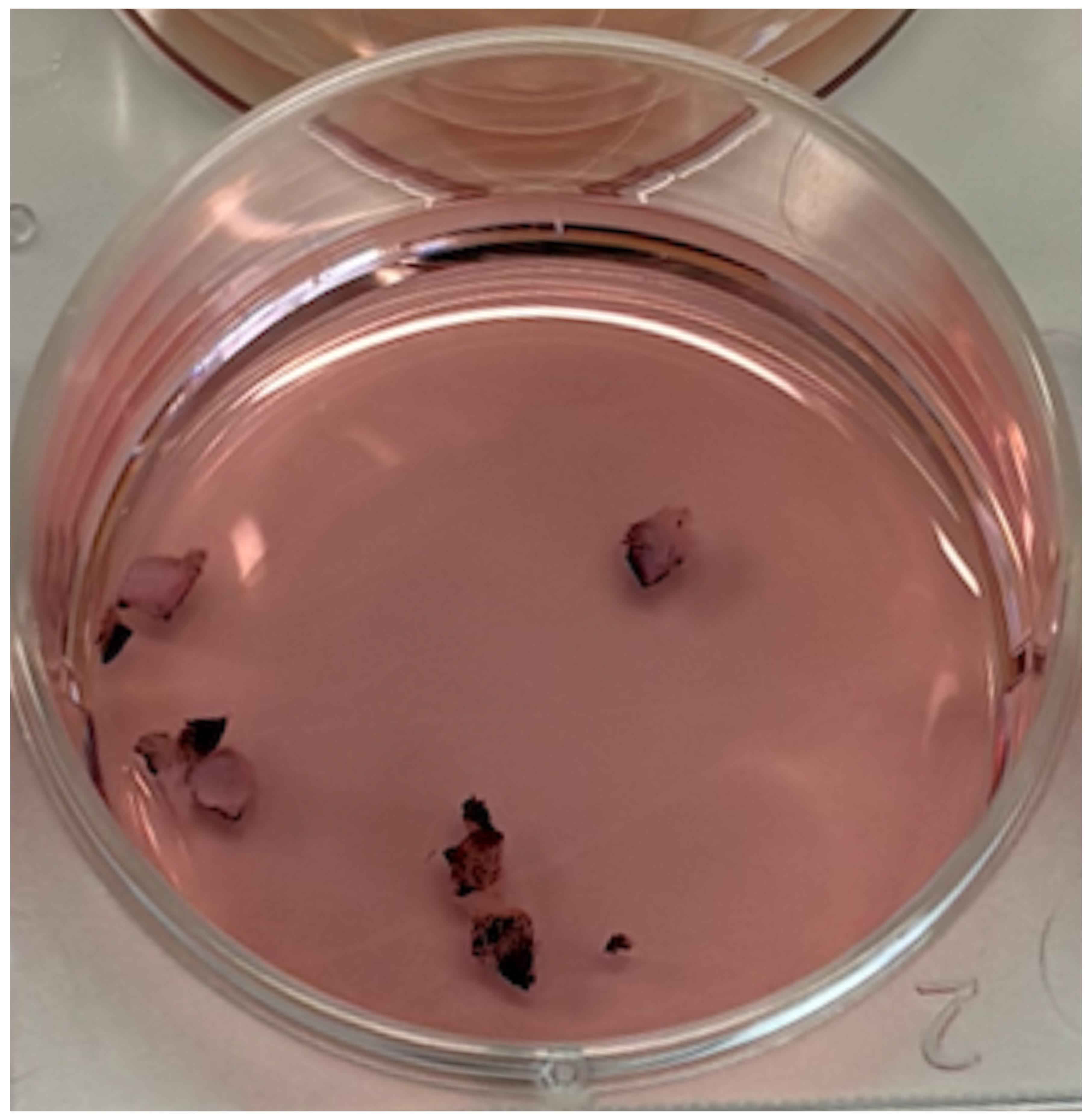
Figure 2. Expected sample appearance following preparation
11. Incubate the plate at 37–38 °C and 5% CO2.
C. Establishment of primary fibroblast culture from nilgai tissue seeds
1. After 24 h of incubation, check the culture plate for signs of contamination such as media changes, putrid scents, or fungal growth. If there are no signs of contamination, allow the culture to incubate for 48–72 h undisturbed. Excessively moving the plate jostles growing tissue seeds and may limit outgrowth.
2. After three days of culture, gently add 1 mL of prewarmed RFM-20% to each well. Try not to disturb the seeds.
3. Continue to monitor for contamination or infection every 24–48 h until outgrowth is visible. If viable cells are not visible by day 6, remove 1 mL of media from each well and replace with 1 mL of prewarmed RFM-20%. Continue to replace 1 mL of media every 72 h until cells are visible. Cells should initially appear as small circular structures (Figure 3) that elongate into spindle-like cells (Figure 4) (see General note 3).
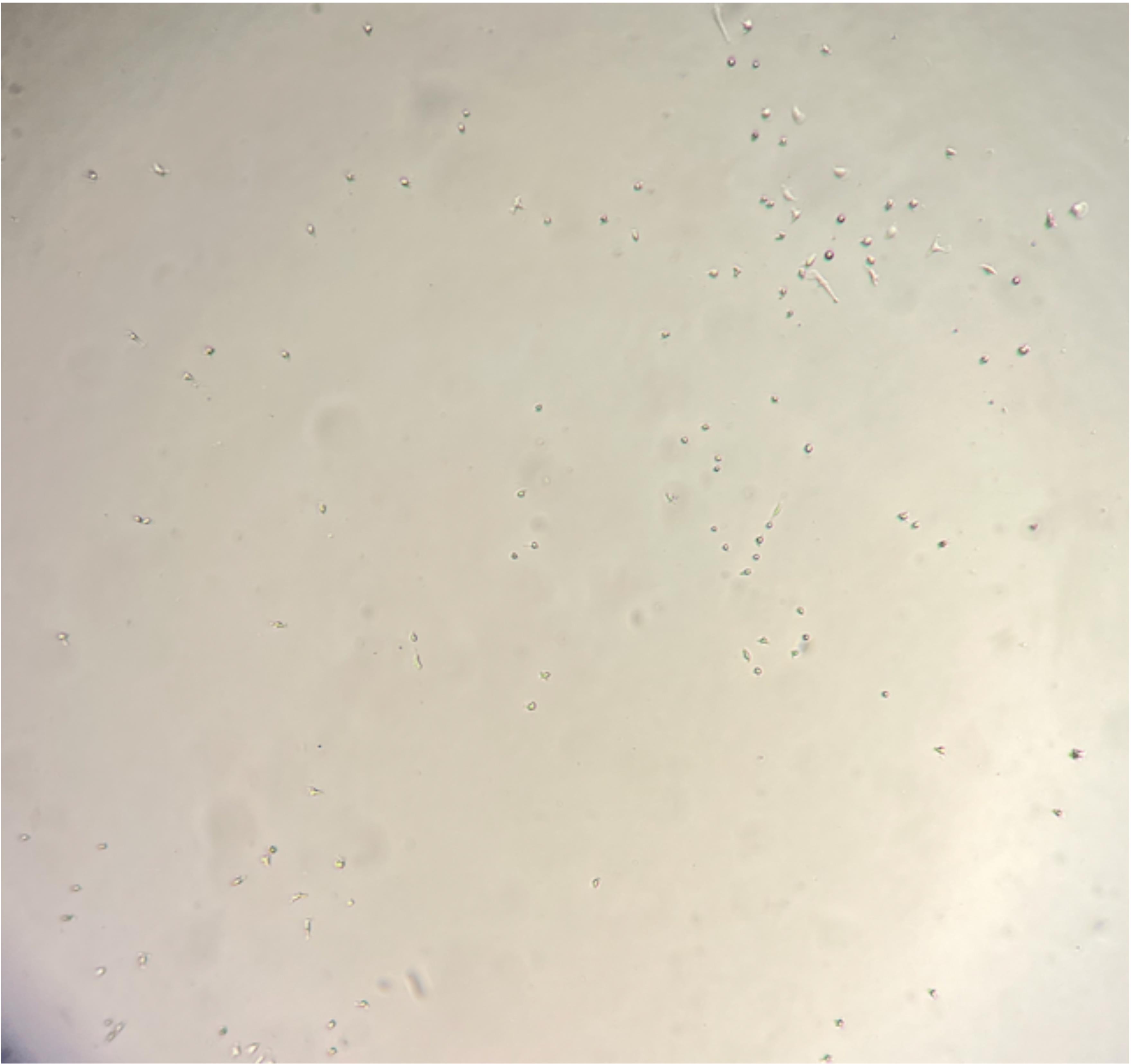
Figure 3. Expected appearance of emerging immature fibroblast cells viewed with brightfield illumination. Magnification: 60×.
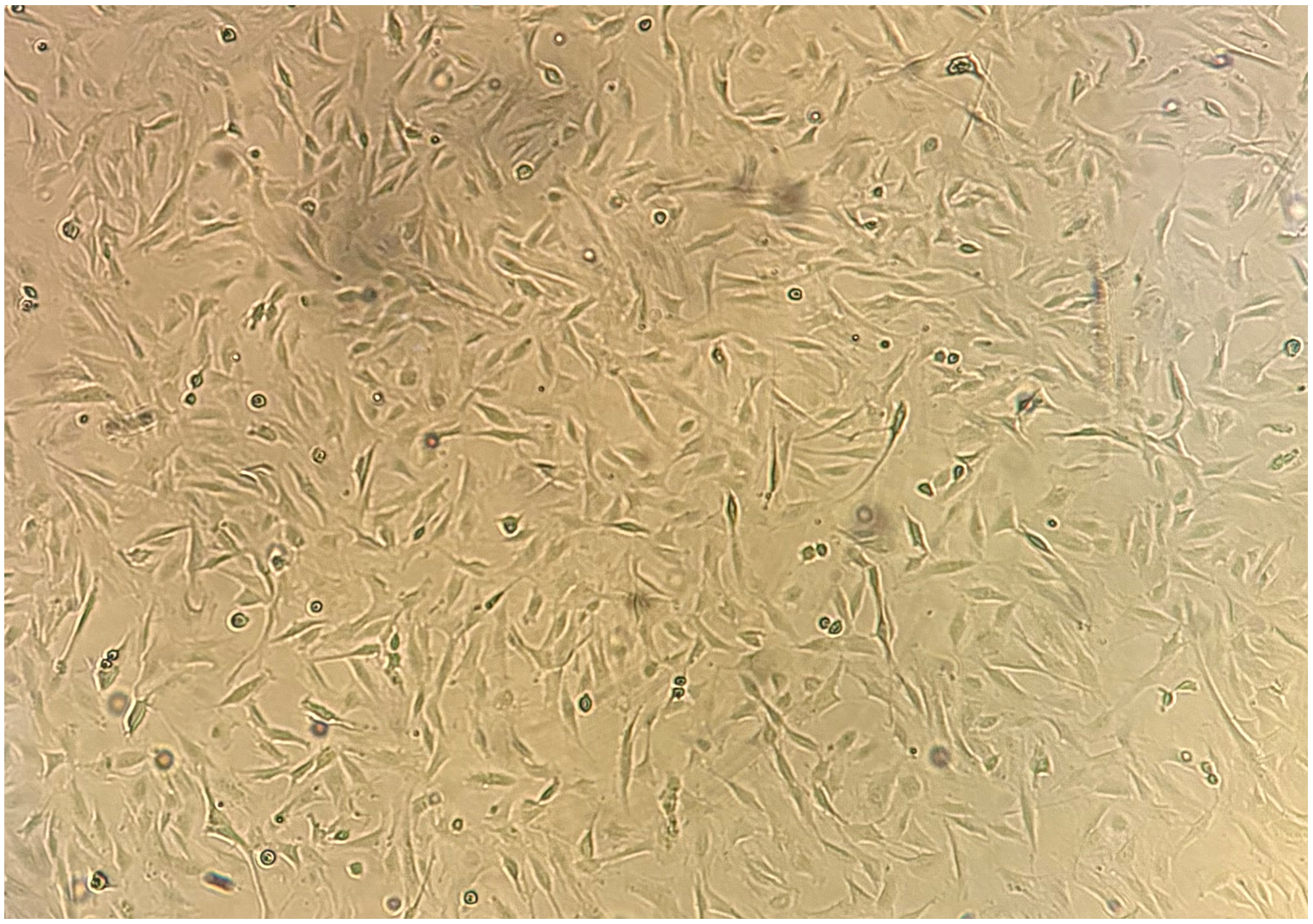
Figure 4. Expected morphology of healthy nilgai fibroblasts viewed with brightfield illumination. Magnification: 60×.
4. Forty-eight to seventy-two hours after cells are visible, gently remove all tissue seeds with sterile tweezers and discard. Avoid touching the bottom of the culture wells.
5. Remove all media and gently wash each well twice with 1 mL of warm DPBS wash. Remove all residual wash and replace with 2 mL of prewarmed RFM-20%.
6. Repeat step C5 every 48 h until cells reach confluence.
Note: Before you proceed, prewarm RFM-20%, FBS, 0.05% trypsin-EDTA, and DPBS wash in a 10 L water bath for at least 30 min.
7. Remove RFM-20% from each well. Gently wash each well twice with 1 mL of DPBS wash. Ensure that all residual wash is removed and discarded.
8. Add 1 mL of 0.05% trypsin-EDTA to each well. Incubate the plate at 37 °C and 5% CO2 for 2–3 min until fibroblast cells release from the plate.
Critical: Incubating for longer than 5 min will result in other cell types releasing into your passage and may induce cellular damage to the nilgai fibroblasts.
9. Add 0.56 mL of FBS to each well and gently swirl to mix.
Alternative: DPBS or PBS may be used in place of FBS. DPBS is cheaper but can decrease cell viability.
10. Tilting the plate at a 45° angle, use the cell/trypsin mixture in each well to gently rinse excess cells to one side of the well. Transfer the mixture into a 15 mL conical centrifugation tube.
11. Centrifuge at 250× g for 5 min at 20 °C.
12. Remove the supernatant and resuspend the pellet in 4 mL of warmed RFM-20%. Transfer resuspended cells to a sterile T25 culture flask and incubate at 37 °C and 5% CO2.
D. Maintenance and proliferation of primary nilgai cell culture
1. Check culture for contamination, adhesion, and proliferation every 24–48 h. Change the RFM-20% every 48–72 h.
2. To refresh RFM-20%, repeat step C5 with modified volumes as in Table 1.
Table 1. Volumes for culture media refreshing
| Reagent | 6-well plate | T25 | T75 |
|---|---|---|---|
| DPBS wash | 1 mL/well | 3 mL | |
| RFM-20% | 2 mL/well |
3. Once cells reach 70%–80% confluence, pass using steps C7–12 with modified volumes and a 1:2 or 1:3 split ratio as in Table 2.
Table 2. Volumes for culture passage
| Reagent | 6-well plate | T25 | T75 |
|---|---|---|---|
| DPBS wash | 1 mL/well | 3–5 mL | 7–10 mL |
| 0.05% trypsin-EDTA | 1 mL/well | 3 mL | 5 mL |
| FBS | 0.56 mL/well | 1 mL | 1.7 mL |
| RFM-20% | 2 mL/well | 4 mL | 10–12 mL |
4. To cryo-preserve cells, follow steps C7–11, using volumes from step D3. Resuspend cells in 1.5 mL of cold freezing medium and transfer to a 2 mL cryovial. Store at -80 °C for 24–48 h and then move to the liquid nitrogen storage tank or other long-term storage.
5. To thaw cells, quickly remove the stored cryovial from the liquid nitrogen tank and add 0.5 mL of prewarmed RFM-20%. Incubate the vial in an incubator or water bath for approximately 1–2 min or until barely thawed. Transfer the thawed cell suspension into a 15 mL Falcon tube. Rinse the cryovial 3× with 1 mL of warm RFM-20% to rinse excess cells from the vial. Transfer all RFM-20% rinse into the 15 mL Falcon tube with the cell suspension. Proceed with steps C11–12 to revive the culture and remove any excess DMSO from the cell suspension.
Validation of protocol
This protocol was used to grow nilgai cells that are currently undergoing extensive sequencing. Preliminary sequencing results positively identified them as nilgai cells.
During passage 3, optimization trials were conducted to identify optimal CO2 and FBS concentrations (Figure 5). After passage, cells were allowed to acclimate for 5 h in 10% RFM and 2.5% or 5.0% CO2. Medium was then replaced with fresh 10% RFM or 20% RFM. Cultures were imaged and counted daily, and medium was refreshed every 48 h. The highest growth increase occurred with 5% CO2 and 20% FBS, despite all cultures being the same passage and similar seeding densities. After establishment, these nilgai fibroblast cultures demonstrated high post-thaw viability (85%–100%) and a doubling time of approximately 48–72 h.
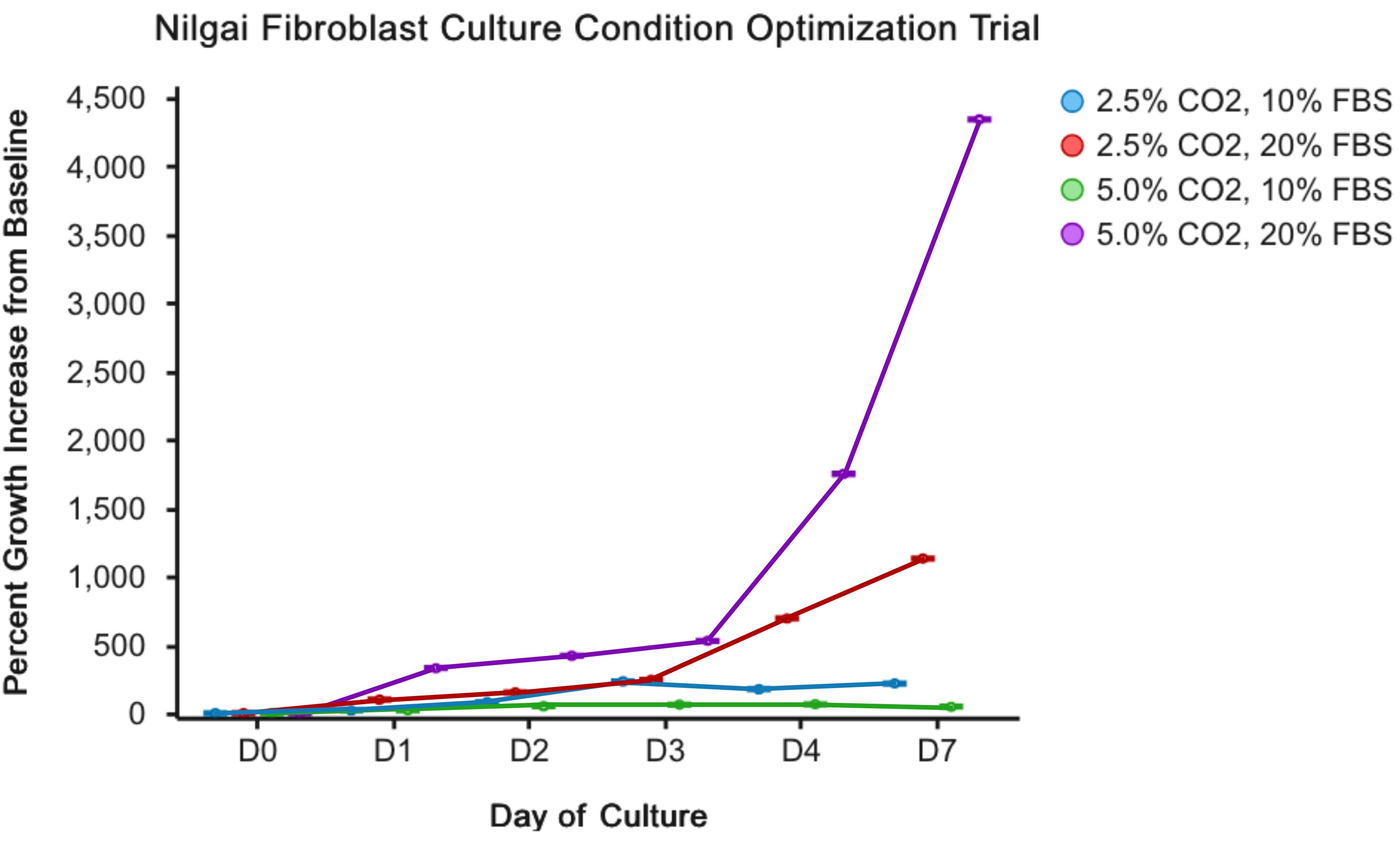
Figure 5. Growth curve of nilgai fibroblasts to determine optimal CO2 and FBS concentrations
Immunofluorescent antibody staining revealed vimentin expression and an absence of cytokeratin expression (Figure 6). Vimentin is expected to exhibit a filamentous staining pattern within the cell, and cytokeratin is expected to be absent in fibroblasts. Together, these results confirm the identity of the culture as primary fibroblast cells.
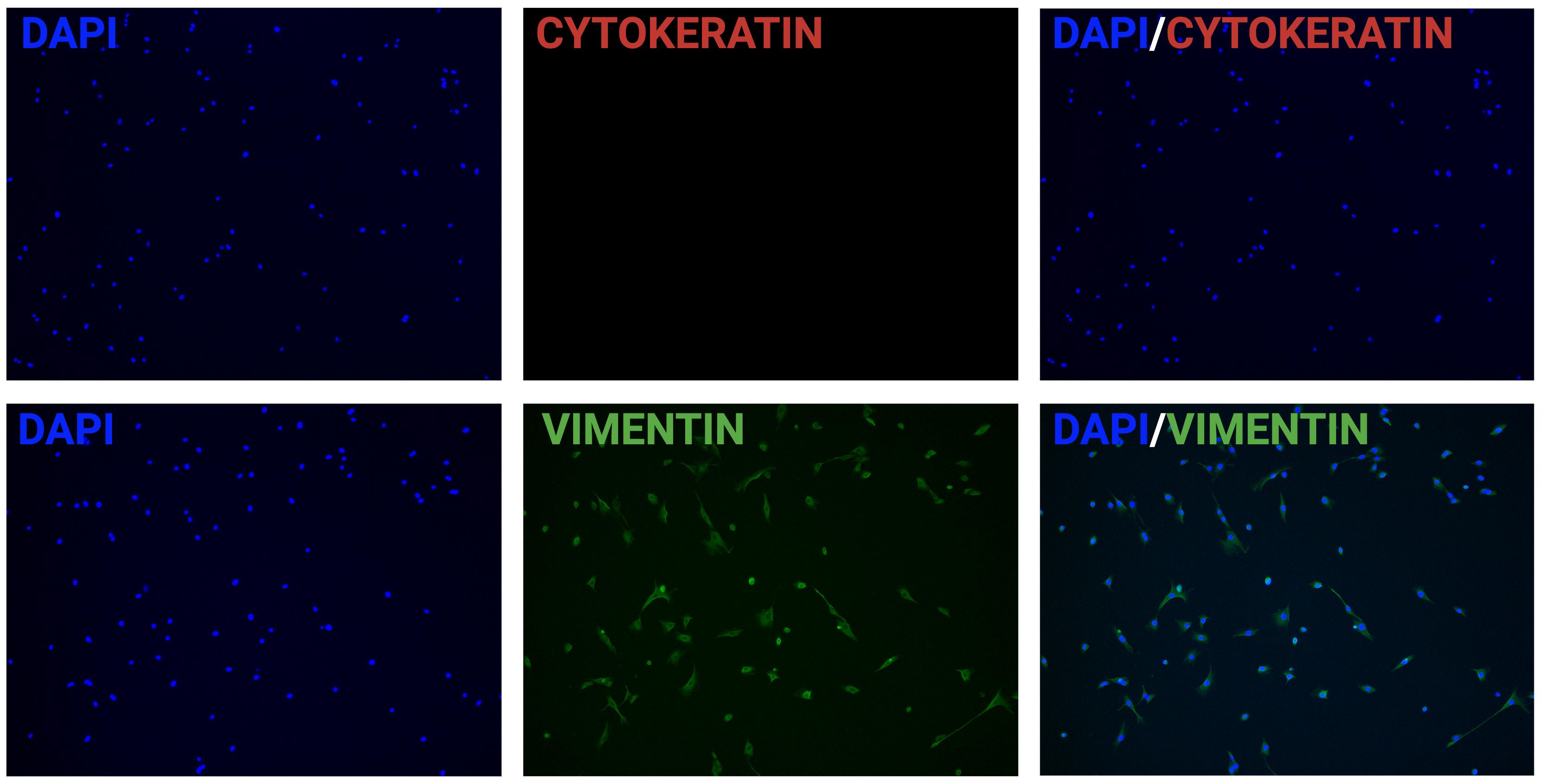
Figure 6. Representative fluorescent immunostaining demonstrating vimentin-positive and cytokeratin-negative staining in nilgai fibroblast culture. Magnification: 100×.
General notes and troubleshooting
General notes
1. Ears can be stored for a maximum of two weeks after collection if properly washed. For best results, use within two days of collection. Do not use samples if a decomposition odor is present or if hair falls out with gentle washing and handling.
2. Nilgai samples, like other wild animals, have many various contaminants. These washing steps are crucial to culture success. If desired, washing steps may be repeated multiple times to maximize bacterial and fungal removal. However, repeated washing in ethanol can result in tissue damage, so keep ethanol washes to a minimum.
3. Cells and outgrowth are usually visible by day 7. Depending on the age of the animal and storage time before processing, initial growth of nilgai cells may take longer. If growth is not seen by day 14, discard the culture plate and begin again.
Troubleshooting
Problem 1: Fungal contamination observed.
Possible cause: Fungal spores are present on initial nilgai samples.
Solution: If minimal, remove fungal growths with a pipette and rinse heavily with DPBS wash. Replace with fresh RFM-20% and closely observe for further growth. If culture is repeated, increase washes (see General note 2).
Note: Unless the sample is irreplaceable, it is best practice to dispose of any fungal-contaminated culture and re-sterilize the incubator.
Problem 2: Bacterial contamination is observed.
Possible cause: Bacteria are present on initial nilgai samples.
Solution: Discard contaminated samples and cultures, re-sterilize the incubator, and begin again with an increased number of washes.
Problem 3: Growth is not observed by day 7.
Solution: See General note 3.
Acknowledgments
This work was supported by the Agriculture and Food Research Initiative’s Animal Breeding, Genetics and Genomics program (A1201) project award nos. 2021-67016-33417 and 2022-08267, from the U.S. Department of Agriculture’s National Institute of Food and Agriculture.
The following figures were created using BioRender: Graphical overview and Figures 1, 5, and 6: https://BioRender.com/hl3pt3w.
This protocol was adapted from our previously established methodology [1] and has been optimized for use in nilgai antelope.
Author contributions
Conceptualization, D.H.; Methodology, M.M.; Investigation, M.M., C.M.; Writing—Original Draft, M.M.; Writing—Review & Editing, M.M., C.M., D.H.; Funding acquisition, D.H.; Supervision, D.H. All authors have read and agreed to the published version of the manuscript.
Competing interests
The authors declare no conflicts of interest.
References
- McKendrick, M. and Hagen, D. (2026). Establishment and Cryopreservation of Primary Fibroblast Cultures From Livestock Ear-Tissue Explants. Bio Protoc. 16(21): e5856. https://doi.org/10.21769/bioprotoc.5856
- David M. (2008). Leslie, Boselaphus Tragocamelus (Artiodactyla: Bovidae). Mammalian Species. 813: 1–16. https://doi.org/10.1644/813.1
- Jimenez, A. G. and Harper, J. M. (2023). Exploring the Role of Primary Fibroblast Cells in Comparative Physiology: A Historical and Contemporary Overview. Am J Physiol Regul Integr Comp Physiol. e2023. https://doi.org/10.1152/ajpregu.00025.2023
- LEE, P. C. and COMIZZOLI, P. (2026). Biobanking biodiversity: recent strategies, challenges, and opportunities. J Reprod Dev. 72(3): 210–224. https://doi.org/10.1262/jrd.2025-114
- Ghildiyal, K., Nayak, S. S., Rajawat, D., Sharma, A., Chhotaray, S., Bhushan, B., Dutt, T. and Panigrahi, M. (2023). Genomic insights into the conservation of wild and domestic animal diversity: A review. Gene. 886: 147719. https://doi.org/10.1016/j.gene.2023.147719
- Chitko‐McKown, C. G. and MacNeil, M. D. (2010). Development of fibroblast cell lines from the cow used to sequence the bovine genome. Anim Genet. 41(4): 445–445. https://doi.org/10.1111/j.1365-2052.2009.02013.x
Article Information
Publication history
Received: Aug 24, 2026
Accepted: Oct 28, 2026
Available online: Nov 18, 2026
Published: Nov 20, 2026
Copyright
© 2026 The Author(s); This is an open access article under the CC BY-NC license (https://creativecommons.org/licenses/by-nc/4.0/).
How to cite
McKendrick, M., Moulder, C. and Hagen, D. (2026). Establishment and Proliferation of Primary Nilgai Fibroblast Cultures from Adult Tissue. Bio-protocol 16(22): e5888. DOI: 10.21769/BioProtoc.5888.

 Download Citation in RIS Format
Category
Cell Biology > Cell isolation and culture > Monolayer culture
Cell Biology > Cell isolation and culture > Cryopreservation
Do you have any questions about this protocol?
Post your question to gather feedback from the community. We will also invite the authors of this article to respond.
 Post a Question


