


- Protocols

- Articles and Issues
- For Authors
- About
- Become a Reviewer


A New PCR-Based Method for the Diagnosis of Leishmania tropica, Leishmania infantum, and Leishmania major Infections

Published: Vol 16, Iss 23, Dec 5, 2026 DOI: 10.21769/BioProtoc.5883 Views: 44
Reviewed by: Anonymous reviewer(s)
 PDF
 Ask a question

Original research article
The authors used this protocol in:

Jan 2026

Protocol Collections
Comprehensive collections of detailed, peer-reviewed protocols focusing on specific topics




Abstract
Leishmaniases are neglected vector-borne diseases caused by protozoan parasites of the genus Leishmania, affecting both humans and animals in endemic regions worldwide. Beyond confirming the presence or absence of the parasite, accurate species identification is further essential for patient management, epidemiological surveillance, and monitoring of parasite circulation in natural reservoirs and vectors. Current molecular diagnostic approaches mainly rely on conventional PCR or qPCR assays, which frequently require additional post-amplification procedures, such as amplicon sequencing or RFLP analysis, to identify the Leishmania species. Moreover, species-identification methods based solely on amplicon size discrimination can lack sufficient resolution to reliably distinguish the diverse Leishmania species circulating in endemic regions, particularly those found in Morocco. The TIM assay (an acronym for tropica, infantum, major) provides a sensitive, robust, and flexible approach based on conventional PCR for the detection and identification of Leishmania parasites. It offers three complementary and adaptable strategies: enhanced sensitivity through an optional nested pre-amplification step (TIM1), rapid screening using a single multiplex reaction (TIM2), and accurate species discrimination through species-specific amplification (TIM3). Although initially developed to address human cutaneous leishmaniasis (CL) diagnostic needs in Morocco, the assay is broadly applicable to other endemic regions and can also be used for the detection of visceral leishmaniasis (VL) caused by L. infantum species. Its adaptability to field applications further supports its use in parasite surveillance and One Health strategies involving both wild and domestic animal reservoirs.
Key features
• Method developed to detect and identify endemic Leishmania species present in Morocco: L. tropica, L. infantum, and L. major.
• Primers used in this protocol are based on conserved kDNA minicircle sequences, which opens up their utilization in other endemic areas.
• This method allows detection of mixed infections/hybrid species.
• This method can detect L. infantum parasites in canine blood samples from different endemic countries.
Keywords: Leishmania PCR
Diagnostic
Species identification
Hybrids
Coinfections
Graphical overview
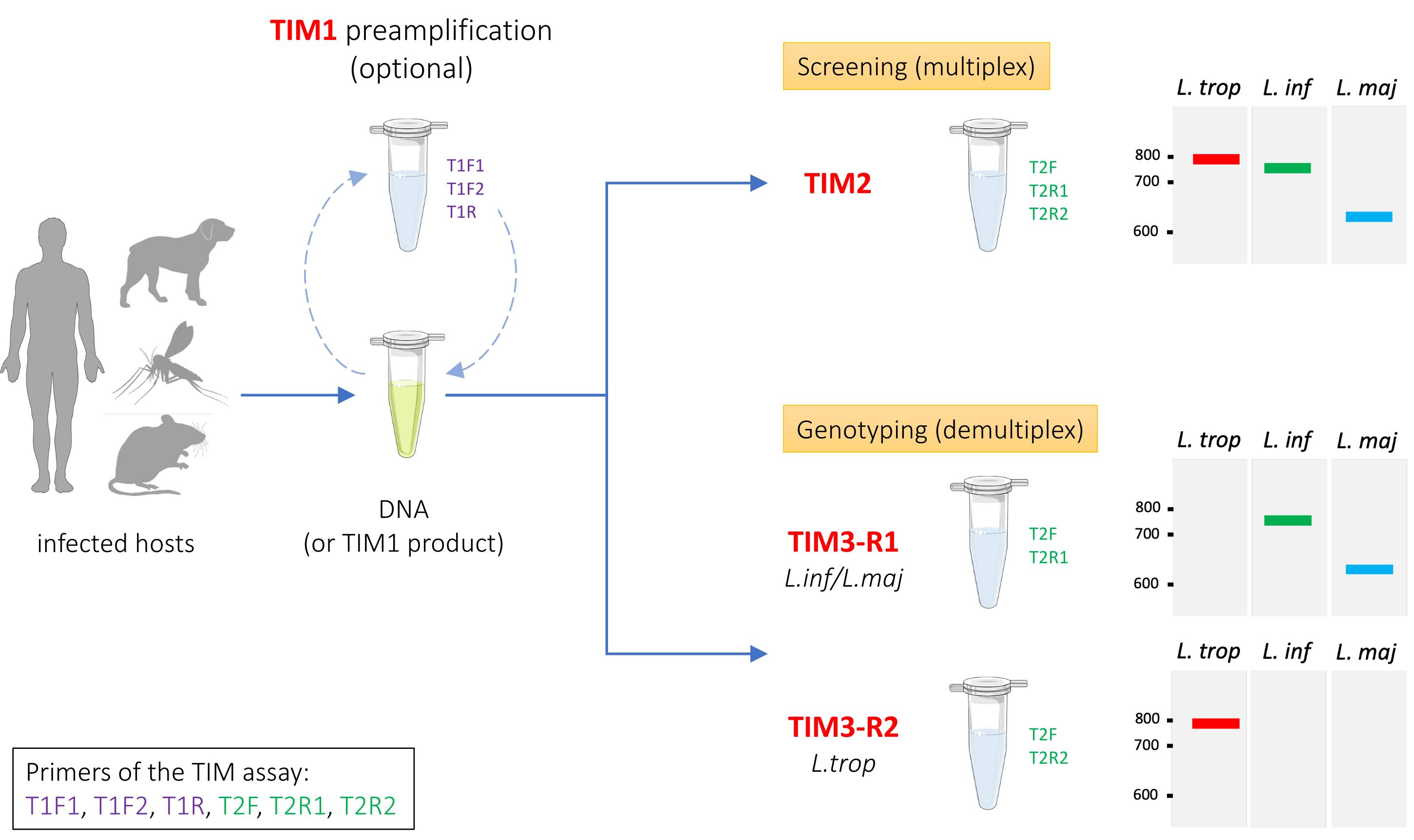
Graphical overview of the TIM assay
Background
Cutaneous leishmaniasis (CL) is one of the most widespread vector-borne parasitic diseases worldwide, with up to one million new cases reported annually. In Morocco, CL is caused by three endemic Leishmania species (Leishmania major, Leishmania tropica, and Leishmania infantum), each presenting with distinct epidemiological and clinical characteristics [1]. From 2023 to 2024, a 28% increase in the number of cases of CL was documented by the Moroccan Ministry of Health and Social Welfare, while the number of visceral leishmaniasis (VL) cases remained stable [2]. On the whole, L. tropica has the widest geographical distribution and is still considered anthroponotic despite recordings of L. tropica infection in animal hosts [3,4]. On the other hand, L. infantum is responsible for both CL and VL cases, primarily reported in the northern regions [5]. Domestic and stray dogs are its main reservoirs; the latter being difficult to monitor or treat. The epidemiology of leishmaniases in Morocco is therefore complex, significantly influenced by human mobility and climate change [6], which will further complicate efforts to control and manage the disease. In this context, accurate parasite detection and species identification are therefore essential for epidemiological surveillance, outbreak monitoring, and the implementation of effective control strategies.
Molecular diagnosis of leishmaniasis has largely relied on PCR-based approaches targeting either ribosomal DNA (rDNA) regions, such as the internal transcribed spacer 1 (ITS1), or kinetoplast DNA (kDNA) minicircles. Among the most commonly used methods, ITS1-PCR followed by restriction fragment length polymorphism analysis (ITS1-RFLP) enables species discrimination but requires an additional enzymatic digestion step and may provide limited resolution when restriction profiles are poorly amplified [7]. Likewise, the Noyes nested PCR assay targeting kDNA minicircles offers high analytical sensitivity but involves a two-step amplification procedure, increasing turnaround time and the risk of contamination [8]. Furthermore, species identification with the Noyes assay relies solely on amplicon size differences, which may be difficult to distinguish when species such as L. tropica and L. infantum produce similar-sized amplicons.
The TIM (Tropica–Infantum–Major) molecular toolbox was developed to overcome these limitations by providing a flexible PCR-based framework for the detection and identification of the three Leishmania species circulating in Morocco [9]. TIM targets specific motifs within kDNA minicircle sequences and can be implemented in three complementary formats: TIM1, an optional nested amplification strategy designed to maximize sensitivity; TIM2, a single multiplex PCR assay for rapid screening; and TIM3, a species-discrimination assay enabling precise identification and genotyping through two parallel reactions (TIM3-R1 and TIM3-R2). Compared with conventional ITS1-RFLP and Noyes PCR methods, TIM offers improved sensitivity, simplified workflows, and enhanced discriminatory power while requiring only standard PCR equipment and a limited set of six primers. Importantly, TIM can also detect mixed interspecific profiles directly from clinical samples, a feature that may facilitate the identification of coinfections or natural Leishmania hybrids that are often missed by traditional diagnostic approaches.
Although this protocol still requires access to molecular biology basic infrastructure and trained personnel, TIM represents a practical, robust, and cost-effective alternative for routine diagnosis in large-scale epidemiological investigations. Beyond its application to human CL, we demonstrate here that TIM is also suitable for monitoring VL caused by L. infantum in canine reservoirs from different endemic countries. Overall, the versatility of TIM broadens its potential use in surveillance programs targeting animal reservoirs, vector monitoring, investigations of parasite population dynamics, and the detection of mixed/hybrid species within a One Health framework.
Materials and reagents
Reagents
1. Molecular-weight size marker, such as the 1 kb DNA ladder 500 μg/mL (New England Biolabs, catalog number: N3232S)
2. 6× gel loading dye (New England Biolabs, catalog number: B7025) supplied with the 1 kb DNA ladder (New England Biolabs, catalog number: N3232S)
3. Standard agarose–type LE for routine gel electrophoresis (BioConcept Ltd, Amimed, catalog number: 7-01P02-R) stored at room temperature
4. Nuclease-free water (NFW) (New England Biolabs, catalog number: B1500S)
5. TAE 50× (1 L) (Euromedex, catalog number: EU0201-A)
6. DNA staining dye, such as GelGreen® nucleic acid stain (10,000× in water; 0.5 mL) (Millipore, catalog number: SCT125), stored at room temperature and compatible with the myGelTM InstaView LED blue-light transilluminator (Accuris instruments, model: E1201)
Specific reagents
1. Q5® Hot Start High-Fidelity 2× Master Mix (New England Biolabs, catalog number: M0494S/L); any other Taq polymerase or master mix can be used but will require further optimization
2. TIM primer stocks (supplied or prepared at 100 μM in H2O or TE buffer); primer sequences include degenerate nucleotides (Y, S, R, N, M) designated according to the IUPAC nucleotide code.
T1F1: 5′ GTGCAGAAAYCCCGTTCA 3′
T1F2: 5′ SATACAGAAACCCCGTTCA 3′
T1R: 5′ GCAGAACGCCCCTACC 3′
T2F: 5′ CTRGGGGTTGGTGTAAAATAG 3′
T2R1: 5′ CGNRGGACCAGAAAAGTTTG 3′
T2R2: 5′ TACCCGGAGGGCTACTM 3′
Laboratory supplies
1. 0.2 mL PCR tubes or PCR tube strips (nuclease-free)
2. Filtered pipette tips (nuclease-free)
3. 1.5 mL microcentrifuge tubes (nuclease-free)
4. Nitrile gloves
Equipment
1. PCR thermocycler (Eppendorf Mastercycler EP Gradient 5341)
2. Horizontal gel electrophoresis system with power supply (myGelTM InstaView Mini Electrophoresis System, Accuris instruments, model: E1201)
3. Gel imaging system (UV or blue-light transilluminator with camera) (myGelTM InstaView Mini Electrophoresis System, Accuris instruments, model: E1201)
4. Precision balance (Ohaus® Scout®)
5. Microwave for agarose gel preparation
6. Micropipettes (P10, P20, P200, P1000 (Eppendorf Reasearch®)
7. Mini centrifuge and vortex mixer
8. Refrigerator (4 °C), freezer (-20 °C)
9. PCR workstation or clean bench for PCR setup, if available
Procedure
A. Sampling and DNA extraction
The TIM assay has been successfully tested and validated on various types of cutaneous samples, including lesion swabs, skin biopsies, dermal aspirates, and lesion scrapings, using either phenol/chloroform-based or kit-based DNA extraction methods. While all tested sample types were suitable for TIM analysis, we selected lesion swabs combined with kit-based DNA extraction as the preferred approach, as this strategy offers greater convenience for both practitioners and patients, while ensuring improved inter-sample consistency and standardization. We observed that highly concentrated DNA extracts may generate additional nonspecific signals (background), particularly following phenol/chloroform extraction. In such cases, dilution of DNA samples to concentrations below 100 ng/μL is recommended prior to performing the TIM assay.
B. Preparation of primer mixes
For convenience, we recommend preparing a primer mix for each TIM assay. The volume of each primer mix can be adjusted according to the number of reactions required and the user's experimental needs. However, we suggest preparing a relatively small volume of primer mix (50 or 100 μL) or making aliquots to reduce the number of freeze/thaw cycles and contamination risks. Primer mix preparation is a sensitive step that must be performed in a controlled environment (PCR hood, room without amplified DNA) with nuclease-free water.
The four primer mixes (TIM1, TIM2, TIM3-R1, and TIM3-R2) are prepared from the six stock primers (100 μM) and nuclease-free water (NFW) according to the procedure described below (Tables 1–4). A primer mix of 100 μL is sufficient for 100 PCR reactions.
Table 1. Primer mix TIM1
| Reagent | Volume | Final concentration |
|---|---|---|
| T1F1 | 10 μL | 10 μM |
| T1F2 | 10 μL | 10 μM |
| T1R | 10 μL | 10 μM |
| H20 (NFW) | 70 μL | |
| Total | 100 μL |
Table 2. Primer mix TIM2
| Reagent | Volume | Final concentration |
|---|---|---|
| T2F | 10 μL | 10 μM |
| T2R1 | 10 μL | 10 μM |
| T2R2 | 10 μL | 10 μM |
| H2O (NFW) | 70 μL | |
| Total | 100 μL |
Table 3. Primer mix TIM3-R1
| Reagent | Volume | Final concentration |
|---|---|---|
| T2F | 10 μL | 10 μM |
| T2R1 | 10 μL | 10 μM |
| H2O (NFW) | 80 μL | |
| Total | 100 μL |
Table 4. Primer mix TIM3-R2
| Reagent | Volume | Final concentration |
|---|---|---|
| T2F | 10 μL | 10 μM |
| T2R2 | 10 μL | 10 μM |
| H2O (NFW) | 80 μL | |
| Total | 100 μL |
All primer mixes are stored at -20 °C and constitute the only working primer solutions required for performing the different TIM assays.
C. TIM concept and procedures
The TIM assay is a molecular diagnostic approach based on the amplification of kinetoplast DNA minicircles, which are present in thousands of copies within each Leishmania parasite. The primers are designed to target the conserved ~120 bp region of the minicircle sequence encompassing the three canonical conserved sequence blocks (CSB1-3), allowing sensitive detection of Leishmania parasites from clinical samples (Figure 1A). T1R and T2F primers are conserved across the three targeted Leishmania species, whereas T1F1 and T2R1 are specific to L. major and L. infantum, while T1F2 and T2R2 are specific to L. tropica. The primers are oriented in opposite directions, enabling amplification of the entire circular DNA template. Because the TIM1 primers (T1F1, T1F2, and T1R) target the central region of the conserved minicircle domain, they generate larger amplicons encompassing the regions subsequently amplified by TIM2/TIM3 primers (T2F, T2R1, T2R2). The external positioning of the TIM2/TIM3 primers therefore results in shorter, nested amplification products (Figure 1B). The TIM1 and TIM2 assays are designed to amplify minicircle sequences from all three targeted Leishmania species, whereas TIM3-R1 and TIM3-R2 specifically amplify minicircles from L. major/L. infantum and L. tropica, respectively (Figure 1B).
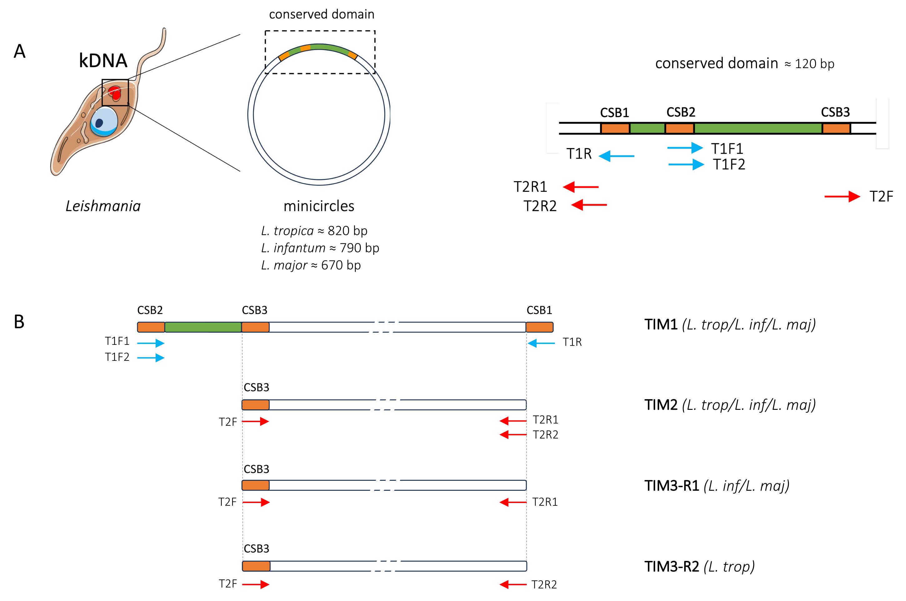
Figure 1. Schematic representation of the TIM assay. (A) Schematic representation of the conserved domain of a Leishmania minicircle, showing the positions of the conserved sequence blocks (CSB1-3) and the six TIM primers in their respective orientations (arrows). Primers used in the TIM1 assay are shown in blue, whereas those used in the TIM2/TIM3 assays are shown in red. (B) Schematic representation of the amplified products and primers used in the TIM1, TIM2, TIM3-R1, and TIM3-R2 assays.
D. TIM1 nested for pre-amplification of low signals
TIM1 is an optional pre-amplification step targeting the internal conserved region of Leishmania kDNA minicircles. It generates a larger amplicon that will be used for downstream TIM2/TIM3 assays (Figure 1). TIM1 pre-amplification is recommended for samples with low expected parasite loads or when TIM2 or TIM3 yields a negative result and can therefore help distinguish true-negative samples from false-negative results due to very low parasite loads.
1. Defrost the primer mix TIM1 (Table 1) and the Q5 Hot Start High-Fidelity 2× master mix and keep on ice.
Note: The Q5 Hot Start High-Fidelity 2× master mix is inhibited at room temperature, allowing flexible reaction setup (room temperature or ice).
2. Prepare the TIM1 reaction mix in a clean 1.5 mL tube by combining the components in Table 5. Multiply the volume of each reaction component (excluding the DNA template) by N + 0.5 to prepare a master mix for N samples, providing a small excess to compensate for pipetting losses. The example provided below is based on the use of 1 μL of DNA template per sample. DNA input volume can be increased up to 5 μL per reaction by adjusting the nuclease-free water volume accordingly to maintain a final reaction volume of 20 μL.
Table 5. TIM1 reaction mix
| Components | Volume for 1 reaction (20 μL final) | Volume necessary for N reactions |
|---|---|---|
| Q5 Hot Start master mix | 10 μL | N.5 × 10 μL |
| Primer mix TIM1 | 1 μL | N.5 × 1 μL |
| H2O (NFW) | 8 μL | N.5 × 8 μL |
| DNA | 1 μL |
3. Prepare the required number of PCR tubes (0.2 mL) and reaction mix volume and dispense 19 μL in each tube.
Note: A positive control should be included in each experiment, using either a confirmed positive sample or Leishmania genomic DNA. A no-template control (NTC) should also always be included to monitor for potential contamination.
4. Add 1 μL of DNA template to each tube, or 1 μL of water in the case of the negative control.
5. Close the tubes and briefly vortex and spin the PCR tubes.
6. Set the thermal cycler with the following program (TIM1 condition):
a. Initial denaturation at 98 °C for 30 s.
b. Denaturation at 98 °C for 10 s.
c. Annealing at 62 °C for 20 s.
d. Extension at 72 °C for 20 s.
e. Repeat steps D6b–d for 35 cycles.
f. Final extension at 72 °C for 2 min.
g. Hold at 4 °C.
Note: All annealing temperatures reported in this protocol were calculated using the NEB Tm Calculator (https://tmcalculator.neb.com/).
7. TIM1 amplification products will serve as template for subsequent TIM2 or TIM3 assays. Use 1 μL of TIM1 amplification product as the template in place of genomic DNA. We have observed, however, that diluting TIM1 amplification products 1:10 in nuclease-free water prior to TIM2 or TIM3 analyses may reduce potential background and improve signal quality.
Note: TIM1 is particularly susceptible to carry-over contamination because the second-round amplification is performed on an already amplified product. Therefore, implementing a unidirectional workflow is the most effective measure to minimize the risk of contamination (strict separation of pre- and post-amplification areas).
E. TIM2 assay for rapid detection of the three Leishmania species in a single PCR reaction
TIM2 is primarily intended for high-throughput screening, allowing multiple samples to be analyzed simultaneously in a single multiplex PCR reaction. Although it can provide species information, discrimination between L. infantum and L. tropica may be difficult based solely on agarose gel profiles. For unequivocal species identification, TIM3 should be used preferentially.
1. Defrost the primer mix TIM2 (Table 2) and the Q5 Hot Start High-Fidelity 2× master mix and keep on ice.
2. Prepare the TIM2 reaction mix in a clean 1.5 mL tube by combining the components in Table 6. Multiply the volume of each reaction component (excluding the DNA template) by N + 0.5 to prepare a master mix for N samples, providing a small excess to compensate for pipetting losses. The example provided below is based on the use of 1 μL of DNA template per sample. DNA input volume can be increased up to 5 μL per reaction by adjusting the nuclease-free water volume accordingly to maintain a final reaction volume of 20 μL.
Table 6. TIM2 reaction mix
| Components | Volume for 1 reaction (20 μL final) | Volume necessary for N reactions |
|---|---|---|
| Q5 Hot Start master mix | 10 μL | N.5 × 10 μL |
| Primer mix TIM2 | 1 μL | N.5 × 1 μL |
| H2O (NFW) | 8 μL | N.5 × 8 μL |
| DNA | 1 μL |
3. Prepare the required number of PCR tubes (0.2 mL) and reaction mix volume and dispense 19 μL in each tube.
Note: A positive control should be included in each experiment, using either a confirmed positive sample or Leishmania genomic DNA. A no-template control (NTC) should also always be included to monitor for potential contamination.
4. Add 1 μL of DNA template to each tube, or 1 μL of water in the case of the negative control.
5. Close the tubes and briefly vortex and spin the PCR tubes.
6. Set the thermal cycler with the following program (TIM2/TIM3 conditions):
a. Initial denaturation at 98 °C for 30 s.
b. Denaturation at 98 °C for 10 s.
c. Annealing at 61 °C for 20 s.
d. Extension at 72 °C for 20 s.
e. Repeat steps E6b–d for 35 cycles.
f. Final extension at 72 °C for 2 min.
g. Hold at 4 °C.
7. Proceed to sample analysis on an agarose gel (see section G).
F. TIM3 assay for accurate species discrimination and interspecific hybrids/coinfection detection
The TIM3 assay consists of replacing the TIM2 assay with two parallel reactions, TIM3-R1 and TIM3-R2, to achieve specific discrimination of the target species. TIM3-R1 specifically amplifies L. major and L. infantum at sufficiently different sizes to allow straightforward species discrimination, whereas TIM3-R2 specifically amplifies L. tropica (Figure 1B).
Notes:
1. For routine diagnosis of cutaneous leishmaniasis, when species identification is clinically or epidemiologically relevant, the TIM3 assay can be used as the first-line approach. It simultaneously detects Leishmania DNA and accurately identifies the infecting species in a single PCR assay.
2. The TIM3 assay can identify mixed DNA profiles (observed in approximately 10% of clinical samples from cutaneous leishmaniasis cases), which may reflect either coinfections involving different Leishmania species or naturally occurring interspecific hybrids [9].
1. Thaw the TIM3-R1 (Table 3) and TIM3-R2 (Table 4) primer mixes and the Q5 Hot Start High-Fidelity 2× master mix and keep on ice.
2. Prepare the TIM3-R1 and TIM3-R2 reaction mixes in two clean 1.5 mL tubes by adding the components in Table 7. Multiply the volume of each reaction component (excluding the DNA template) by N + 0.5 to prepare a master mix for N samples, providing a small excess to compensate for pipetting losses. The example provided below is based on the use of 1 μL of DNA template per sample. DNA input volume can be increased up to 5 μL per reaction by adjusting the nuclease-free water volume accordingly to maintain a final reaction volume of 20 μL.
Table 7. TIM3-R1 and TIM3-R2 reaction mixes
| Components | Volume for 1 reaction TIM3-R1 (20 μL final) | Volume for 1 reaction TIM3-R2 (20 μL final) |
|---|---|---|
| Q5 Hot Start master mix | 10 μL | 10 μL |
| Primer mix TIM3-R1 | 1 μL | - |
| Primer mix TIM3-R2 | - | 1 μL |
| H2O (NFW) | 8 μL | 8 μL |
| DNA | 1 μL | 1 μL |
3. Prepare the required number of 0.2 mL PCR tubes and the corresponding reaction mix volume, then dispense 19 μL of the reaction mix into each tube. In this case, each sample will be analyzed in duplicate using both the TIM3-R1 and TIM3-R2 assays.
Note: A positive control should be included in each experiment, using either a confirmed positive sample or Leishmania genomic DNA. A no-template control (NTC) for TIM3-R1 and TIM3-R2 assays should also always be included to monitor for potential contamination.
4. Add 1 μL of DNA template to each tube, or 1 μL of water in the case of the negative control.
5. Close the tubes and briefly vortex and spin the PCR tubes.
6. Set the thermal cycler with the following program (TIM2/TIM3 conditions):
a. Initial denaturation at 98 °C for 30 s.
b. Denaturation at 98 °C for 10 s.
c. Annealing at 61 °C for 20 s.
d. Extension at 72 °C for 20 s.
e. Repeat steps F6b–d for 35 cycles.
f. Final extension at 72 °C for 2 min.
g. Hold at 4 °C.
7. Proceed to sample analysis on an agarose gel (section G).
Note: Loading TIM3-R1 and TIM3-R2 products side by side for each sample, as illustrated in Figure 2, facilitates result interpretation and makes the amplification profiles easier to read.
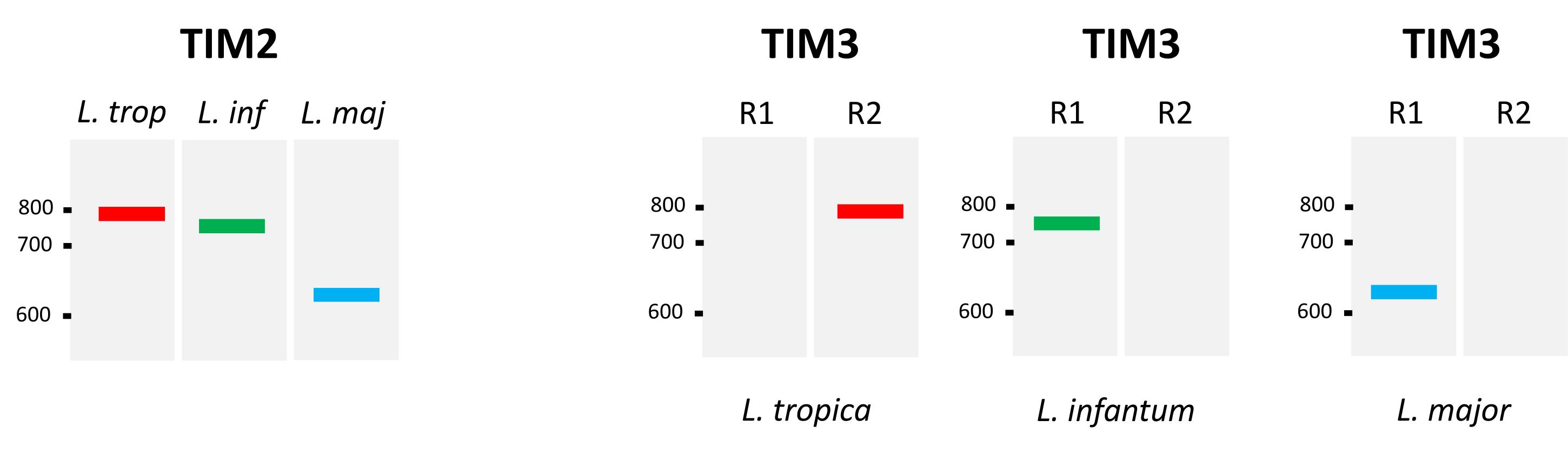
Figure 2. Expected amplification products and their respective sizes for the TIM2 and TIM3 assays across different Leishmania species. The TIM2 and TIM3 assays produce a single amplification band of approximately 820 bp for L. tropica, 790 bp for L. infantum, and 670 bp for L. major. TIM3-R1 amplifies minicircles from L. infantum and L. major, whereas TIM3-R2 amplifies minicircles specific to L. tropica.
G. Gel electrophoresis and interpretation
G1. Gel preparation
1. Weigh 1.5 g of agarose powder and dissolve it in 100 mL of 0.5× TAE buffer using a microwave to make a 1.5% agarose gel (w/v).
Note: We found that 1.5% agarose gels provided better resolution in the 650–800 bp range, corresponding to the expected sizes of the amplification products generated by the different TIM assays.
2. Add 10 μL of GelGreen® nucleic acid staining solution (10,000×) and mix thoroughly.
Note: Other DNA staining dyes may also be used, depending on the gel imaging system available.
3. Pour the dissolved agarose solution into a gel tray with a well comb. Allow the poured gel to solidify fully at room temperature for 20–30 min.
G2. Loading samples and running
1. Place the solidified agarose gel into the electrophoresis unit and submerge it entirely in 0.5× TAE buffer.
2. Mix 10 μL of the PCR products with 2 μL of 6× gel loading dye solution.
3. Load your samples and a DNA ladder into the gel wells.
4. Perform gel migration at 100 V for 30–40 min (adjust the voltage and running time depending on gel concentration and PCR product size).
5. After gel running, carefully remove the gel from the unit and place it under a gel imaging system to visualize DNA fragments.
Note: A schematic representation of the expected amplification product sizes for the TIM2 and TIM3 assays is shown in Figure 2.
Validation of protocol
This protocol has been used and validated in the following research article:
• El Idrissi Saik, I. et al. [9]. TIM, a novel molecular toolbox for the detection and identification of Leishmania species in Morocco. Microbiol Spectrum. 14(1): e03447–25. https://doi.org/10.1128/spectrum.03447-25
Although TIM was initially developed for the diagnosis and species identification of Leishmania parasites causing CL in Morocco, we further explored its applicability for the diagnosis of mammalian infection in the context of VL. Specifically, we evaluated the ability of the TIM3-R1 assay (which targets L. infantum and L. major species) to detect L. infantum infection in canine blood samples collected from endemic areas outside Morocco. Given the typically low parasite loads expected in blood, we also assessed whether the optional TIM1 pre-amplification step could improve detection efficiency. Twelve blood samples from dogs with suspected canine leishmaniasis (CanL) from the Cévennes region (France) and provided by the French National Reference Center for Leishmaniasis (CNRL) were analyzed using either the TIM3-R1 assay alone or the combined TIM1 + TIM3-R1 approach. As shown in Figure 3, TIM1 pre-amplification clearly improved detection sensitivity compared with TIM3-R1 alone, with 5/12 samples producing clear amplicons of the expected size characteristic of L. infantum. All five samples were subsequently confirmed as positive by the CNRL reference qPCR method, with samples 1, 10, and 11 exhibiting higher parasite loads (data not shown). These results are consistent with the stronger amplification bands observed for these samples using TIM3-R1 alone (Figure 3). Conversely, four canine blood samples collected from the Pantanal and Cerrado biomes in Brazil and previously identified as positive for L. infantum by the CNRL reference method were also positive using the combined TIM1 + TIM3-R1 approach (data not shown). Overall, these results highlight the robustness of this assay beyond its initial development for CL and support its potential use for the molecular surveillance of L. infantum transmission across different mammalian hosts, sample types, and endemic regions worldwide.
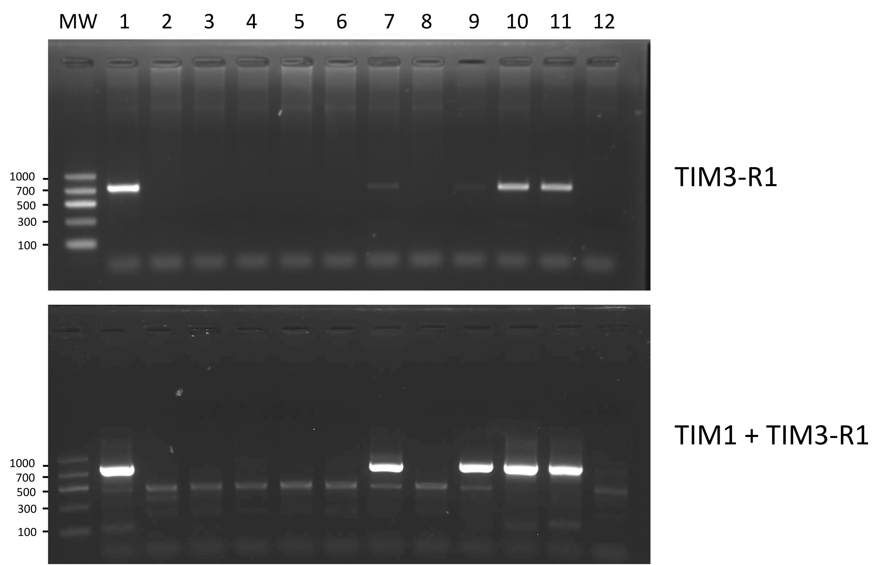
Figure 3. Benefits of TIM1 pre-amplification for screening canine blood samples with TIM assay. Comparison of the results obtained using either the TIM3-R1 assay alone or the combined TIM1 + TIM3-R1 approach on 12 canine blood samples from a CanL endemic region (Cévennes, France). Gel profiles were obtained using 10 μL of PCR products per lane. MW: molecular weight marker (Clearline DNA ladder 100–1,000 bp, 257738).
General notes and troubleshooting
Troubleshooting
Problem 1: Smearing or unexpected banding pattern on the gel.
Possible cause: Excessive DNA template concentration.
Solution: Dilute the DNA template 1:10 and repeat the TIM assay.
Problem 2: Contamination of negative controls after TIM1 pre-amplification.
Possible cause: Carry-over contamination during the transfer of TIM1 amplification products.
Solution: Maintain a unidirectional workflow and strict physical separation of pre- and post-amplification areas.
Acknowledgments
This study was supported by the EU-funded project LeiSHield-MATI RISE "A multi-disciplinary international effort to identify clinical, molecular and social factors impacting cutaneous leishmaniasis" (Grant Agreement No. 778298), the Occitanie Region as part of the RIVOC Key Challenge led by the University of Montpellier (Leishtrack project), the LabEx CeMEB, an ANR "Investissements d'avenir" program (ANR-10-LABX-04-01), and the Moroccan Ministry of Higher Education Scientific Research and Innovation (National Center for Scientific and Technical Research/CNRST, PPR/2015/27). We thank Danilo Kluyber, Idris Mhaidi, and Julio Benavides for providing canine blood samples from Brazil as part of the ZAMBA project supported by the French Government through the Agence Nationale de la Recherche (ANR) under the France 2030 programme, the European Union’s NextGeneration EU programme (grant no. ANR-21-EXES-0005), the Occitanie Region, and the ExposUM Institute of the University of Montpellier.
This protocol was used in [9].
Author contributions
Protocol Development, K.L., I.EIS., M.R., B.V.; Protocol Optimization or Validation, K.L., I.EIS., M.R., B.V.; Writing—Original Draft, K.L., M.R., B.V.; Writing—Review & Editing, K.L., I.EIS., AL.B., M.L., C.R., M.R., B.V.; Funding acquisition, AL.B., M.L., M.R., B.V.; Supervision, M.R., B.V.
Competing interests
The authors declare no conflicts of interest.
Ethical considerations
The canine blood samples from France were collected under an experimental protocol approved by an animal experimentation ethics committee (reference number APAFIS #33307-2021070114534565 v4). The owners were informed of the research protocol, the samples to be taken, and the ethics committee’s approval. Their informed consent was obtained verbally.
References
- Baghad, B., Razanapinaritra, R., Maksouri, H., El Bouri, H., Outlioua, A., Fellah, H., Lemrani, M., Akarid, K., Martin-Sanchez, J., Chiheb, S., et al. (2020). Possible introduction of Leishmania tropica to urban areas determined by epidemiological and clinical profiles of patients with cutaneous leishmaniasis in Casablanca (Morocco). Parasite Epidemiol Control. 9: e00129. https://doi.org/10.1016/j.parepi.2019.e00129
- Ministère de la Santé et de la Protéction sociale. (2024). Santé en chiffres 2024 (p. 122). Royaume du Maroc: Ministère de la Santé et de la Protéction sociale. Retrieved from https://www.sante.gov.ma/Documents/2026/06/Sante%20en%20chiffre%202024%20VF.pdf
- Kholoud, K., Bounoua, L., Sereno, D., El Hidan, M. and Messouli, M. (2020). Emerging and Re-Emerging Leishmaniases in the Mediterranean Area: What Can Be Learned from a Retrospective Review Analysis of the Situation in Morocco during 1990 to 2010? Microorganisms. 8(10): 1511. https://doi.org/10.3390/microorganisms8101511
- El Idrissi Saik, I., Benlabsir, C., Fellah, H., Lemrani, M. and Riyad, M. (2022). Transmission patterns of Leishmania tropica around the Mediterranean basin: Could Morocco be impacted by a zoonotic spillover? PLoS NeglTrop Dis. 16(1): e0010009. https://doi.org/10.1371/journal.pntd.0010009
- El mazini, S., Ejghal, R., Bekhti, K. and Lemrani, M. (2022). The Sporadic cutaneous leishmaniasis due to Leishmania infantum in Morocco: A presumably trend towards endemicity. Acta Trop. 227: 106288. https://doi.org/10.1016/j.actatropica.2021.106288
- Daoui, O., Bennaid, H., Kbaich, M. A., Mhaidi, I., Aderdour, N., Rhinane, H., Bouhout, S., Akarid, K. and Lemrani, M. (2022). Environmental, Climatic, and Parasite Molecular Factors Impacting the Incidence of Cutaneous Leishmaniasis Due to Leishmania tropica in Three Moroccan Foci. Microorganisms. 10(9): 1712. https://doi.org/10.3390/microorganisms10091712
- Schönian, G., Nasereddin, A., Dinse, N., Schweynoch, C., Schallig, H. D., Presber, W. and Jaffe, C. L. (2003). PCR diagnosis and characterization of Leishmania in local and imported clinical samples. Diagn Microbiol Infect Dis. 47(1): 349–358. https://doi.org/10.1016/s0732-8893(03)00093-2
- Noyes, H. A., Reyburn, H., Bailey, J. W. and Smith, D. (1998). A Nested-PCR-Based Schizodeme Method for Identifying Leishmania Kinetoplast Minicircle Classes Directly from Clinical Samples and Its Application to the Study of the Epidemiology of Leishmania tropica in Pakistan. J Clin Microbiol. 36(10): 2877–2881. https://doi.org/10.1128/jcm.36.10.2877-2881.1998
- El Idrissi Saik, I., Lemkhayar, K., Mhaidi, I., El Mazini, S., Chiheb, S., Bañuls, A. L., Lemrani, M., Riyad, M. and Vergnes, B. (2026). TIM, a novel molecular toolbox for the detection and identification of Leishmania species in Morocco. Microbiol Spectrum. 14(1): e03447–25. https://doi.org/10.1128/spectrum.03447-25
Article Information
Publication history
Received: Sep 11, 2026
Accepted: Oct 25, 2026
Available online: Nov 11, 2026
Published: Dec 5, 2026
Copyright
© 2026 The Author(s); This is an open access article under the CC BY-NC license (https://creativecommons.org/licenses/by-nc/4.0/).
How to cite
Lemkhayar, K., El Idrissi Saik, I., Bañuls, A. L., Lemrani, M., Ravel, C., Riyad, M. and Vergnes, B. (2026). A New PCR-Based Method for the Diagnosis of Leishmania tropica, Leishmania infantum, and Leishmania major Infections. Bio-protocol 16(23): e5883. DOI: 10.21769/BioProtoc.5883.

 Download Citation in RIS Format
Do you have any questions about this protocol?
Post your question to gather feedback from the community. We will also invite the authors of this article to respond.
 Post a Question


