


- Protocols

- Articles and Issues
- For Authors
- About
- Become a Reviewer


Producing High-Purity, Immobilized Enzymes on Inactivated Bacillus subtilis Using the Coated Bacterial Enzyme (CBE) Platform

Published: Vol 16, Iss 23, Dec 5, 2026 DOI: 10.21769/BioProtoc.5880 Views: 15
Reviewed by: Mirela Tkalcic CavuzicAnonymous reviewer(s)
 PDF
 Ask a question

Original research article
The authors used this protocol in:
Oct 2026

Protocol Collections
Comprehensive collections of detailed, peer-reviewed protocols focusing on specific topics




Abstract
The purification of industrial enzymes, such as β-galactosidase for lactose-reduced milk production, typically requires costly and time-consuming multi-step chromatography. Most purification protocols use two or more column fractionation steps and involve techniques such as gel filtration, ion exchange, and affinity chromatography, which are expensive and generally result in low yields. To address this limitation, we developed the Coated Bacterial Enzymes (CBEs) platform, which enables one-step purification and immobilization of recombinant enzymes fused to the SlpA cell wall-binding domain using chemically inactivated Bacillus subtilis as a low-cost biological carrier. This protocol describes the expression of the chimeric enzyme BbgII-SlpA (a β-galactosidase from Bifidobacterium bifidum) in Escherichia coli, the preparation of the bacterial carrier, the direct capture of the target enzyme from crude cell lysate onto the carrier surface, and the functional characterization of the resulting immobilized biocatalyst. The entire purification and immobilization process is completed in approximately 2 h after cell lysis. The procedure also includes an optional elution step to recover the purified enzyme in soluble form. The CBE platform offers a one-step, cost-effective, and scalable alternative for obtaining high-purity, immobilized enzymes with enhanced thermal stability and tolerance to inhibitory cations, making it suitable for industrial applications such as lactose hydrolysis.
Key features
• This protocol requires basic experience in recombinant protein expression in E. coli for successful implementation.
• The protocol enables simultaneous purification and immobilization in a single incubation step, reducing processing time to ~2 h after cell lysis.
• This protocol delivers high-purity immobilized enzyme with enhanced thermal stability, retaining ~80% activity after 390 min at 45 °C.
• The bacterial carrier can be prepared in bulk and stored at 4 °C up to 6 months, allowing flexible scheduling and repeated use.
Keywords: β-galactosidase Enzyme purification
Enzyme immobilization
Lactose hydrolysis
Coated Bacterial Enzymes
SlpA domain
Inactivated Bacillus subtilis
Low-cost biological carrier
Graphical overview
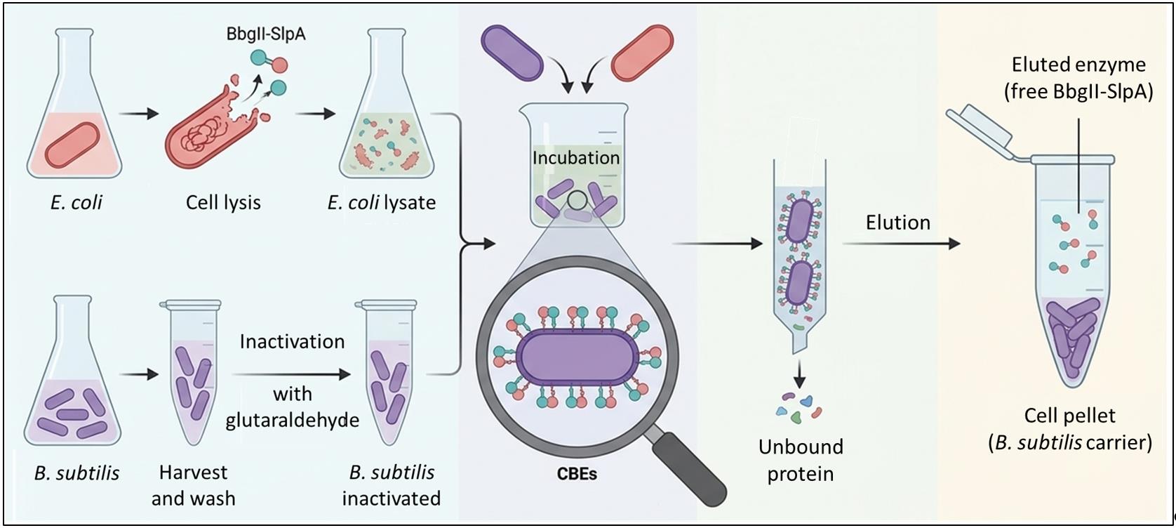
Schematic of Coated Bacterial Enzyme (CBE) production. The recombinant BbgII-SlpA enzyme is expressed in E. coli, and the cell lysate is incubated with chemically inactivated B. subtilis to immobilize the enzyme on its surface. Optionally, the enzyme can be eluted from the carrier to obtain purified enzyme.
Background
The enzymatic hydrolysis of lactose is the most widely adopted strategy for producing lactose-reduced milk, a product that addresses the dietary needs of the estimated 70% of the global population with some degree of lactose intolerance [1,2]. The process requires high-purity β-galactosidase to avoid contaminating proteases, which can generate bitter off-flavors and undesirable Maillard reaction products [3]. Conventional purification protocols rely on two or more chromatographic steps, such as gel filtration, ion exchange, and affinity chromatography, that are time-consuming, expensive, and generally result in low overall yields [4,5]. To overcome these limitations, one-step affinity purification strategies have been developed, typically involving the fusion of a polypeptide tag to the target protein, enabling selective capture onto a specific ligand matrix. Commonly used tags include poly-histidine for Ni2+-affinity chromatography, as well as chitin-binding, cellulose-binding, and starch-binding domains [6–8]. While effective, these methods still require costly affinity matrices or specialized supports. For instance, Gennari et al. achieved one-step purification and immobilization of a β-galactosidase using a cellulose-binding domain (CBD) tag on magnetic cellulose supports, which enhanced thermal stability but required the preparation of functionalized magnetic particles [9]. Similarly, immobilization on live bacteria using peptidoglycan-binding motifs such as LysM, or via surface display systems, has also been reported [10,11], yet these approaches rely on recombinant microorganisms that must maintain enzyme expression while alive, introducing metabolic burden and regulatory concerns regarding genetically modified organisms. To address these challenges, we recently developed the Coated Bacterial Enzymes (CBEs) platform, which builds upon the SlpA cell wall–binding domain from Lactobacillus acidophilus [12]. This tag enables specific and high-affinity binding to the cell wall of Bacillus subtilis, mediated by recognition of teichoic and lipoteichoic acids [13,14]. The CBE platform employs chemically inactivated B. subtilis as a low-cost, non-recombinant biological carrier for the simultaneous purification and immobilization of recombinant enzymes fused to the SlpA domain. This approach decouples enzyme production from the final biocatalyst, avoiding GMO-related limitations and providing a stable, ready-to-use affinity matrix. The primary advantage of the CBE platform lies in its operational simplicity and time-saving ability—purification and immobilization are achieved in a single step, eliminating the need for separate chromatographic and immobilization procedures—rather than in achieving a higher final purity than in optimized conventional methods. Our protocol is based on a simple two-step workflow: expression of the chimeric enzyme BbgII-SlpA (a β-galactosidase from Bifidobacterium bifidum) in Escherichia coli, and direct capture of the target enzyme from crude lysate onto the inactivated B. subtilis carrier. The entire purification and immobilization process is completed in approximately 2 h after cell lysis. After incubation and washing, the resulting CBE biocatalyst retains high catalytic activity and exhibits enhanced thermal stability, retaining ~80% activity after 390 min at 45 °C. Importantly, the CBEs platform is adaptable to other SlpA-tagged proteins and includes an optional elution step for recovering the purified enzyme in soluble form, making it a versatile tool for diverse biotechnological applications where high-purity, immobilized enzymes are required.
Materials and reagents
Biological materials
1. Escherichia coli BL21-CodonPlus (DE3)-RIPL competent cells (Agilent Technologies, catalog number: 230280)
2. Bacillus subtilis var. natto (ATCC, catalog number: 15245)
3. pET-29c(+) plasmid (EMD Biosciences, catalog number: 69873-3)
4. pET-BbgII-SlpA expression plasmid (custom synthesized)
Note: The BbgII-SlpA DNA sequence was codon-optimized for E. coli expression using J-Cat software, chemically synthesized by General Biosystems, Inc., and subsequently cloned into the pET-29c(+) plasmid using NdeI and XhoI restriction enzymes to yield the expression plasmid (pET-BbgII-SlpA). The SlpA sequence comprises amino acids 310–470 of the carboxy-terminal fragment of the SlpA protein from L. helveticus (GenBank ID: MBW7998749.1), and the BbgII amino acid sequence (GenBank ID: WP_270548944.1) was used. The construct has been described in detail in Harguindeguy et al. [15].
Reagents
1. Tryptone (Thermo Fisher Scientific, catalog number: LP0042B)
2. Yeast extract (Merck Millipore, catalog number: 92144)
3. Sodium chloride (NaCl) (Sigma-Aldrich, catalog number: S9888)
4. Agar (Merck Millipore, catalog number: 05040)
5. Kanamycin sulfate (Genbiotech, catalog number: K-120-25)
6. Isopropyl β-D-1-thiogalactopyranoside (IPTG) (Sigma-Aldrich, catalog number: I6758)
7. Tris base (Sigma-Aldrich, catalog number: T1503)
8. Ethylenediaminetetraacetic acid (EDTA) (Sigma-Aldrich, catalog number: E9884)
9. Lysozyme from chicken egg white (Inbio Highway, catalog number: E1405)
10. Sodium phosphate dibasic (Na2HPO4) (Sigma-Aldrich, catalog number: S9763)
11. Potassium phosphate monobasic (KH2PO4) (Sigma-Aldrich, catalog number: P0662)
12. Potassium chloride (KCl) (Sigma-Aldrich, catalog number: P3911)
13. TritonTM X-100 (Sigma-Aldrich, catalog number: T8787); store at room temperature; irritant: handle with gloves and eye protection
14. Urea (Sigma-Aldrich, catalog number: U1250)
15. Glutaraldehyde solution, 25% (v/v) (Sigma-Aldrich, catalog number: G4004); store at 4 °C; toxic and irritant: handle in a fume hood with appropriate personal protective equipment
16. Sodium bicarbonate (NaHCO3) (Sigma-Aldrich, catalog number: S6014)
17. Sodium carbonate (Na2CO3) (Sigma-Aldrich, catalog number: S7795)
18. Citric acid (Sigma-Aldrich, catalog number: C0759)
19. Glycerol (Sigma-Aldrich, catalog number: G5516)
20. Sodium dodecyl sulfate (SDS) (Sigma-Aldrich, catalog number: L3771)
21. Bromophenol blue (Sigma-Aldrich, catalog number: B0126)
22. 2-Mercaptoethanol (Sigma-Aldrich, catalog number: M6250)
23. Bovine serum albumin (BSA) (Sigma-Aldrich, catalog number: A9647)
24. PierceTM BCA Protein Assay kit (Thermo Fisher Scientific, catalog number: 23225)
25. Goat anti-mouse IgG antibody Alexa FluorTM 488 (Thermo Fisher Scientific, catalog number: A11001)
26. Propidium iodide (Thermo Fisher Scientific, catalog number: P1304MP)
27. Anti-SlpA mouse serum (generated in-house [15])
28. Coomassie Brilliant Blue R-250 (Sigma-Aldrich, catalog number: 27816)
29. Methanol (Sigma-Aldrich, catalog number: 34860); toxic and flammable: handle in a fume hood
30. Glacial acetic acid (Sigma-Aldrich, catalog number: 695092); corrosive and flammable: handle in a fume hood
31. SOC medium (Thermo Fisher Scientific, catalog number: 15544034)
Note: All other reagents are stored according to the manufacturer’s instructions.
Solutions
1. Luria-Bertani (LB) broth (see Recipes)
2. LB agar (see Recipes)
3. Kanamycin 50 mg/mL (1,000× solution) (see Recipes)
4. IPTG 1 M (10,000× solution) (see Recipes)
5. Tris-HCl 1 M, pH 7.5 (see Recipes)
6. NaCl 5 M (see Recipes)
7. EDTA 0.5 M (see Recipes)
8. Lysis buffer (see Recipes)
9. Phosphate-buffered saline (PBS) 10× (see Recipes)
10. Wash buffer (see Recipes)
11. Solubilization buffer (see Recipes)
12. Glutaraldehyde 2.5% v/v (see Recipes)
13. Carbonate-bicarbonate buffer 400 mM, pH 9.0 (see Recipes)
14. Citric acid 0.1 M (see Recipes)
15. Sodium phosphate dibasic 0.1 M (see Recipes)
16. Citrate-phosphate buffer 100 mM, pH 6.0 (see Recipes)
17. Elution buffer (see Recipes)
18. Tris-HCl 1 M, pH 6.8 (see Recipes)
19. SDS-PAGE loading buffer 5× (see Recipes)
20. Coomassie Brilliant Blue dye (see Recipes)
21. Destaining solution (see Recipes)
22. BSA 0.1 μg/μL (see Recipes)
23. SDS-PAGE running buffer 1× (see Recipes)
Recipes
1. LB broth
| Reagent | Final concentration | Quantity or volume |
|---|---|---|
| Tryptone | 10 g/L | 10 g |
| Yeast extract | 5 g/L | 5 g |
| NaCl | 10 g/L | 10 g |
| Distilled water | n/a | Up to 1,000 mL |
Dissolve the reagents in 800 mL of distilled water. Adjust pH to 7.0 with 5 M NaOH. Bring the volume up to 1 L with distilled water. Autoclave at 121 °C for 20 min. Store at room temperature (15–25 °C) for up to 6 months. Ensure the medium is clear and free of precipitates before use.
2. LB agar
| Reagent | Final concentration | Quantity or volume |
|---|---|---|
| LB broth | n/a | 200 mL |
| Agar | 15 g/L | 3 g |
Prepare LB broth as described in Recipe 1. Add 15 g/L of agar, autoclave at 121 °C for 20 min, and cool to ~55 °C. When necessary, add kanamycin to a final concentration of 50 μg/mL from a stock solution of 1,000× (Recipe 3). Mix gently to avoid bubbles and pour approximately 20–25 mL into sterile Petri dishes (100 mm). Allow plates to solidify at room temperature for 30 min. Store plates at 4 °C for up to 4 weeks, sealed in plastic bags to prevent drying.
3. Kanamycin 50 mg/mL (1,000× solution)
| Reagent | Final concentration | Quantity or volume |
|---|---|---|
| Kanamycin sulfate | 50 mg/mL | 250 mg |
| Milli-Q water | n/a | Up to 5 mL |
Weigh 250 mg of kanamycin sulfate on an analytical balance, add approximately 4 mL of Milli-Q water, and gently mix with a magnetic stirrer or by vortexing until the antibiotic is completely dissolved. Bring the volume up to 5 mL with Milli-Q water. Sterilize the solution by passing it through a 0.22 μm syringe filter into a sterile container. Divide the solution into small volumes (e.g., 1 mL aliquots) to avoid repeated freeze/thaw cycles. Store the aliquots at -20 °C for up to one year.
4. IPTG 1 M (10,000× solution)
| Reagent | Final concentration | Quantity or volume |
|---|---|---|
| IPTG | 1 M | 1.19 g |
| Distilled water | n/a | Up to 5 mL |
Weigh 1.19 g of IPTG on an analytical balance, add approximately 4 mL of distilled water, and gently mix with a magnetic stirrer until the IPTG is completely dissolved. Bring the volume up to 5 mL with distilled water. Sterilize the solution by passing it through a 0.22 μm syringe filter into a sterile container. Divide the solution into small volumes (e.g., 1 mL aliquots) to avoid repeated freeze/thaw cycles. Store the aliquots at -20 °C for up to one year.
5. Tris-HCl 1 M, pH 7.5
| Reagent | Final concentration | Quantity or volume |
|---|---|---|
| Tris base | 1 M | 24.22 g |
| Distilled water | n/a | Up to 200 mL |
Adjust pH to 7.5 with HCl.
6. NaCl 5 M
| Reagent | Final concentration | Quantity or volume |
|---|---|---|
| NaCl | 5 M | 58.44 g |
| Distilled water | n/a | Up to 200 mL |
7. EDTA 0.5 M
| Reagent | Final concentration | Quantity or volume |
|---|---|---|
| EDTA | 0.5 M | 18.61 g |
| Distilled water | n/a | Up to 100 mL |
8. Lysis buffer
| Reagent | Final concentration | Quantity or volume |
|---|---|---|
| Tris-HCl 1 M, pH 7.5 | 50 mM | 5 mL |
| NaCl 5 M | 150 mM | 3 mL |
| EDTA 0.5 M | 1 mM | 0.2 mL |
| Lysozyme | 0.3 mg/mL | 30 mg |
| Distilled water | n/a | Up to 100 mL |
Add lysozyme just before use, as the enzyme loses activity over time in solution. Store at 4 °C.
9. PBS, 10×
| Reagent | Final concentration | Quantity or volume |
|---|---|---|
| Na2HPO4 | 100 mM | 14.4 g |
| NaCl | 1.37 M | 80 g |
| KCl | 27 mM | 2 g |
| KH2PO4 | 18 mM | 2.45 g |
| Distilled water | n/a | Up to 1,000 mL |
Dissolve reagents in 800 mL of distilled water. Adjust pH to 7.4 with 1 M NaOH or HCl. Bring the volume up to 1,000 mL with distilled water. Autoclave at 121 °C for 20 min or filter-sterilize through a 0.22 μm filter. Store at room temperature (15–25 °C) for up to 6 months.
10. Wash buffer
| Reagent | Final concentration | Quantity or volume |
|---|---|---|
| PBS 10× | 1× | 10 mL |
| TritonTM X-100 | 2% (v/v) | 2 mL |
| Urea | 1 M | 6.0 g |
| Distilled water | n/a | Up to 100 mL |
Dissolve and mix reagents in 50 mL of distilled water. Adjust pH to 7.4 with 1 M NaOH or HCl. Bring the volume up to 100 mL with distilled water. Store at room temperature (15–25 °C) for up to 1 month. If precipitates form, warm the buffer to 37 °C and mix until dissolved.
11. Solubilization buffer
| Reagent | Final concentration | Quantity or volume |
|---|---|---|
| PBS 10× | 1× | 10 mL |
| Urea | 8 M | 48.0 g |
| Distilled water | n/a | Up to 100 mL |
Dissolve and mix reagents in 30 mL of distilled water. Adjust pH to 7.4 with 1 M NaOH or HCl. Bring the volume up to 100 mL with distilled water. Store at room temperature (15–25 °C) for up to 1 month. Urea may require heating to 37 °C to dissolve completely.
12. Glutaraldehyde 2.5% (v/v)
| Reagent | Final concentration | Quantity or volume |
|---|---|---|
| Glutaraldehyde 25% (stock) | 2.5% (v/v) | 10 mL |
| PBS 10× | 1 × | 10 mL |
| Distilled water | n/a | Up to 100 mL |
Prepare fresh immediately before use. Handle in a fume hood with appropriate personal protective equipment (gloves, goggles, lab coat). Use a 25% glutaraldehyde stock solution.
13. Carbonate-bicarbonate buffer 400 mM, pH 9.0
| Reagent | Final concentration | Quantity or volume |
|---|---|---|
| NaHCO3 | 382 mM | 3.21 g |
| Na2CO3 | 18 mM | 189 mg |
| Distilled water | n/a | Up to 100 mL |
Dissolve reagents in 80 mL of distilled water. Adjust pH to 9.0 with 1 M NaOH or HCl. Bring the volume up to 100 mL with distilled water. Store at 4 °C for up to 1 month. Bring to room temperature before use.
14. Citric acid 0.1 M
| Reagent | Final concentration | Quantity or volume |
|---|---|---|
| Citric acid | 0.1 M | 1.92 g |
| Distilled water | n/a | Up to 100 mL |
15. Sodium phosphate dibasic 0.1 M
| Reagent | Final concentration | Quantity or volume |
|---|---|---|
| Na2HPO4 | 0.1 M | 1.42 g |
| Distilled water | n/a | Up to 100 mL |
16. Citrate-phosphate buffer 100 mM, pH 6.0
| Reagent | Final concentration | Volume |
|---|---|---|
| Citric acid 0.1 M | 22.6 mM | 22.6 mL |
| Sodium phosphate dibasic 0.1 M | 77.4 mM | 77.4 mL |
| Total | 100 mM | 100 mL |
Adjust pH to 6.0 with 1 M NaOH or HCl. Store at room temperature (15–25 °C) for up to 1 month. If precipitates form, warm to 37 °C and stir until dissolved.
17. Elution buffer
| Reagent | Final concentration | Volume |
|---|---|---|
| Carbonate-bicarbonate buffer 400 mM, pH 9.2 | 200 mM | 100 mL |
| Citrate-phosphate buffer 100 mM, pH 6.0 | 50 mM | 100 mL |
Prepare fresh by mixing equal volumes of both buffers (Recipes 13 and 16) immediately before use. The pH of the final mixture should be approximately 6.0; adjust with NaOH or HCl if necessary.
18. Tris-HCl 1 M, pH 6.8
| Reagent | Final concentration | Quantity or volume |
|---|---|---|
| Tris base | 1 M | 12.11 g |
| Distilled water | n/a | Up to 100 mL |
Adjust pH to 6.8 with HCl.
19. SDS-PAGE loading buffer 5×
| Reagent | Final concentration | Quantity or volume |
|---|---|---|
| Tris-HCl 1 M, pH 6.8 | 250 mM | 2.5 mL |
| Glycerol | 50% (v/v) | 5 mL |
| SDS | 10% (w/v) | 1 g |
| Bromophenol blue | 0.1% (w/v) | 10 mg |
| 2-Mercaptoethanol | 5% (v/v) | 0.5 mL |
| Distilled water | n/a | Up to 10 mL |
Dissolve SDS and bromophenol blue in 5 mL of distilled water. Add Tris-HCl, glycerol, and 2-mercaptoethanol. Bring the volume up to 10 mL with distilled water. Mix until all components are dissolved. Store in aliquots at -20 °C for up to 6 months. Avoid repeated freeze/thaw cycles. Handle 2-mercaptoethanol in a fume hood.
20. Coomassie Brilliant Blue dye
| Reagent | Final concentration | Quantity or volume |
|---|---|---|
| Coomassie Brilliant Blue R-250 | 0.25% (w/v) | 1.25 g |
| Methanol | 50% (v/v) | 250 mL |
| Glacial acetic acid | 10% (v/v) | 50 mL |
| Distilled water | n/a | Up to 500 mL |
Dissolve 1.25 g of Coomassie Brilliant Blue R-250 in 250 mL of methanol, 50 mL of glacial acetic acid, and 200 mL of distilled water. Stir with a magnetic stirrer until the dye is completely dissolved. Filter the solution through Whatman No. 1 filter paper to remove any undissolved particles. Store in a dark glass bottle at room temperature (15–25 °C) for up to 6 months. Protect from light.
Note: Methanol and acetic acid are toxic and flammable. Prepare the solution in a fume hood and wear appropriate personal protective equipment (gloves, safety goggles, and lab coat). If precipitates form, filter the solution again before use.
21. Destaining solution
| Reagent | Final concentration | Quantity or volume |
|---|---|---|
| Methanol | 20% (v/v) | 200 mL |
| Glacial acetic acid | 10% (v/v) | 100 mL |
| Distilled water | n/a | Up to 1,000 mL |
Mix 200 mL of methanol, 100 mL of glacial acetic acid, and 700 mL of distilled water. Store at room temperature (15–25 °C) for up to 6 months.
22. BSA 0.1 μg/μL
| Reagent | Final concentration | Quantity or volume |
|---|---|---|
| BSA | 0.1 μg/μL | 0.5 mg |
| Distilled water | n/a | Up to 5 mL |
Weigh 0.5 mg of BSA on an analytical balance and transfer to a 15 mL conical tube. Add approximately 4 mL of distilled water and gently mix by vortexing until the BSA is completely dissolved. Bring the volume up to 5 mL with distilled water. Store the aliquots at -20 °C for up to 6 months. Avoid repeated freeze/thaw cycles, as they may cause protein aggregation.
23. SDS-PAGE running buffer 1×
| Reagent | Final concentration | Quantity or volume |
|---|---|---|
| Glycine | 192 mM | 14.4 g |
| Tris base | 25 mM | 3 g |
| SDS | 0.1% (w/v) | 1 g |
| Distilled water | n/a | Up to 1,000 mL |
Dissolve reagents in 800 mL of distilled water. Adjust pH to 8.3 with 1 M NaOH or HCl. Bring the volume up to 1,000 mL with distilled water. Store at 4 °C for up to 6 months. Bring to room temperature before use.
Laboratory supplies
1. Erlenmeyer flasks, 50 mL (Corning, catalog number: 4980-50)
2. Erlenmeyer flasks, 500 mL (Corning, catalog number: 4995-500)
3. Petri dishes, sterile, 100 mm (Greiner Bio-One, catalog number: 664161)
4. Polycarbonate centrifuge tube with sealing cap, 30 mL (Thermo Fisher Scientific, catalog number: 3138-0030)
5. Polypropylene centrifuge tubes, 50 mL (Corning, catalog number: 430829)
6. Polypropylene centrifuge tubes, 15 mL (Corning, catalog number: 430790)
7. Microcentrifuge tubes, 1.5 mL (Eppendorf, catalog number: 0030120086)
8. Serological pipettes, 10 mL (Greiner Bio-One, catalog number: 607180)
9. Pipette tips, sterile, various volumes (10, 20, 200, 1,000 μL)
10. Cellulose dialysis tubing, 12.4 kDa cutoff (Sigma-Aldrich, catalog number: D9777)
11. Poly-L-lysine-coated coverslips (Corning, catalog number: 354085)
12. Syringe filters, 0.22 μm pore size, PES membrane (Thermo Fisher Scientific, catalog number: 15206869)
13. Syringes, 50 mL (Chirana T. Injecta, catalog number: CH03050LL)
14. Precast SDS-PAGE gels, 15% (Bio-Rad, catalog number: 4561084); contain a 4% stacking gel and a 15% resolving gel
15. Cuvettes for spectrophotometry, polystyrene, 1.5 mL (Brand, catalog number: 759015)
16. Cryovials, 2 mL (Maxxline, catalog number: MLC2B)
17. WhatmanTM filter paper, Grade No. 1 (Cytiva, catalog number: 1001-125)
Equipment
1. Biological safety cabinet, Class II (ESCO, catalog number: NC2-L)
2. New Brunswick Innova 44 shaking incubator with temperature control (Eppendorf, catalog number: M1282-0004)
3. Sorvall Legend XTR refrigerated centrifuge with rotors for 15 and 50 mL conical tubes, used for low-speed steps (up to 5,000× g) (Thermo Fisher Scientific, catalog number: 75004521)
4. Sorvall RC 6 Plus refrigerated centrifuge with SS-34 rotor, required for high-speed centrifugation steps (up to 32,000× g) (Thermo Fisher Scientific, catalog number: 46915)
5. Microcentrifuge for 1.5 mL tubes (Eppendorf, model 5424R, catalog number: 5404000014)
6. Ultrasonic homogenizer equipped with a No. 6 tip (Numak model HUZ-JY96, catalog number: EQL-01780)
7. Mini-PROTEAN Tetra Cell electrophoresis system (Bio-Rad, catalog number: 1658004)
8. PowerPacTM Basic power supply for electrophoresis (Bio-Rad, catalog number: 1645050)
9. Flatbed scanner (Epson Stylus, model: CX3700)
10. Olympus BX51 epifluorescence microscope with appropriate filter sets (Olympus, catalog number: BX51TRF)
11. Dry bath with temperature control (Benchmark Scientific, catalog number: BSH1000)
12. Water bath with agitation and temperature control (Thermo Fisher Scientific, catalog number: TSSWB15)
13. Orion Star A111 pH meter (Thermo Fisher Scientific, catalog number: 1115000)
14. Analytical balance (Ohaus, model PA213C, catalog number: 30421610)
15. Magnetic stirrer with heating capability (IKA, C-MAG HS7, catalog number: 3581925)
16. Spectrophotometer (Shimadzu, UV-1600, catalog number: 206-70840-92)
17. Freezer (-80 °C)
18. Freezer (-20 °C)
19. Refrigerator (2-8 °C)
20. Racks (for tubes, pipettes, and microcentrifuge tubes)
21. Automatic pipettes, adjustable volume, covering 0.2–20 μL, 20–200 μL, 100–1,000 μL, and 1,000–5,000 μL (Eppendorf Research plus, catalog numbers: 3123000020; 3123000055; 3123000063; and 3123000071, respectively)
22. Rotator (Grant Bio, PTR-35, catalog number: PTR-35)
23. Vortex mixer (VWR, catalog number: 97043-566)
Software and datasets
1. J-Cat software (online codon adaptation tool; https://www.jcat.de); used for codon optimization of the BbgII-SlpA DNA sequence for expression in E. coli. Free to use; no license required.
2. ImageJ (National Institutes of Health, USA; https://imagej.nih.gov/ij/); used for densitometric analysis of SDS-PAGE gels to quantify protein yields and binding capacity. Version 1.54. Free to use; open-source software.
Procedure
A. Production of BbgII-SlpA and cell lysis
1. Transform E. coli BL21-CodonPlus (DE3)-RIPL cells with the pET-BbgII-SlpA expression plasmid following a standard heat-shock protocol. Briefly, incubate 50 μL of competent cells with 100 ng of plasmid on ice for 30 min, heat-shock at 42 °C for 45 s, and immediately place on ice for 2 min. Add 1000 μL of SOC medium and recover at 37 °C for 1 h. Plate the transformed cells on LB agar plates containing 50 μg/mL kanamycin and incubate at 37 °C overnight (12–16 h). See [16] for a detailed protocol.
2. Inoculate a single colony into 10 mL of LB broth containing 50 μg/mL kanamycin in a 50 mL Erlenmeyer flask. Incubate at 37 °C with shaking at 200 rpm for 16 h (overnight culture).
3. Use the overnight culture to inoculate 100 mL of fresh LB broth with 50 μg/mL kanamycin in a 500 mL Erlenmeyer flask to an initial OD600 of ~0.1. Grow the culture at 37 °C with shaking at 200 rpm until the OD600 reaches ~0.5 (typically 2–3 h).
Note: The culture volume can be scaled up proportionally if larger amounts of enzyme are required. Ensure the flask volume is at least 5 times the culture volume for adequate aeration.
4. Cool the culture to 22 °C with shaking at 200 rpm for 15–20 min. Once the temperature has stabilized, add IPTG to a final concentration of 0.1 mM from a 1 M stock solution (10,000×, see Recipes) to induce protein expression.
Critical: The induction temperature (22 °C) and low IPTG concentration (0.1 mM) are essential for obtaining BbgII-SlpA in the soluble fraction. Higher temperatures or IPTG concentrations increase the formation of inclusion bodies (see Troubleshooting 1).
Note: Before adding IPTG, take a 40 μL aliquot of the culture for SDS-PAGE analysis of the non-induced control (NI) (see step A16).
5. Incubate the induced culture at 22 °C with shaking at 200 rpm for 16 h.
6. To determine the OD600 of the culture, measure the OD600 of a 1:100 dilution in PBS using a spectrophotometer and multiply the result by 100.
7. Harvest the cells by centrifuging the culture at 5,000× g for 20 min at 4 °C in a refrigerated centrifuge using pre-weighed 50 mL polypropylene centrifuge tubes. Discard the supernatant using a pipette.
Pause point: The induced cell pellet can be stored at -20 °C for up to 6 months before proceeding to cell lysis, without significant loss of soluble protein. For long-term storage (up to 12 months), flash-freeze in liquid nitrogen and store at -80 °C.
8. Resuspend the cell pellet in lysis buffer at a ratio of 15 mL of lysis buffer per 1 g of wet cell weight (or approximately 1 mL per 40 OD600 units of the original culture). Calculate the resuspension volume from the OD600 of the culture before harvesting, using the formula: Volume (mL) = (OD600 of culture × culture volume (mL)) / 40. For example, for a 200 mL culture at OD600 = 3.0 (600 OD600 units), use 15 mL of lysis buffer.
9. Incubate the cell suspension at 37 °C with gentle agitation in a shaking water bath for 30 min. Mix manually by inversion every 5 min.
Note: The agitation speed is not critical, but periodic mixing prevents cell settling and helps ensure uniform lysozyme activity.
10. Place the cell suspension on ice and disrupt the cells by sonication using an ultrasonic homogenizer equipped with a No. 6 tip. Sonicate with 2-s pulses and 2-s pauses at 30% amplitude for a total of 30 cycles.
Notes:
1. Keep the sample on ice throughout the sonication process to prevent protein denaturation. If the sample becomes warm, pause and cool on ice for 5 min before resuming.
2. For larger volumes (>5 mL), increase the number of cycles proportionally (e.g., 60 cycles for 10 mL).
3. Take a 40 μL aliquot of the total cell lysate (T) for SDS-PAGE analysis to confirm protein expression (see step A16).
11. Transfer the homogenate to a 30 mL polycarbonate centrifuge tube with a sealing cap and centrifuge at 32,000× g for 30 min at 4 °C to separate the soluble and insoluble fractions.
12. Carefully collect the supernatant (soluble fraction, containing soluble BbgII-SlpA) and transfer it to a fresh 50 mL conical tube. Keep the supernatant on ice.
Note: Take a 40 μL aliquot of the soluble fraction (SF) for SDS-PAGE analysis to confirm the expression of soluble protein (see step A16).
13. Wash the pellet (insoluble fraction, containing inclusion bodies) three times with one volume of wash buffer, centrifuging at 32,000× g for 10 min at 4 °C between washes, and then solubilize it in one volume of solubilization buffer (See Recipes). “One volume” refers to the volume of lysis buffer used in step A7.
14. Centrifuge the inclusion bodies at 32,000× g for 30 min at 4 °C to recover the solubilized inclusion body fraction (IB-S) from the supernatant.
Note: Take a 40 μL aliquot of the insoluble fraction (IF) for analysis by SDS-PAGE (see step A16).
15. Determine the total protein concentration of the soluble fraction (SF) using the Pierce BCA Protein Assay kit according to the manufacturer’s instructions.
Pause point: The soluble fraction (SF) can be stored at -20 °C for up to 1 month. For longer storage, add glycerol to a final concentration of 10% (v/v) and store at -80 °C. Avoid repeated freeze/thaw cycles.
16. Prepare four separate samples for SDS-PAGE analysis by mixing 20 μL of each of the following fractions with 5 μL of 5× SDS-PAGE loading buffer: non-induced (NI, from step A4), total cell lysate (T, from step A10), soluble fraction (SF, from step A12), and insoluble fraction (IF, from step A14). Heat the samples at 90 °C for 10 min.
17. Load the samples onto a 15% SDS-PAGE gel (4% stacking gel) alongside a protein molecular weight marker. Run the gel at 150 V for 60 min in a Mini-PROTEAN Tetra Cell electrophoresis system. Stain the gel with Coomassie Brilliant Blue dye and destain with destaining solution following standard procedures (see Figure 1).
Note: BbgII-SlpA appears as a prominent band at approximately 98 kDa (see Figure 1).
18. Quantify the yields of soluble and insoluble protein by densitometric analysis using ImageJ (see Data analysis, Section A).
Note: Include a BSA standard curve (5, 10, and 15 μL of BSA 0.1 μg/μL solution) on each gel for quantification.
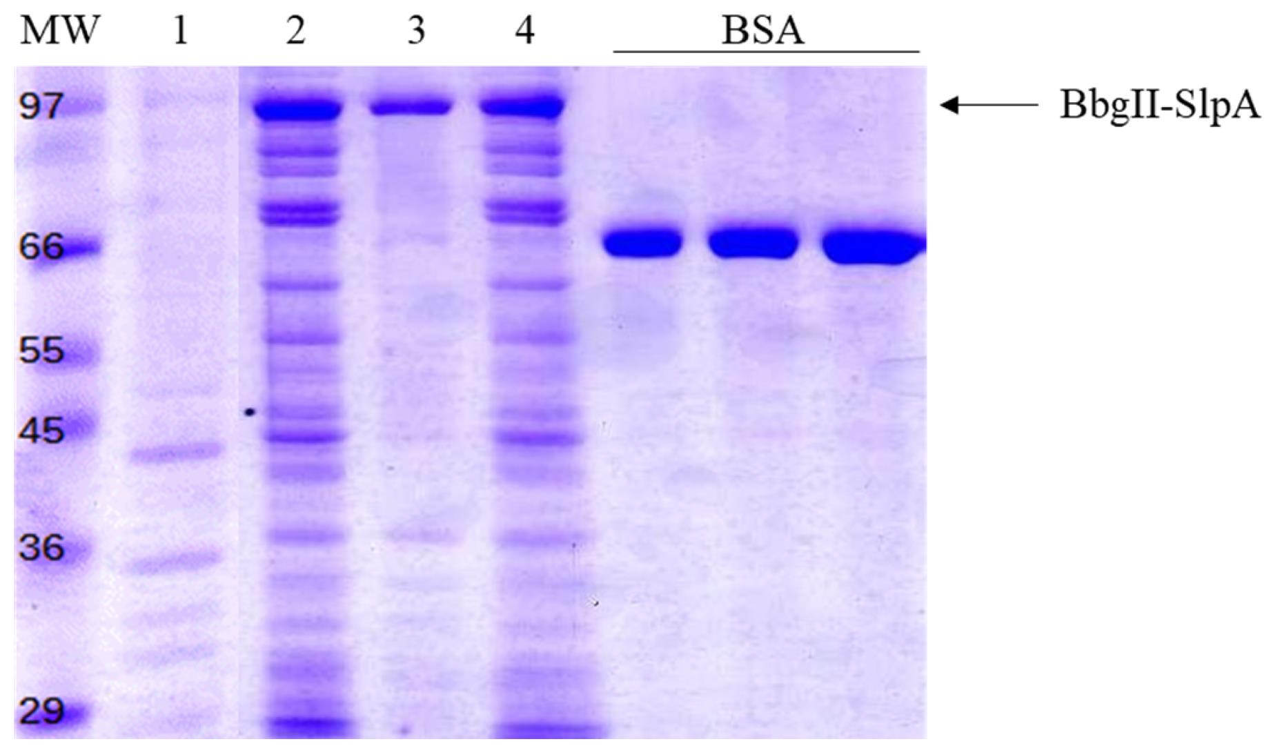
Figure 1. SDS-PAGE analysis of BbgII-SlpA production. Coomassie Blue–stained 15% SDS-PAGE gel showing the expression of BbgII-SlpA (98 kDa band, arrow). Lane 1, total protein from E. coli lysate before induction (NI); Lane 2, total cell lysate (T); Lane 3, insoluble fraction (IF); Lane 4, soluble fraction (SF) after induction with 0.1 mM IPTG at 22 °C for 16 h. BSA standard curve (0.5, 1, and 1.5 μg). MW, molecular weight marker (kDa).
B. Preparation of the bacterial carrier
1. Inoculate B. subtilis var. natto from a frozen stock (-80 °C) into 100 mL of LB broth in a 500 mL Erlenmeyer flask. Incubate at 37 °C with shaking at 200 rpm for 16 h (overnight culture).
Note: Ensure the culture reaches stationary phase (OD600 > 2.0) before proceeding.
2. Harvest the cells by centrifugation at 5,000× g for 10 min at 4 °C in 50 mL polypropylene centrifuge tubes. Discard the supernatant.
3. Wash the bacterial pellet three times with 50 mL of PBS each time, resuspending the pellet by pipetting or gentle vortexing, and centrifuging at 5,000× g for 10 min at 4 °C between washes.
4. After the final wash, resuspend the pellet in 20 mL of glutaraldehyde 2.5% (v/v) in a 50 mL conical tube.
Caution: Glutaraldehyde is toxic and an irritant. Perform all steps involving glutaraldehyde in a fume hood using appropriate personal protective equipment (gloves, safety goggles, and lab coat).
5. Incubate the cell suspension at 4 °C overnight (12–16 h) with gentle stirring on a rotator to allow complete inactivation of the bacterial cells.
Note: The inactivation process cross-links cell wall proteins and kills the bacteria, preventing any metabolic activity or growth.
6. Wash the inactivated bacteria five times with 50 mL of PBS, centrifuging at 5,000× g for 10 min at 4 °C between washes, to remove residual glutaraldehyde.
Critical: Complete removal of glutaraldehyde is essential, as residual glutaraldehyde can cross-link the target enzyme and reduce its activity (see Troubleshooting 2).
7. After the final wash, resuspend the inactivated cells in PBS to a final concentration of 40 OD600/mL (based on the original culture OD600 before inactivation). Calculate the resuspension volume using the formula: Volume (mL) = (OD600 of culture × culture volume (mL)) / 40. For example, if the original culture was 100 mL with a final OD600/mL of 2.0, resuspend in 5 mL of PBS. Determine the OD600 of a 1:100 dilution in PBS using a spectrophotometer and adjust the volume accordingly.
Note: After the inactivation step, plate 100 μL of the carrier suspension on LB agar and incubate at 37 °C for 24–48 h to verify inactivation. If no growth is observed, the carrier is considered inactivated and can be used. This inactivation protocol has been validated in [17].
Pause point: The inactivated B. subtilis carrier can be stored at 4 °C for up to 6 months. If the suspension becomes turbid or develops an odor, discard and prepare fresh.
Critical: Store inactivated carrier at 4 °C. Do not freeze, as freezing may disrupt the cell wall structure and reduce binding efficiency.
C. CBE production (simultaneous purification and immobilization)
1. In a 1.5 mL microcentrifuge tube, centrifuge 10 OD600 units of the inactivated B. subtilis carrier at 5,000× g for 10 min at room temperature (from step B8).
Note: For example, if the carrier suspension is at 40 OD600/mL, use 250 μL.
2. Discard the supernatant and resuspend the bacterial pellet in 20 OD600 units of the E. coli soluble fraction (SF) containing soluble BbgII-SlpA (from step A11), in a final volume of 500 μL with PBS.
Note: For example, if the soluble fraction (SF) is at 40 OD600/mL, use 500 μL. Adjust the volume to 500 μL with PBS if necessary.
3. Incubate the mixture at room temperature (20–25 °C) for 1 h with gentle rotation on a rotator to allow the SlpA domain of BbgII-SlpA to bind specifically to the surface of the B. subtilis carrier.
4. Centrifuge the mixture at 5,000× g for 3 min at room temperature.
5. Collect the supernatant (unbound fraction) and transfer it to a fresh 1.5 mL microcentrifuge tube.
Note: Take a 40 μL aliquot of the supernatant (unbound fraction) for SDS-PAGE analysis (see step C9).
6. Wash the pellet (CBEs) three times with 500 μL of PBS, centrifuging at 5,000× g for 3 min at room temperature between washes. Discard the supernatants after each wash.
Note: Take a 40 μL aliquot of the final wash for SDS-PAGE analysis (see step C9).
7. After the final wash, resuspend the CBE pellet in 500 μL of PBS. This suspension contains the immobilized enzyme and is ready for use in activity assays or further characterization (see Troubleshooting 2).
Pause point: CBEs can be stored at 4 °C for up to 2 weeks without significant loss of activity.
Critical: Store CBEs at 4 °C and use them within 2 weeks. Freezing is not recommended.
Note: Take a 40 μL aliquot of the CBE suspension for SDS-PAGE analysis to confirm binding efficiency (see step C9).
8. Prepare four separate samples for SDS-PAGE analysis: soluble fraction (from step A11), unbound fraction (from step C5), wash fractions (from step C6), and bound protein fraction (CBEs, from step C7). Mix 20 μL of each fraction with 5 μL of 5× SDS-PAGE loading buffer. Heat the samples at 90 °C for 10 min.
9. Load the samples onto a 15% SDS-PAGE gel (4% stacking gel) alongside a protein molecular weight marker. Run the gel at 150 V for 60 min in a Mini-PROTEAN Tetra Cell electrophoresis system. Stain the gel with Coomassie Brilliant Blue dye and destain with destaining solution following standard procedures (see Figure 2A, B).
Notes:
1. Confirm the presence of BbgII-SlpA (98 kDa band) on the bacterial carrier (bound protein fraction) (see Figure 2A, lane 6).
2. The presence of BbgII-SlpA on the bacterial carrier surface can also be verified by immunofluorescence microscopy (see Figure 2B).
10. Determine the amount of BbgII-SlpA bound to B. subtilis, expressed as protein amount per OD600 unit of the bacterial carrier by densitometric analysis using ImageJ (see Data analysis, Section B).
Note: Include a BSA standard curve (5, 10, and 15 μL of BSA 0.1 μg/μL solution) on each gel for quantification.
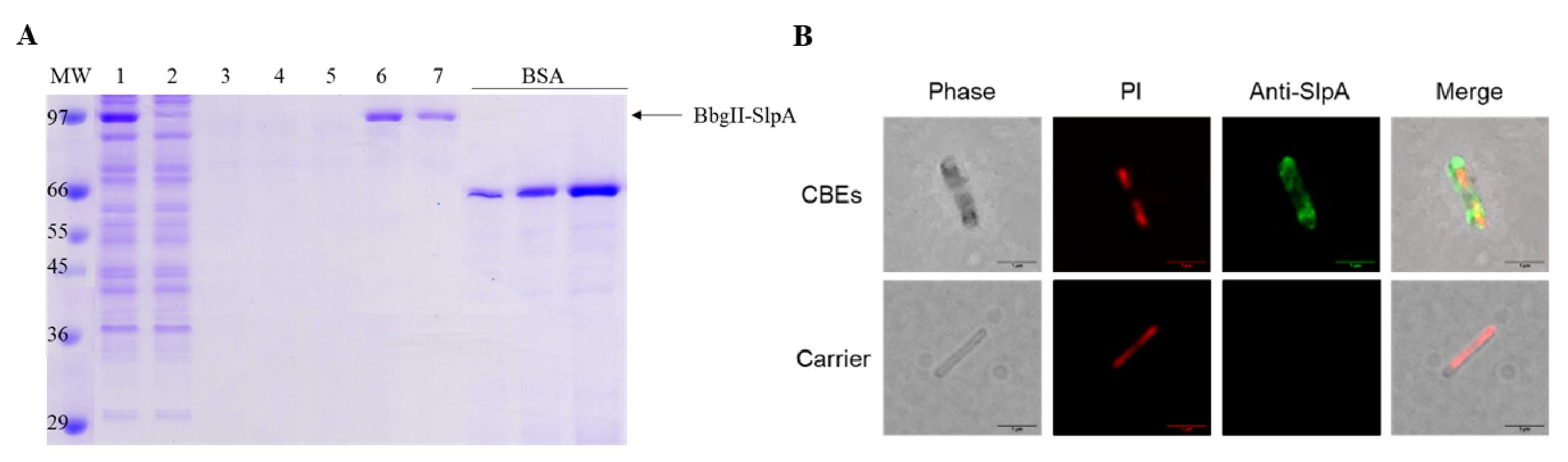
Figure 2. BbgII-SlpA binding to B. subtilis carrier. (A) SDS-PAGE analysis of BbgII-SlpA purification and immobilization. Coomassie Blue–stained 15% SDS-PAGE gel showing the efficient binding and purification of BbgII-SlpA (98 kDa band, arrow). Lane 1, input soluble fraction of E. coli lysate containing BbgII-SlpA; Lane 2, unbound fraction after incubation with the bacterial carrier; Lanes 3, 4, and 5, bacterial pellet washes with PBS; Lane 6, bound protein fraction (BbgII-SlpA immobilized on B. subtilis carrier); Lane 7, eluted purified BbgII-SlpA. BSA standard curve (0.5, 1, and 1.5 μg). MW, molecular weight marker (kDa). (B) Immunofluorescence microscopy of CBEs. Inactivated B. subtilis cells (carrier) were incubated with soluble BbgII-SlpA and labeled with anti-SlpA primary antibody followed by Alexa FluorTM 488-conjugated secondary antibody (green, Anti-SlpA panel). Bacterial cells were stained with propidium iodide (red, PI panel). Phase contrast, propidium iodide staining (red, bacteria), anti-SlpA antibody (green, BbgII-SlpA), and merge showing uniform surface distribution of the enzyme on the bacterial carrier. Scale bars: 1 μm.
D. Optional elution of purified enzyme
1. If purified soluble enzyme is required, resuspend the CBE pellet (from step C7) in 500 μL of BbgII-SlpA elution buffer in a 1.5 mL microcentrifuge tube.
Note: Elution buffer is prepared fresh by mixing equal volumes of carbonate-bicarbonate buffer (pH 9.0, 400 mM) and citrate-phosphate buffer (pH 6.0, 100 mM) immediately before use.
2. Incubate the suspension at room temperature for 10–15 min with gentle agitation on a rotator to release the bound BbgII-SlpA from the carrier.
3. Centrifuge at 5,000× g for 3 min at room temperature to pellet the bacterial carrier. Collect the supernatant (first eluate) and transfer it to a fresh 1.5 mL tube.
4. Repeat the elution (steps D1–3) two more times with fresh elution buffer, collecting the second and third eluates separately.
Note: Typically, more than 80% of the bound enzyme is recovered in the first eluate. The second and third eluates can be pooled with the first if high concentration is not required.
5. Pool the eluates if desired and dialyze against PBS overnight at 4 °C using cellulose dialysis tubing with a 12.4 kDa cutoff (see Troubleshooting 3).
Note: Ensure the dialysis tubing is properly prepared by rinsing with distilled water and soaking in PBS for 10 min before use. Use at least 100-fold excess of PBS relative to the sample volume.
6. After dialysis, transfer the purified enzyme to a fresh 1.5 mL tube. Determine the protein concentration using the Pierce BCA Protein Assay kit.
Note: Take a 40 μL aliquot of the purified enzyme for SDS-PAGE analysis to confirm purity and integrity of the eluted enzyme (see Figure 2A, lane 7).
Pause point: The purified enzyme can be stored at 4 °C for up to 1 week. For longer storage (up to 6 months), store at -20 °C. Avoid repeated freeze/thaw cycles.
Data analysis
A. Quantify the yields of soluble and insoluble BbgII-SlpA by densitometric analysis (see Procedure, step A18)
1. Digitize the gels using a flatbed scanner.
2. Open the image files in ImageJ.
3. Use the Gel analysis tool to measure the intensity of the BbgII-SlpA band (~98 kDa) in the soluble and insoluble fractions.
4. Quantify the amount of protein in each fraction using the BSA standard curve.
5. Calculate the percentage of soluble BbgII-SlpA as:
% Soluble = (Amount of soluble protein / (Amount of soluble + insoluble protein)) × 100
Note: Perform densitometry on at least three independent expression experiments to ensure reproducibility. The results are summarized in Table 1.
B. Quantify the amount of BbgII-SlpA bound to the bacterial carrier (CBEs) using densitometry (see Procedure, step C10)
1. Digitize the gels using a flatbed scanner.
2. Open the image files in ImageJ.
3. Measure the intensity of the BbgII-SlpA band in the bound fraction (CBEs) and interpolate the protein amount from the BSA standard curve.
4. Express the binding capacity as μg of BbgII-SlpA per OD600 unit of bacterial carrier.
Note: Perform the binding assay in triplicate (three independent CBE preparations) to calculate mean ± standard deviation. The results are summarized in Table 1.
Table 1. Quantification of BbgII-SlpA solubility and binding efficiency
| Parameter | Value (mean ± SD) |
|---|---|
| Soluble BbgII-SlpA (%) | 72.3% ± 5.1% |
| Insoluble BbgII-SlpA (%) | 27.7% ± 4.3% |
| Binding efficiency (%) | 85.4% ± 3.8% |
| Binding capacity (μg/OD600) | 56.2 ± 2.3 μg/OD600 |
Validation of protocol
This protocol has been used and validated in the following research article:
Harguindeguy et al. [15]. Coated bacterial enzymes: a one-step approach for enzymatic purification and immobilization. Preparative Biochemistry & Biotechnology.
The published study provides a comprehensive functional and comparative characterization of the CBE platform, including expression optimization, binding parameters, catalytic properties, thermal stability, pH and temperature profiles, cation tolerance, reusability, and lactose hydrolysis in milk.
Expression optimization and purity
Soluble expression of BbgII-SlpA in E. coli BL21-CodonPlus (DE3)-RIPL was optimized by a one-factor-at-a-time approach. The highest proportion of soluble protein was obtained at 22 °C with 0.1 mM IPTG, yielding approximately 70% of the expressed BbgII-SlpA in the soluble fraction. The fusion protein migrated as a single major band at ~98 kDa by SDS-PAGE, consistent with its calculated molecular mass (695 amino acids). After one-step purification and immobilization, the eluted enzyme showed a highly purified band with minimal contaminants (Figure 2A, lane 7), confirming the efficiency of the SlpA-based capture.
Binding characterization
The interaction between BbgII-SlpA and the inactivated B. subtilis carrier was quantified by a saturation binding assay. Nonlinear regression analysis yielded a dissociation constant (Kd) of 16.2 μM and a maximum binding capacity (Bmax) of 144 μmol per gram of dry cell weight. These values indicate a stable and specific interaction, comparable to those reported for other SlpA-based and cellulose-binding domain systems. Immunofluorescence microscopy confirmed a uniform distribution of the enzyme on the bacterial surface (Figure 2B).
Catalytic properties
Kinetic parameters of the immobilized enzyme (CBEs) were determined for both ONPG and lactose. The Vmax for ONPG was 9459.7 ± 504.9 U/mg with a Km of 5.90 ± 0.77 mM, while the Vmax for lactose was 28.8 ± 2.3 U/mg with a Km of 106.97 ± 15.91 mM. The Km for lactose is comparable to values reported for wild-type BbgII. The catalytic efficiency (kcat/Km) for ONPG was 2.62 × 10⁶ M-1 s-1, more than double that of the free enzyme reported in the literature, while for lactose, it was 4.40 × 10² M-1 s-1 (see Table 1 in Harguindeguy et al. [15]).
pH and temperature profiles
Both free and immobilized BbgII-SlpA exhibited an optimal pH of 6.0. However, the immobilized preparation displayed a broader pH-activity profile, retaining more than 60% of maximal activity between pH 5.5 and 8.0, whereas the free enzyme showed a sharper decline outside the optimum. The optimal temperature for CBEs was 60 °C for ONPG and 50 °C for lactose, compared with 50 °C and 40 °C, respectively, for the free enzyme. At 50 °C with lactose, CBEs retained >80% of maximal activity, while the free enzyme retained <10% (see Figure 3 in Harguindeguy et al. [15]).
Thermal stability
Thermal stability was evaluated by pre-incubating free enzyme or CBEs at 45 °C for up to 390 min. The free enzyme lost activity rapidly, retaining only ~20% of its initial activity. In contrast, the CBEs maintained ~80% of their original activity under identical conditions, demonstrating a significant protective effect conferred by immobilization on the bacterial cell wall (see Figure 4 in Harguindeguy et al. [15]). Inactivation constants (ki) were calculated using first-order rate equations, and half-lives (t1/2) were derived from these values.
Cation tolerance
The effect of various cations was examined at 1, 10, and 100 mM. At 100 mM Ca2+, the free enzyme lost all activity, whereas the CBEs retained 36% activity. At 100 mM Fe2+, the free enzyme retained only ~5% activity, while the CBEs retained 73% (see Table 2 in Harguindeguy et al. [15]). These results indicate that immobilization protects the enzyme against inhibitory cations naturally present in milk, such as Ca2+ and Fe2+.
Reusability
The operational stability of the CBEs was evaluated through five consecutive cycles of lactose hydrolysis in buffer (5% w/v lactose), skim milk, and whey. Residual activity after five cycles was 35% in buffer, 40% in skim milk, and 45% in whey, indicating that complex matrices may exert a stabilizing effect (see Figure 5 in Harguindeguy et al. [15]). These values are competitive with other one-step immobilized systems (see Table 3 in Harguindeguy et al. [15]).
Milk-lactose hydrolysis at low temperature
Lactose hydrolysis in skim milk at 7 °C reached approximately 52% after 20 h, with a glucose release of ~12.6 g/L. This demonstrates that the CBEs remain active under refrigeration conditions typical of lactose-reduced milk production.
Validation of carrier inactivation
The inactivation of B. subtilis was validated by plating 100 μL of the carrier suspension on LB agar and incubating at 37 °C for 24–48 h. No growth was observed, confirming the absence of viable cells.
Statistical analysis
All experiments were performed in triplicate. Data are presented as mean ± standard deviation. Kinetic parameters were obtained by nonlinear fitting of the Michaelis–Menten equation, and errors were propagated quadratically.
Summary of validation data
A comprehensive summary of all validation parameters is presented in Table 2.
Table 2. Summary of validation parameters for the CBE platform. Data from Harguindeguy et al. [15].
| Parameter | Value |
|---|---|
| Expression | |
| Optimal induction temperature | 22 °C |
| Optimal IPTG concentration | 0.1 mM |
| Soluble fraction | ~70% of expressed BbgII-SlpA |
| Binding | |
| Dissociation constant (Kd) | 16.2 μM |
| Maximum binding capacity (Bmax) | 144 μmol/g DCW |
| Kinetics (ONPG) | |
| Vmax | 9459.7 ± 504.9 U/mg |
| Km | 5.90 ± 0.77 mM |
| kcat/Km | 2.62 × 10⁶ M-1 s-1 |
| Kinetics (lactose) | |
| Vmax | 28.8 ± 2.3 U/mg |
| Km | 106.97 ± 15.91 mM |
| kcat/Km | 4.40 × 10² M-1 s-1 |
| pH optimum | 6.0 (both free and immobilized) |
| Temperature optimum | 60 °C (ONPG), 50 °C (lactose) for CBEs |
| Thermal stability (45 °C, 390 min) | ~80% residual activity (CBEs) vs. ~20% (free) |
| Cation tolerance (100 mM) | Ca2+: 36% (CBEs) vs. 0% (free); Fe2+: 73% (CBEs) vs. ~5% (free) |
| Reusability (5 cycles) | 35% (buffer), 40% (skim milk), 45% (whey) |
| Milk hydrolysis (7 °C, 20 h) | ~52% lactose hydrolysis |
Limitations and future work
A direct side-by-side purification yield comparison with conventional multi-step chromatography was not performed in the published study. However, the one-step method yields highly purified enzyme in a single step (Figure 2A, lane 7), and the kinetic parameters are consistent with those reported for purified BbgII. Future work should include a comprehensive economic analysis and pilot-scale evaluation under real processing conditions.
General notes and troubleshooting
General notes
1. Although this protocol does not require strict aseptic conditions for all steps, since B. subtilis is inactivated and E. coli lysate is used, it is recommended to use sterile solutions during the preparation of the bacterial carrier and CBEs to avoid microbial contamination, which could affect enzyme activity or interfere with downstream assays. If contamination is observed (e.g., turbidity or off-odor in the carrier suspension), discard the preparation and start fresh.
2. The protocol is described for small-scale CBE production. The culture volumes and amounts of carrier can be scaled up proportionally if larger quantities of CBEs are required. When scaling up, ensure the flask volume is at least five times the culture volume to maintain adequate aeration and adjust the sonication parameters proportionally (see Procedure, step A9, Note) to maintain efficient cell lysis.
3. This protocol was developed and validated for BbgII-SlpA. However, the CBE platform is adaptable to other recombinant proteins fused to the SlpA cell wall–binding domain, provided that the fusion protein is expressed in a soluble and active form in E. coli. For new target proteins, the expression conditions (temperature, IPTG concentration, and induction time) may need to be re-optimized to maximize soluble protein yield (see Procedure, Section A). For detailed optimization data on temperature, IPTG concentration, and induction time, please refer to [15]. Readers are encouraged to consult that article to design their own optimization approaches. The binding and elution conditions (incubation time, buffer composition, pH) may also require adjustment depending on the stability and properties of the target protein.
4. Always prepare fresh lysis buffer (Recipe 8) and glutaraldehyde 2.5% v/v (Recipe 12) on the day of use, as their stability is limited. For other buffers (e.g., PBS, wash buffer), prepare in advance and store appropriately.
5. A protein that is soluble in its native form may not be soluble when fused to SlpA. This could occur due to improper folding, interaction with the SlpA domain amino acids, or covalent interactions. If this problem arises, consider testing different linkers between the target protein and the SlpA domain, lowering the induction temperature, or using solubility-enhancing tags. Computational tools such as AlphaFold or other structure prediction software may be used to predict the structure, solubility, and/or stability of the enzyme-SlpA chimeric protein before applying the CBE platform to a new target protein.
Troubleshooting
Problem 1: Low yield of soluble BbgII-SlpA in the E. coli lysate (Procedure, Section A).
Possible cause: The induction temperature (37 °C) or IPTG concentration (≥0.5 mM) is too high, leading to the formation of inclusion bodies.
Solution: Ensure that the culture is cooled to 22 °C before adding IPTG and use a final IPTG concentration of 0.1 mM (see Procedure, step A4, Critical). If the problem persists, optimize the induction conditions further by testing temperatures between 18 and 25 °C and IPTG concentrations between 0.05 and 0.2 mM. For difficult-to-express proteins, consider reducing the shaking speed (e.g., 150 rpm) during induction to slow down protein synthesis and promote proper folding. Additionally, verify that the lysozyme concentration (0.3 mg/mL) and sonication conditions (30% amplitude, 30 cycles) are adequate for complete cell lysis; insufficient lysis may result in lower protein recovery (see General note 2).
Problem 2: Low binding efficiency of BbgII-SlpA to the B. subtilis carrier (Procedure, Section C).
Possible cause: Residual glutaraldehyde in the carrier preparation cross-links the enzyme, preventing binding or inactivating it.
Solution: Ensure that the inactivated B. subtilis carrier is washed thoroughly (five times with PBS; see Procedure, step B7, Critical) to remove residual glutaraldehyde. Test a small aliquot of the carrier by incubating it with purified BbgII-SlpA and checking binding by SDS-PAGE. If binding is still low, prepare a fresh batch of carrier, ensuring that the glutaraldehyde inactivation solution is prepared fresh and that the incubation time (overnight, 4 °C) is strictly followed.
Problem 3: Protein precipitation or aggregation during dialysis (Procedure, Section D).
Possible cause: The eluted enzyme may be unstable in the elution buffer, or the dialysis buffer (PBS) may not be compatible with the protein’s stability requirements.
Solution: Reduce the dialysis time (e.g., 2–4 h at 4 °C instead of overnight) and replace the dialysis buffer once during this period to maintain the concentration gradient. Add stabilizing agents to the dialysis buffer, such as 10% glycerol or 1 mM DTT, if the protein is prone to aggregation. If precipitation persists, test a different elution buffer (e.g., 50 mM Tris-HCl, pH 8.0, with 500 mM NaCl) or perform buffer exchange using a desalting column (e.g., PD-10, Sephadex G-25) instead of dialysis.
Problem 4: The target protein is soluble in its native form but becomes insoluble when fused to the SlpA domain (Procedure, Section A).
Possible cause: The fusion with the SlpA domain may interfere with proper folding, cause steric hindrance, or promote aggregation.
Solution: First, use computational tools such as AlphaFold or RoseTTAFold to predict the structure, solubility, and/or stability of the enzyme-SlpA chimeric protein before experimental work. If the prediction suggests potential issues, consider the following strategies:
1. Optimize the linker: Use a longer or more flexible linker [e.g., (GGGGS)3 or (GGGGS)4] between the target protein and the SlpA domain to reduce steric hindrance.
2. Lower the induction temperature: Reduce the induction temperature to 18 °C and/or lower the IPTG concentration to 0.05 mM to slow down protein synthesis and promote proper folding.
3. Use solubility-enhancing tags: Consider adding a solubility-enhancing tag (e.g., MBP, SUMO, or NusA) in addition to the SlpA domain.
4. Co-expression with chaperones: Co-express the chimeric protein with molecular chaperones (e.g., GroEL/GroES or DnaK/DnaJ/GrpE) to assist with proper folding.
5. Test alternative hosts: If E. coli fails to produce soluble protein, consider using other expression hosts such as Pichia pastoris or mammalian cells.
Acknowledgments
Funding: This work was supported by the Agencia Nacional de Promoción de la Investigación, el Desarrollo Tecnológico y la Innovación (Agencia I+D+i), Argentina, through grants PICT 2019-3123 (issued by Dr. SF Cavalitto) and PICT 2017-3123 (issued by Dr. GE Ortiz).
This protocol has been described and validated in the following research article:
Harguindeguy, I., Homse, M. S., Ramírez, A. C., Sabetta, A. E., Cavalitto, S. F. and Ortiz, G. E. (2026). Coated bacterial enzymes: a one-step approach for enzymatic purification and immobilization. Prep Biochem Biotechnol, 1-13. Advance online publication. https://doi.org/10.1080/10826068.2026.2736623. PMID: 42823390.
The graphical overview was created with the assistance of Google Gemini and Microsoft PowerPoint.
Author contributions
Harguindeguy, I.: Investigation, Writing—Original Draft, Visualization. Ortiz, G. E.: Conceptualization, Funding acquisition, Project administration, Writing—Review & Editing, Supervision.
Competing interests
The authors declare that they have no competing interests.
References
- LOMER, M. C. E., PARKES, G. C. and SANDERSON, J. D. (2007). Review article: lactose intolerance in clinical practice – myths and realities. Aliment Pharmacol Ther. 27(2): 93–103. https://doi.org/10.1111/j.1365-2036.2007.03557.x
- Storhaug, C. L., Fosse, S. K. and Fadnes, L. T. (2017). Country, regional, and global estimates for lactose malabsorption in adults: a systematic review and meta-analysis. Lancet Gastroenterol Hepatol. 2(10): 738–746. https://doi.org/10.1016/s2468-1253(17)30154-1
- Troise, A. D., Bandini, E., De Donno, R., Meijer, G., Trezzi, M. and Fogliano, V. (2016). The quality of low lactose milk is affected by the side proteolytic activity of the lactase used in the production process. Food Res Int. 89: 514–525. https://doi.org/10.1016/j.foodres.2016.08.021
- Chen, W., Chen, H., Xia, Y., Zhao, J., Tian, F. and Zhang, H. (2008). Production, Purification, and Characterization of a Potential Thermostable Galactosidase for Milk Lactose Hydrolysis from Bacillus stearothermophilus. J Dairy Sci. 91(5): 1751–1758. https://doi.org/10.3168/jds.2007-617
- Yu, S. Q. and Liu, Y. H. (2012). Expression and one-step purification of a β-galactosidase by fusion with elastin-like polypetides. Process Biochem. 47(7): 1108–1114. https://doi.org/10.1016/j.procbio.2012.04.003
- Terpe, K. (2003). Overview of tag protein fusions: from molecular and biochemical fundamentals to commercial systems. Appl Microbiol Biotechnol. 60(5): 523–533. https://doi.org/10.1007/s00253-002-1158-6
- Oliveira, C., Guimarães, P. M. and Domingues, L. (2011). Recombinant microbial systems for improved β-galactosidase production and biotechnological applications. Biotechnol Adv. 29(6): 600–609. https://doi.org/10.1016/j.biotechadv.2011.03.008
- Duan, F., Sun, T., Zhang, J., Wang, K., Wen, Y. and Lu, L. (2022). Recent innovations in immobilization of β-galactosidases for industrial and therapeutic applications. Biotechnol Adv. 61: 108053. https://doi.org/10.1016/j.biotechadv.2022.108053
- Gennari, A., Simon, R., Sperotto, N. D. d. M., Bizarro, C. V., Basso, L. A., Machado, P., Benvenutti, E. V., Da Cas Viegas, A., Nicolodi, S., Renard, G., et al. (2022). One-step purification of a recombinant beta-galactosidase using magnetic cellulose as a support: Rapid immobilization and high thermal stability. Bioresour Technol. 345: 126497. https://doi.org/10.1016/j.biortech.2021.126497
- Pham, M. L., Tran, A. M., Kittibunchakul, S., Nguyen, T. T., Mathiesen, G. and Nguyen, T. H. (2019). Immobilization of β-Galactosidases on the Lactobacillus Cell Surface Using the Peptidoglycan-Binding Motif LysM. Catalysts. 9(5): 443. https://doi.org/10.3390/catal9050443
- Lozančić, M., Sk. Hossain, A., Mrša, V. and Teparić, R. (2019). Surface Display—An Alternative to Classic Enzyme Immobilization. Catalysts. 9(9): 728. https://doi.org/10.3390/catal9090728
- Muruaga, E. J., Uriza, P. J., Eckert, G. A. K., Pepe, M. V., Duarte, C. M., Roset, M. S. and Briones, G. (2023). Adaptation of the binding domain of Lactobacillus acidophilus S-layer protein as a molecular tag for affinity chromatography development. Front Microbiol. 14: e1210898. https://doi.org/10.3389/fmicb.2023.1210898
- Fina Martin, J., Palomino, M. M., Cutine, A. M., Modenutti, C. P., Fernández Do Porto, D. A., Allievi, M. C., Zanini, S. H., Mariño, K. V., Barquero, A. A., Ruzal, S. M., et al. (2019). Exploring lectin-like activity of the S-layer protein of Lactobacillus acidophilus ATCC 4356. Appl Microbiol Biotechnol. 103(12): 4839–4857. https://doi.org/10.1007/s00253-019-09795-y
- Sagmeister, T., Gubensäk, N., Buhlheller, C., Grininger, C., Eder, M., Ðordić, A., Millán, C., Medina, A., Sánchez-Murcia, P., Berni, F., et al. (2024). The molecular architecture of Lactobacillus S-Layer: Assembly and attachment to teichoic acids. Research Square. ers–3689109/v1. https://doi.org/10.21203/rs.3.rs-3689109/v1
- Harguindeguy, I., Homse, M. S., Ramírez, A. C., Sabetta, A. E., Cavalitto, S. F. and Ortiz, G. E. (2026). Coated bacterial enzymes: a one-step approach for enzymatic purification and immobilization. Prep Biochem Biotechnol. 1–13. https://doi.org/10.1080/10826068.2026.2736623
- Sambrook, J.; Russell, D. W. Molecular Cloning: A Laboratory Manual. 3rd ed. Cold Spring Harbor Laboratory Press: Cold Spring Harbor, NY, 2001.
- Harguindeguy, I., Assandri, M. H., Gorgojo, J. P., Hiriart, Y., Alarcón, V., Huergo, M. A., Cavalitto, S. F., Serradell, M. d. l. A. and Ortiz, G. E. (2025). Coated Bacterial Vaccine: A New Approach for Antigen Display on Bacterial Surface. Biotechnol J. 20(7): e70015. https://doi.org/10.1002/biot.70015
Article Information
Publication history
Received: Sep 1, 2026
Accepted: Oct 26, 2026
Available online: Nov 15, 2026
Published: Dec 5, 2026
Copyright
© 2026 The Author(s); This is an open access article under the CC BY-NC license (https://creativecommons.org/licenses/by-nc/4.0/).
How to cite
Harguindeguy, I., Cavalitto, S. F. and Ortiz, G. E. (2026). Producing High-Purity, Immobilized Enzymes on Inactivated Bacillus subtilis Using the Coated Bacterial Enzyme (CBE) Platform. Bio-protocol 16(23): e5880. DOI: 10.21769/BioProtoc.5880.

 Download Citation in RIS Format
Do you have any questions about this protocol?
Post your question to gather feedback from the community. We will also invite the authors of this article to respond.
 Post a Question


