


- Protocols

- Articles and Issues
- For Authors
- About
- Become a Reviewer


Measuring Functional and Structural Muscle Properties in Drosophila Larvae for Testing Genetic Variants Involved in Neuromuscular Disorders

Published: Vol 16, Iss 23, Dec 5, 2026 DOI: 10.21769/BioProtoc.5875 Views: 26
Reviewed by: Abigail WilsonAnonymous reviewer(s)
 PDF
 Ask a question

Original research article
The authors used this protocol in:

May 2026

Protocol Collections
Comprehensive collections of detailed, peer-reviewed protocols focusing on specific topics



Abstract
Drosophila melanogaster has emerged as a powerful model for investigating the genetic control of muscle development and muscle function in both normal and pathological conditions. To date, several Drosophila models of human neuromuscular disorders (NMDs) have been generated and characterized, allowing a better understanding of disease mechanisms, improving diagnosis, and testing of new therapeutic strategies. Conserved genes encoding large muscle proteins such as Titin or Ryanodine receptor (RyR) harbor hundreds of variant mutations, many of which remain variants of unknown significance (VUS). Here, we describe procedures for generating a CRISPR-engineered Drosophila model of a VUS and an integrated workflow for quantitative assessment of functional and structural muscle properties, providing an indication of VUS pathogenicity. The protocol combines larval locomotor assays, muscle morphometry, and structural muscle properties assessment. The workflow enables systematic evaluation of muscle structure and function and provides a versatile platform for studying genetic variants and their involvement in NMDs.
Key features
• Generation of CRISPR-engineered Drosophila models of human muscular disease mutations.
• Quantitative evaluation of larval muscle performance using three complementary locomotor assays.
• Standardized workflow for skeletal muscle morphometric analysis.
• Applicable to characterization of neuromuscular disease-associated genetic variants.
Keywords: Drosophila Skeletal muscle
Larval locomotion
Sarcomere analysis
CRISPR engineering
Human neuromuscular disease
Ryanodine receptor
Background
Muscle development depends on an evolutionarily conserved genetic cascade triggering myogenic differentiation and formation of functional multinucleated muscle fibers that attach to tendon cells, build a sophisticated contractile machinery, and get innervated to ensure guided muscle activity [1]. Coordinated regulation of cytoskeletal organization, nuclear number and positioning, calcium signaling, mitochondrial homeostasis, and excitation-contraction coupling are required for a muscle to fulfill its functions and are affected in diseased and aging muscles [2–6]. Drosophila possesses highly conserved genetic pathways regulating muscle differentiation and function and offers powerful tools that facilitate mechanistic investigations of healthy and diseased muscle [7].
Among the conserved players involved in both muscle development and muscle function are genes encoding Ryanodine receptors (RyRs), the calcium release channels essential to maintaining correct Ca2+ dynamics across biological membranes [8]. RYR gene mutations underlie an array of muscular diseases, including one of the most frequent congenital myopathies, central core disease (CCD), and malignant hyperthermia susceptibility (MHS), both of which could be fatal [9]. Of the numerous mutations identified to date in the RYR genes, most are missense mutations with single amino acid substitutions [10], with a majority that remain of unknown significance [8].
Considering extensive structural and functional dRyR conservation, we generated Drosophila models of undiagnosed human RYR1 variant mutations and analyzed their impact on muscle structure and function. Our protocol was developed for investigating the function of the Drosophila ryanodine receptor gene (RyR) and was subsequently optimized as a platform for quantitative analysis of structural and functional muscle phenotypes in Drosophila models of genetic RYR1 variants involved in NMDs. The workflow can be applied not only to CRISPR-generated disease models but also to investigate the impacts of gene loss-of-function, gain-of-function, or RNA interference [8].
Materials and reagents
Biological materials
1. w1118 (white null mutant; Bloomington Drosophila Stock Center, BDSC# 3605) used as a control
2. CRISPR-generated human RYR1 VUS p.Ile3345Phe line (available upon request)
3. nos-Cas9 transgenic line (Bloomington Drosophila Stock Center, stock number: 54591)
4. Competent E. coli cells (e.g., DH5α NEB) (New England Biolabs, catalog number: C2987H)
5. Expression vector: pCFD5 (Addgene ID #73914)
Reagents
1. Primary antibodies:
a. Guinea pig anti-dRyR (1:200) (kindly provided by Robert Scott and Benjamin White from NIH/NIMH Institute and previously described in Gao et al. [11]) (dRyR pattern in Figure 1)
b. Mouse anti-Kettin (1:50) (Developmental Studies Hybridoma Bank 1B8-3D9); RRID: AB_2721939
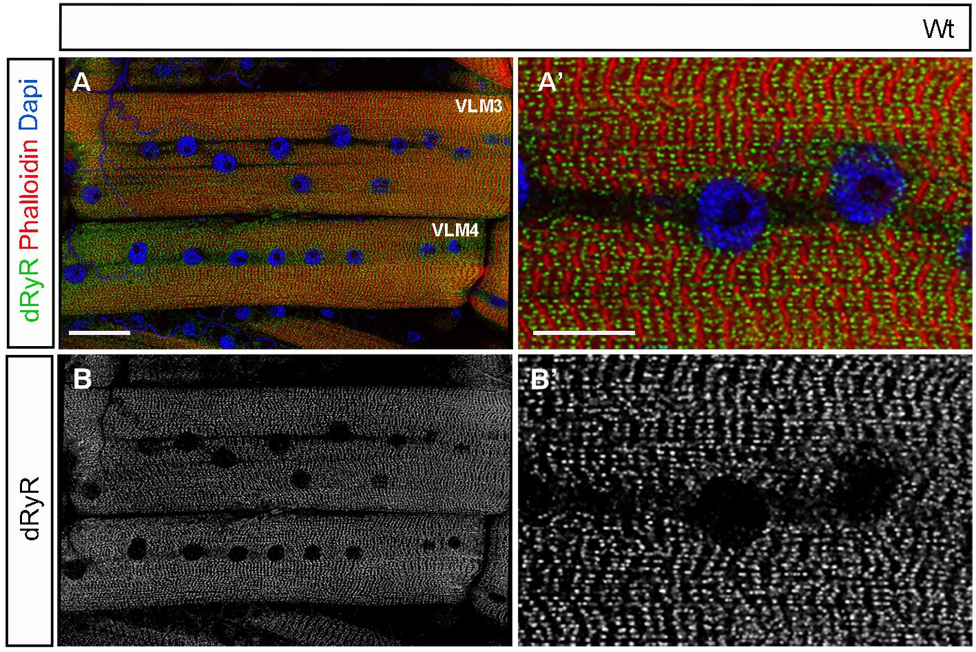
Figure 1. Representative views of ventral longitudinal (VLM3 and VLM4) muscles. (A) and a zoomed view (A’) in control (w1118) larvae stained for dRyR (green), Phalloidin (red), and DAPI (blue). (B, B’) Grayscale views of dRyR antibody staining. Scale bars: 50 μm in A and 20 μm in A’.
2. Phosphate buffer solution (PBS) 10× (Euromedex, catalog number: ET330-A)
3. Triton X-100 (Euromedex, catalog number: 2000-B)
4. Ethylene diamine tetraacetic acid disodium salt dihydrate (EDTA) (Euromedex, catalog number: EU-0007-B)
5. Potassium chloride (KCl) (Sigma-Aldrich, catalog number: P9541)
6. Sodium phosphate buffer (Sigma-Aldrich, catalog number: S0751)
7. Paraformaldehyde (PFA), 16% aqueous solution, EM grade (Electron Microscopy Sciences, catalog number: 15710)
8. Rhodamine phalloidin (Thermo Fisher Scientific, catalog number: A30106)
9. Horse serum (Gibco, catalog number: 16050122) (any lot will be appropriate)
10. Vectashield antifade mounting medium with DAPI (Eurobio Scientific, catalog number: H-1200-10)
11. Fluorescent secondary antibodies: Alexa Fluor 488 donkey anti-guinea pig (Jackson ImmunoResearch, catalog number: 706-545-148) (RRID: AB_2340472) and Alexa Fluor 488 donkey anti-mouse (Jackson ImmunoResearch, catalog number: 715-547-003) (RRID: AB_2340851)
12. Sylgard TM184 (Sylgard)
13. Sterile filtered water (any commercial product)
14. Grape juice (any commercial product)
15. Agar powder (Euromedex, catalog number: EU0031)
16. Halocarbon oil 700 (Sigma-Aldrich, catalog number: H8898)
17. Quick-lysis Genomic DNA Extraction kit (New England Biolabs, catalog number: M0591S)
18. PCR Master Mix & Gel Purification kit (New England Biolabs, catalog number: M0591S; Qiagen, catalog number: 28704)
19. Endotoxin-Free Plasmid Purification kit (Qiagen, catalog number: 12362)
Solutions
1. Larva dissection buffer (see Recipes)
2. Larva washing buffer (see Recipes)
3. Blocking solution (see Recipes)
4. Larva fixation solution (see Recipes)
5. Injection buffer (see Recipes)
Recipes
1. Larva dissection buffer
PBS with 25 mM EDTA
2. Larva washing buffer
PBS with 0.1% Triton
3. Blocking solution
10% horse serum in 0.1% PBT
4. Larva fixation solution
4% PFA in PBS (prepare and manipulate in the hood)
5. Injection buffer
5 mM KCl, 0.1 mM sodium phosphate buffer, pH 6.8
Laboratory supplies
1. Insect pins 0.1 mm diameter (Fine Science Tools)
2. Petri dish 60 mm (Thermo Fischer Scientific, catalog number: 150326)
Equipment
1. P10, P100, P1000 pipettes (Eppendorf)
2. Sharp forceps (Fine Science Tools, model: Dumont 55, catalog number: 11255-20)
3. Sharp scissors (Vannas Spring Scissors, 2.5 mm cutting edge, curved) (Fine Science Tools, catalog number: 15001-08)
4. Metal egg picker (any commercial product, e.g., Dumont, catalog number: 10140-01)
5. Drosophila incubator maintained at 25 °C (Facis)
6. Stereo dissecting microscope (Olympus, model: KL 25000 LCD)
7. Rocking device (Thermoline Scientific)
8. SP8 confocal microscope (Leica)
9. PCR station (Eppendorf)
10. Microinjection station (Leica) on Leica DM IRB microscope (Leica, catalog number: 090-134.010-000)
Software and datasets
1. Fiji/ImageJ (https://imagej.net)
2. GraphPad Prism 9.5.1
3. Adobe Photoshop 25.12.4
Procedure
A. Generation of CRISPR-engineered disease models (timing: 8–10 weeks)
Below, we present the generation of a CRISPR-engineered Drosophila model of human RYR1 variant mutation p.Ile3345Phe. This model represents an example of application of our workflow, different but analogous to that described by Zmojdzian et al. [8].
A1. Bioinformatic identification and sequence alignment
Before designing CRISPR reagents, the human pathogenic variant must be mapped to the orthologous gene in Drosophila melanogaster (dRyR). Align the amino acid sequences of human RYR1 and Drosophila dRyR using Clustal Omega or BLASTp to identify conserved residues and locate the disease-relevant position (example of alignment below with the disease-relevant aa position in bold):
dRyR Dros NHVTMVTADHMNPLLRNVLKMIKKNIGNDNAPWMTRIAAYTQQIIINTSEELLKDPFLPL 3467
RYR1 Hum PPCTAVTSDHLNSLLGNILRIIVNNLGIDEASWMKRLAVFAQPIVSRARPELLQSHFIPT 3361
A2. CRISPR reagent design, annotation, and validation tools
• gRNA target design
a. Target disease variant: RyR1 mutation: Ile >Phe (ATT>TTT)
b. gRNA target sequence (20-nt protospacer + PAM): 5′-CCTATACGCAGCAGATTATC ATC [AGG]-3′
• Single-stranded oligodeoxynucleotide (ssODN) donor design: The donor oligo is designed as a reverse-complement (minus strand) template (119 nt) centered around the target edit:
a. Sense strand sequence (5′-3′): 5′-AGAACATTGGCAACGATAACGCCCCTTGGATGAC TCGCATTGCCGCCTATACGCAACAGTTTATCATCAATACGTCGGAGGAGCTGCTCAAGGATCCGTTCTTGCCGCTGGCGGAGCG-3′
b. Final ssODN oligo sequence (Antisense/Reverse Complement 5′-3′): 5′-CGCTCCGCCA GCGGCAAGAACGGATCCTTGAGCAGCTCCTCCGACGTATTGATGATAAACTGTTGCGTATAGGCGGCAATGCGAGTCATCCAAGGGGCGTTATCGTTGCCAATGTTCT-3′
• Sequence alignment and mutation annotation
a. Wild-type (WT) genomic locus (+ strand): 5′-AGAACATTGGCAACGATAACGCC CCTTGGATGACTCGCATTGCCGCCTAT ACG CAG CAG ATT ATC ATC AAT ACG TCG GAG GAG CTG CTC AAG GAT CCG TTG CTG CCG CTG GCG GAG CG -3'
b. CRISPR-edited allele (+ strand): 5′-AGAACATTGGCAACGATAACGCCCCTTGGAT GACTCGCATTGCCGCCTAT ACG CAA CAG TTT ATC ATC AAT ACG TCG GAG GAG CTG CTC AAG GAT CCG TTG CTG CCG CTG GCG GAG CG-3′
• Mutations introduced
a. Target disease mutation: ATT >TTT (Ile>Phe).
b. Silent blocking mutation: CAG >CAA (Gln>Gln; introduces a synonymous change within the gRNA seed region to prevent Cas9 re-cleavage of the successfully edited allele).
• PCR and validation primers, target gene dRyR (Drosophila melanogaster)
a. Forward primer: 5′-CGATGGCACCCTGACGCATGAC-3′ (Annealing temperature: 62 °C)
b. Reverse primer: 5′-TTCCTGCTCTCGACCCGCGAAC-3′ (Annealing temperature: 62 °C)
c. Sanger sequencing primer: 5′-GCGCCTCATATTATCGACGTGA-3′
A3. Step-by-step procedure
1. Guide RNA selection and design
a. Identify target-specific gRNAs matching the gene region of interest using the Optimal Target Finder platform (http://targetfinder.flycrispr.neuro.brown.edu/).
b. Select candidate 20-nt protospacers (such as the target sequence specified in step A2) with minimal predicted off-target activity and high on-target efficiency scores.
2. Expression vector construction (pCFD5 cloning)
a. Perform PCR assembly or restriction-digest-based cloning to insert the chosen target gRNA sequence into the pCFD5 vector [12,13]; for details, refer to https://crisprflydesign.org/protocols/.
b. Transform competent E. coli cells (e.g., DH5α) via heat shock using a standard procedure (https://www.protocols.io/view/transformation-of-plasmid-into-competent-e-coli-ce-c5kfy4tn.pdf) and verify positive clones by colony PCR using a standard procedure (https://www.zubairkhalid.com/knowledge/diagnostics/molecular/colony-pcr-screening-bacterial-transformants). Then, i) select a positive colony from your saved master plate, inoculate 3–5 mL of LB broth containing the selection antibiotic, and grow overnight at 37 °C with shaking; ii) extract and purify the plasmid DNA using a standard miniprep kit (e.g., Qiagen); and iii) send the purified plasmid along with standard universal vector primers to a commercial Sanger sequencing service to confirm that the entire insert sequence and junction boundaries are error-free.
c. Isolate high-quality, endotoxin-free plasmid DNA suitable for embryo microinjection (we use Qiagen EndoFree Plasmid Maxi kit, but other EndoFree Plasmid purification kits could be used). Plasmid quality and concentration are measured on a TapeStation (Agilent).
3. Donor template preparation: Synthesize the single-stranded oligodeoxynucleotide donor (ssODN, antisense orientation, as specified in step A2) by IDT (Integrated DNA Technologies) containing the desired disease-associated mutation and the silent blocking mutation. We use the “standard desalting” purification option.
4. Preparation of injection mix and embryo microinjection
a. Prepare an injection mix (100 μL per injection session) containing the pCFD5/gRNA plasmid construct at a final concentration of 150 ng/μL and the ssODN donor DNA at 100 ng/μL in injection buffer (see Recipes).
b. Centrifuge the mixture at high speed (13,000× g) for 10–15 min at 4 °C to remove particulates and prevent microinjection needle clogging.
c. Collect and dechorionate freshly laid embryos from nos-Cas9 transgenic flies within 30–45 min of oviposition to target the pre-blastoderm stage. Embryos develop at 25 °C and then are collected, aligned, and injected at 18 °C. Manipulating embryos at 18 °C reduces the developmental rate during the preparation and injection process.
d. Align and desiccate embryos briefly (usually 6–8 min), cover with Halocarbon oil 700, and microinject the DNA mixture directly into the posterior pole (0.1–0.2 μL per embryo). Injection is done with a micromanipulator (Leica).
e. Transfer injected embryos (~200–300 per session) to humidified egg-laying agar plates at 25 °C and allow them to hatch. Collect crawling larvae into vials with growth medium (50 larva/vial).
5. Primary PCR screening
a. Collect surviving adults and individually cross them to suitable balancer flies (e.g., 1 virgin female/male crossed with 3 balancer flies).
b. Once progeny emerge, extract genomic DNA from individual flies using standard quick-lysis buffer.
c. Perform target site amplification via PCR using the specified forward and reverse primers (step A2).
6. Sequence validation (Figure 2)
a. Purify PCR products using the QIAquick PCR Purification kit or perform gel extraction and submit candidate samples for Sanger sequencing using the specified Sanger sequencing primer.
b. Align raw chromatogram trace files against the designed target sequence (step A2) to verify precise incorporation of the desired nucleotide substitution and confirm the absence of unintended indel mutations.
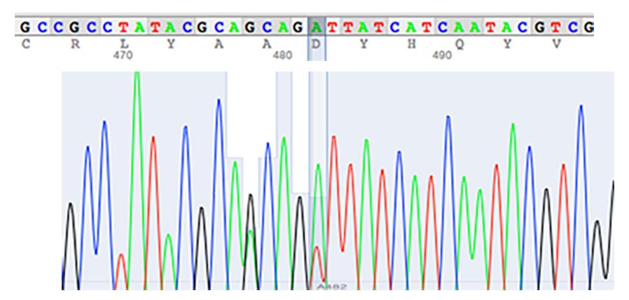
Figure 2. Validation of CRISPR-edited mutation. Double A/T peak (heterozygous context) by sequencing of PCR products encompassing the edited region.
7. Line stabilization: Cross positively identified founder flies with balancer stocks to establish stable, balanced heterozygous or homozygous mutant line stocks. This step takes approximately 4 weeks for autosomal mutation and up to 6 weeks for X chromosome mutant stabilization. Amplify the generated mutant line for locomotor and muscle morphometry analysis. The amplification step takes, in general, 2–3 weeks.
B. Fly husbandry (timing: continuous)
Note: Maintain all fly stocks on standard food at 25 °C.
1. For third-instar larvae immunostaining and mobility tests, put 10–20 young flies (e.g., 10 females and 5 males) in a culture tube with standard medium.
2. Allow the flies to mate and start laying eggs for 1–2 days in a 25 °C incubator with a 12/12 h light/dark cycle. Remove/transfer the flies to a new culture tube and allow the larvae to develop for 4–5 days.
C. Larval locomotor performance assays (Figure 3A–C’) (timing: 1–2 h)
Notes:
1. Prepare grape juice agar medium according to standard protocols, pour it into 60 mm Petri dishes, and allow the medium to solidify at room temperature. The plates with medium can be stored at 4 °C for up to two weeks.
2. To ensure the third-instar larvae used for locomotor assays are age-matched, we collect eggs every 4 h, allow them to develop at 25 °C, and select larvae for tests at the early wandering stage (about 120 h after egg laying), when larvae leave the food to crawl upward.
C1. Peristaltic contraction assay (motility test) (Figure 3A)
1. Place third-instar larva at the border of the grape medium plate.
2. Allow 20 s to acclimatize.
3. Count (manually) complete peristaltic waves for 1 min.
4. Analyze 30 larvae per genotype.
C2. Righting assay (Figure 3B)
1. Place third-instar larva in the center of the grape medium plate.
2. Allow 20 s to acclimatize.
3. Turn the larva on the dorsal side using forceps.
4. Record the time required to return to normal crawling orientation.
5. Repeat for 30 larvae.
C3. Locomotor assay (Figure 3C-C’)
Note: Prepare tracks in the grape medium plates using a surgical scalpel blade and a small ruler. Check and select tracks that are of proper dimensions: width, 2 mm; depth, 5 mm; length, 5 cm.
1. Place third-instar larvae individually on the track.
2. Allow 20 s to acclimatize.
3. Measure the time required to crawl the entire distance.
4. Analyze 30 larvae per genotype.
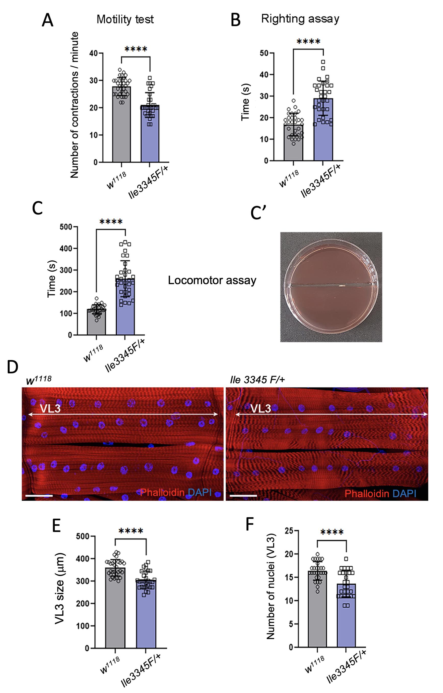
Figure 3. Assessment of larval muscle performance and morphological characteristics. (A) Mobility, (B) righting, and (C) locomotor tests in wild-type and RYR1 mutant conditions. (C’) Grape juice plate with a third-instar larva inside the track, tested in the locomotor assay. (D) Representative VL3 and VL4 ventral longitudinal muscle views from age-matched third-instar larvae in control and RYR1 variant mutant larvae. Muscle pattern was revealed by Phalloidin staining (red), and nuclei were stained with DAPI (blue). Scale bar: 50 μm. (E, F) Statistical representation of VL3 muscle characteristics: (E) VL3 muscle length; (F) number of nuclei. All statistical analyses were performed using Prism, n = 27–32; ****p ≤ 0.0001 (Student’s t-test; data presented as mean ± SD).
D. Quantitative skeletal muscle morphometry (timing: 2 days)
D1. Larval dissection and immunostaining (Figure 3D)
Note: Dissect third-instar larvae according to Lavergne et al. [14].
1. Put the larva on a Sylgard dish.
2. Using the forceps, pin the larva with the small pins at the anterior and posterior ends, keeping the dorsal side up.
3. Cover the larva with a drop of dissection buffer (see Recipes). With sharp scissors, incise the body wall close to the posterior end and open the larva to the anterior end.
4. Using the forceps, gently remove all internal tissue (intestine, fat body, salivary gland, etc.).
5. Incise the body wall close to the pins finely and, using the forceps, pin the body wall, making it flat.
6. Remove the brain and remaining tissues.
7. Rinse the larva in dissection buffer.
8. Remove the remaining dissection buffer and add the fixation solution (4% PFA in PBS) for 20 min at room temperature.
9. Remove the fixation solution and rinse in washing buffer.
10. Transfer the dissected larva into a 1.5 mL Eppendorf tube. Up to eight dissected larvae can be pooled at this step.
11. Rinse three times with 0.1% PBT for 5 min at room temperature.
12. Incubate the tissue in blocking solution (10% horse serum in 0.1% PBT) for 30 min at room temperature on a rocking device.
13. Incubate in primary antibody diluted in PBT solution at 4 °C overnight on a rocking device, with a speed of 15–20 tilts/min.
14. Rinse three times with 0.1% PBT for 10 min at room temperature on a rocking device.
15. Incubate in secondary antibody diluted with 0.1% PBT for 2 h at room temperature on a rocking device in the dark.
16. Rinse three times with 0.1% PBT for 10 min at room temperature on a rocking device in the dark.
17. Incubate with Phalloidin, diluted in 0.1% PBT for 20 min at room temperature on a rocking device in the dark.
18. Rinse three times with 0.1% PBT for 10 min at room temperature on a rocking device in the dark.
19. Mount dissected larva on a slide, using forceps, in a drop of Vectashield mounting medium. Orient cuticle side down and muscle layer side up. Control mounting under stereomicroscope. Use spacers to protect tissue integrity.
D2. Imaging
1. Acquire z-stacks using a Leica SP8 confocal microscope.
2. Maintain identical acquisition settings between genotypes.
D3. Muscle length measurements (Figure 3E)
1. Import the confocal micrographs of ventrally located Drosophila VL3 and VL4 muscles with a channel displaying Actin/Phalloidin associated fluorescence from a series into ImageJ for processing (example shown in Figure 3D).
2. Draw a line with the Straight tool between muscle attachment sites.
3. Choose Analyze and Measure and repeat for 30 muscles.
4. Export measurements to Prism.
D4. Myonuclear quantification (Figure 3F)
1. Open confocal micrographs (z-stack) of Drosophila VL muscle with the channel displaying DAPI and Phalloidin.
2. Count the nuclei in the entire VL3 muscle.
3. Analyze approximately 30 muscles from 6–8 larvae.
D5. Sarcomere analysis (Figure 4)
1. Import the confocal micrographs of Drosophila VL muscle with the channel displaying Kettin-associated (Z-line) fluorescence from the series into ImageJ for processing.
2. Draw a line with the Straight tool at the level of myofibrils. Avoid placing the line over nuclei and ensure it is parallel to the longitudinal muscle axis. Adjust the length of the line to 80–100 μm.
3. Choose Analyze and Plot Profile.
4. Calculate the average size of the sarcomere from the length of the line divided by the number of sarcomeres (Z-line to Z-line space). Repeat for 30 muscles.
5. Export measurements to Prism (example shown in Figure 4).
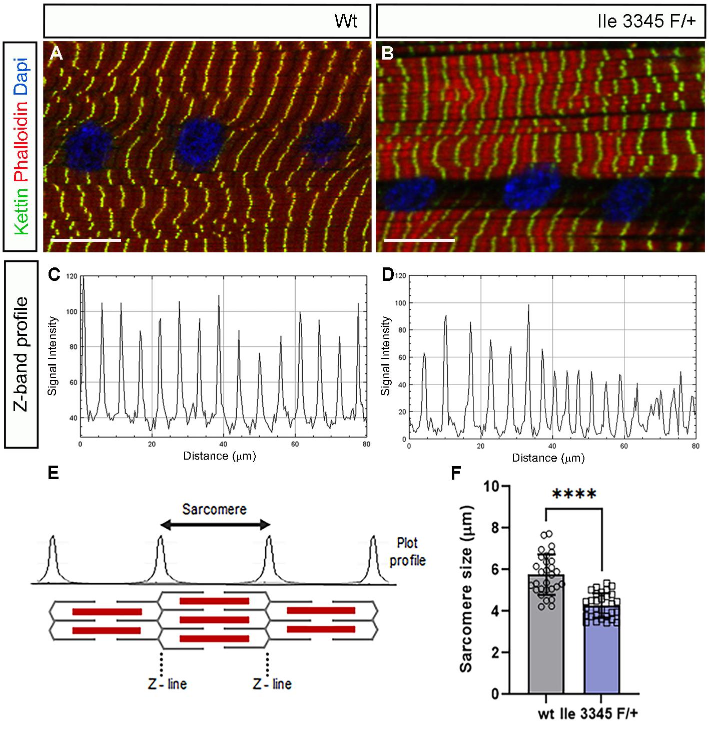
Figure 4. Sarcomere pattern and morphometry. Representative confocal images of ventral longitudinal (VL3) muscles showing sarcomere pattern in wild-type (A) and RYR1 variant mutant larvae (B) (green, anti-kettin-stained Z-bands; red, TRITC-phalloidin-stained thin filaments; blue, DAPI-stained nuclei). Z-band profile generated from the confocal image of a fluorescently labeled Z-line, revealing a reduction in sarcomere length in the RYR1 variant context (B, D) compared to control (A, C). Scale bars, 20 μm. (E) Schematic organization of the sarcomere and Z-band plot profile. (F) Statistical representation of VL3 muscle sarcomere size performed using Prism. n = 29–30; ****p ≤ 0.0001 (Student t-test; data presented as mean ± SD).
Data analysis
1. Perform analyses using GraphPad Prism 9.5.1.
2. Check the normality of data and use Student’s t-test for pairwise comparisons.
3. Present data as mean ± SD.
Validation of protocol
This protocol has been used and validated in the following research article: Zmojdzian et al. [8]. eLife 2026. The workflow was validated through analysis of dRyR loss-of-function conditions, tissue-specific RNAi-mediated depletion, overexpression studies (reported in Figure 3 of [8]), and CRISPR-mediated modeling of the RYR1 p.Met4881Ile variant (reported in Figure 6 of [8]). The protocol reproducibly detected alterations in locomotor performance, muscle morphometry, and sarcomere organization.
General notes and troubleshooting
General notes
1. The quantitative muscle morphometry is performed here on the ventral longitudinal muscle 3 (VL3). After larval dissection, VL3 is at the optimal position for imaging and precise morphometric analyses. The neighboring VL4 muscle is also appropriate for quantitative morphometry.
2. The presented workflow could be applied for assessing functional and structural muscle properties in different genetic contexts. Notably, it is suitable for phenotyping neuromuscular Drosophila disease models, including genome-edited lines carrying variants of unknown significance.
Troubleshooting
Problem 1: Larval muscles appear contracted during imaging.
Possible cause: Incomplete relaxation during fixation.
Solution: Analyze only muscles displaying regular sarcomere spacing and relaxed morphology.
Problem 2: Variable locomotor performance.
Possible cause: Differences in larval age or nutritional status.
Solution: Use synchronized third-instar larvae and identical culture conditions [14].
Problem 3: Low post-injection survival.
Possible cause: Vector- or reagent-associated toxicity.
Solution: To mitigate this, reduce the concentration of both the gRNA and donor template. Additionally, verify gRNA cleavage efficiency prior to injection using a mismatch-specific nuclease assay (e.g., T7E1 or Surveyor) or high-resolution melting analysis (HRMA) to confirm robust on-target endonuclease activity.
Acknowledgments
We thank Drosophila Bloomington Stock Centers for Drosophila lines and L. Mouty for the technical assistance. This study was supported by the Priority Research Programme on Rare Diseases of the French Investments for the Future Program, by the AFM-Telethon strategic grant to MyoNeurAlp consortium, the Reference Centre for Neuromuscular Diseases grant to M.Z. and C.S., and an ANR-iSITE-2025 grant to M.Z.
This protocol was used in [8].
Author contributions
Protocol Development, M.Z., T.J.; Protocol Optimization and Validation, M.Z., T.J.; Writing—Original Draft, M.Z., T.J., K.J., C.S.; Writing—Review & Editing, K.J., C.S.; Funding acquisition, K.J., C.S.; Supervision, K.J., C.S.
Competing interests
The authors declare no conflicts of interest.
References
- Poovathumkadavil, P. and Jagla, K. (2020). Genetic Control of Muscle Diversification and Homeostasis: Insights from Drosophila. Cells. 9(6): 1543. https://doi.org/10.3390/cells9061543
- Daczewska, M., Picchio, L., Jagla, T., Figeac, N. and Jagla, K. (2010). Muscle Development and Regeneration in Normal and Pathological Conditions: Learning from Drosophila. Curr Pharm Des. 16(8): 929–941. https://doi.org/10.2174/138161210790883462
- Plantié, E., Migocka-Patrzałek, M., Daczewska, M. and Jagla, K. (2015). Model Organisms in the Fight against Muscular Dystrophy: Lessons from Drosophila and Zebrafish. Molecules. 20(4): 6237–6253. https://doi.org/10.3390/molecules20046237
- Luis, N. M. and Schnorrer, F. (2021). Mechanobiology of muscle and myofibril morphogenesis. Cells Dev. 168: 203760. https://doi.org/10.1016/j.cdev.2021.203760
- Christian, C. J. and Benian, G. M. (2020). Animal models of sarcopenia. Aging Cell. 19(10): e13223. https://doi.org/10.1111/acel.13223
- Endo, Y. (2026). Molecular genetics of skeletal muscle diseases associated with abnormal excitation–contraction coupling. J Hum Genet. e1038/s10038–026–01499–2. https://doi.org/10.1038/s10038-026-01499-2
- Junion, G. and Jagla, K. (2022). Diversification of muscle types in Drosophila embryos. Exp Cell Res. 410(1): 112950. https://doi.org/10.1016/j.yexcr.2021.112950
- Zmojdzian, M., Jagla, T., Cherik, F., Dubinska-Magiera, M., Migocka-Patrzałek, M., Daczewska, M., Rendu, J., Jagla, K. and Sarret, C. (2026). Drosophila ryanodine receptor gene triggers functional and developmental muscle properties and could be used to assess the impact of human RYR1 mutations. eLife. 15: e111053. https://doi.org/10.7554/elife.111053
- Ogasawara, M. and Nishino, I. (2024). Update on RYR1-related myopathies. Curr Opin Neurol. 37(5): 504–508. https://doi.org/10.1097/wco.0000000000001296
- Lanner, J. T. (2012). Ryanodine Receptor Physiology and Its Role in Disease. Adv Exp Med Biol. 740: 217–234. https://doi.org/10.1007/978-94-007-2888-2_9
- Gao, S., Sandstrom, D. J., Smith, H. E., High, B., Marsh, J. W. and Nash, H. A. (2013). Drosophila Ryanodine Receptors Mediate General Anesthesia by Halothane. Anesthesiology. 118(3): 587–601. https://doi.org/10.1097/aln.0b013e31827e52c6
- Port, F. and Bullock, S. L. (2016). Creating Heritable Mutations in Drosophila with CRISPR-Cas9. Methods Mol Biol. 1478: 145–160. https://doi.org/10.1007/978-1-4939-6371-3_7
- Port, F., Chen, H. M., Lee, T. and Bullock, S. L. (2014). Optimized CRISPR/Cas tools for efficient germline and somatic genome engineering in Drosophila. Proc Natl Acad Sci USA. 111(29): e1405500111. https://doi.org/10.1073/pnas.1405500111
- Lavergne, G., Zmojdzian, M., Da Ponte, J. P., Junion, G. and Jagla, K. (2020). Drosophila adult muscle precursor cells contribute to motor axon pathfinding and proper innervation of embryonic muscles. Development. 147: e183004. https://doi.org/10.1242/dev.183004
Article Information
Publication history
Received: Aug 20, 2026
Accepted: Oct 14, 2026
Available online: Nov 2, 2026
Published: Dec 5, 2026
Copyright
© 2026 The Author(s); This is an open access article under the CC BY license (https://creativecommons.org/licenses/by/4.0/).
How to cite
Readers should cite both the Bio-protocol article and the original research article where this protocol was used:
- Zmojdzian, M., Jagla, T., Jagla, K. and Sarret, C. (2026). Measuring Functional and Structural Muscle Properties in Drosophila Larvae for Testing Genetic Variants Involved in Neuromuscular Disorders. Bio-protocol 16(23): e5875. DOI: 10.21769/BioProtoc.5875.
- Zmojdzian, M., Jagla, T., Cherik, F., Dubinska-Magiera, M., Migocka-Patrzałek, M., Daczewska, M., Rendu, J., Jagla, K. and Sarret, C. (2026). Drosophila ryanodine receptor gene triggers functional and developmental muscle properties and could be used to assess the impact of human RYR1 mutations. eLife. 15: e111053. https://doi.org/10.7554/elife.111053

 Download Citation in RIS Format
Do you have any questions about this protocol?
Post your question to gather feedback from the community. We will also invite the authors of this article to respond.
 Post a Question


