


- Protocols

- Articles and Issues
- For Authors
- About
- Become a Reviewer


Transcriptomic Signature-Guided EGFP Labeling of Alveolar Epithelial Cells via RNA Sensing in Mouse Lungs In Vivo and Human Idiopathic Pulmonary Fibrosis Lung Slices Ex Vivo

Published: Vol 16, Iss 21, Nov 5, 2026 DOI: 10.21769/BioProtoc.5863 Views: 58
Reviewed by: Kirsten A. CoprenGururaj KidiyoorAnonymous reviewer(s)
 PDF
 Ask a question

Original research article
The authors used this protocol in:

Jan 2026

Protocol Collections
Comprehensive collections of detailed, peer-reviewed protocols focusing on specific topics



Abstract
Single-cell RNA sequencing has revealed transient and disease-associated alveolar epithelial states that are difficult to access with conventional transgenic approaches. Here, we describe a programmable RNA-sensing workflow based on CellREADR [cell access through RNA sensing by endogenous adenosine deaminases acting on RNA (ADAR)] to couple a selected cellular RNA signature to enhanced green fluorescent protein (EGFP) translation. The protocol covers sequence-encoded sensing RNA (sesRNA) design and cloning, in vitro validation of sesRNA-dependent EGFP expression in HEK293T cells, production and heparin-column purification of adeno-associated virus variant 6.2FF (AAV6.2FF), delivery to mouse lungs, and application to human idiopathic pulmonary fibrosis (IPF) precision-cut lung slices (PCLS). Sensors targeting Sftpc, Pdpn, or Sprr1a can be used to label alveolar type 2, alveolar type 1, and injury-induced Krt8+ alveolar differentiation intermediate cells in mice and KRT17+KRT5- aberrant basaloid cells in humans, respectively. Because cell specificity is programmed by the sensor sequence rather than a cell type–specific promoter or newly generated mouse line, this approach can be rapidly adapted to emerging transcriptomic targets in both experimental mouse models and human lung tissues, enabling cross-species validation and translational investigation of disease-associated epithelial states.
Key features
• Programmable sesRNAs convert endogenous RNA signatures into ADAR-dependent EGFP translation without requiring a new transgenic mouse line.
• Includes candidate sensor design, in vitro validation of target-dependent EGFP expression in HEK293T cells, and AAV6.2FF production and purification using a heparin column.
• Supports in vivo labeling of AT2, AT1, and Sprr1a-positive transitional alveolar epithelial cells in mice.
• Extends the workflow to human IPF PCLS for ex vivo assessment of SPRR1A-guided reporter expression in KRT17+KRT5- aberrant basaloid cells.
Keywords: RNA sensing CellREADR
EGFP labeling
Alveolar epithelial cells
Pulmonary fibrosis
Adeno-associated virus
Precision-cut lung slices
Heparin-column purification
Graphical overview
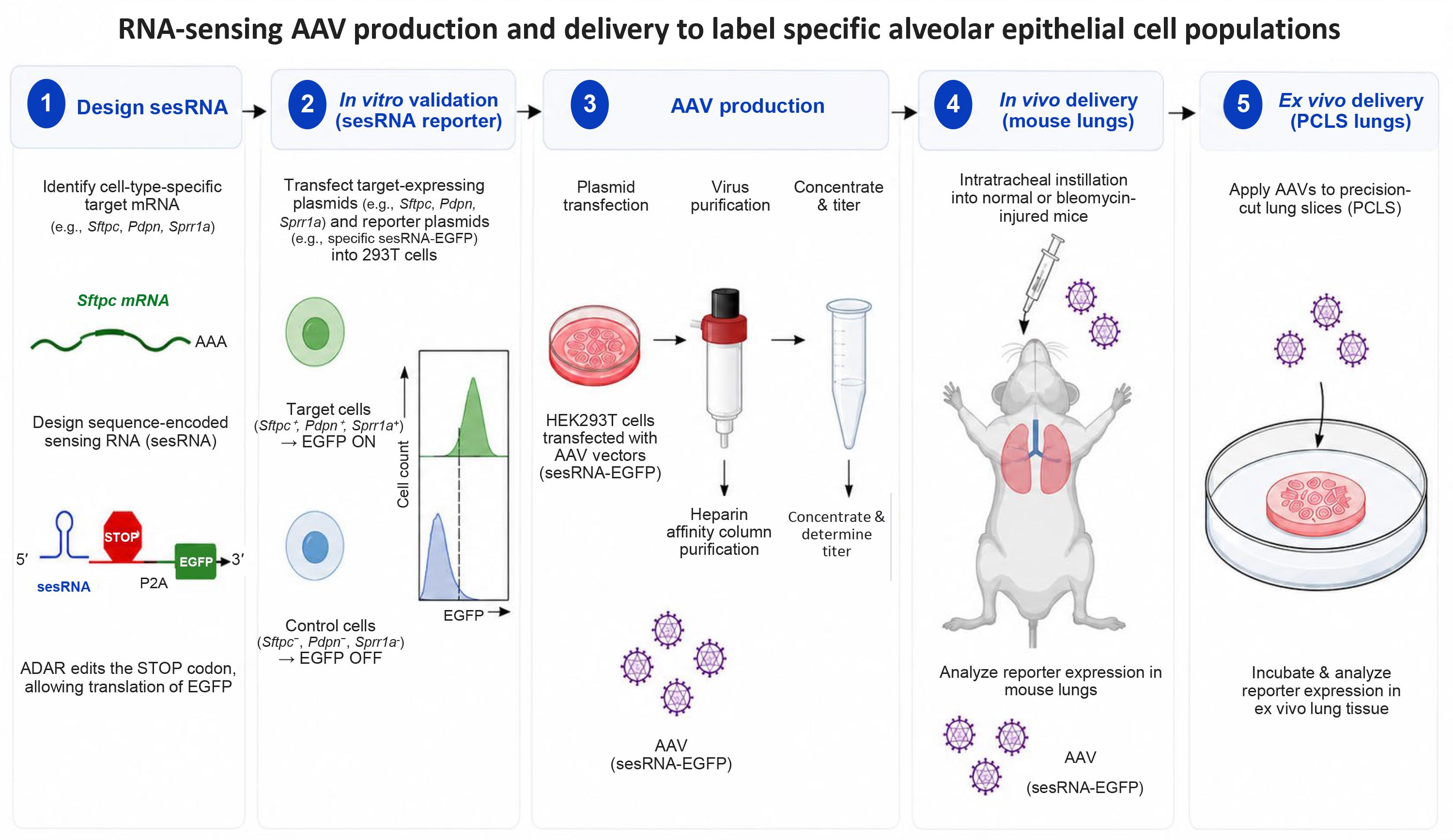
Graphical overview of transcript-guided RNA sensing and AAV delivery. (1) Select a cell-enriched target RNA and design a sequence-encoded sensing RNA (sesRNA). (2) Validate target-dependent reporter translation in HEK293T cells. (3) Produce and purify AAV6.2FF carrying the sesRNA-EGFP cassette. (4) Deliver AAV vectors to normal or bleomycin-injured mouse lungs for in vivo labeling. AAV icons beneath the mouse denote the administered vector; reporter-positive cells are evaluated in lung tissue downstream. (5) Apply AAV vectors to human precision-cut lung slices (PCLS) for ex vivo labeling. Approximate timing: sesRNA design and cloning, 3–7 days; in vitro screening, 3–4 days; AAV production and purification, 5–7 days; in vivo reporter expression, at least 6 days after AAV delivery; human PCLS workflow, 14 days. AAV, adeno-associated virus; ADAR, adenosine deaminase acting on RNA; BFP, blue fluorescent protein; EGFP, enhanced green fluorescent protein; PCLS, precision-cut lung slice.
Background
Alveolar type 2 (AT2) cells function as facultative progenitors that proliferate and differentiate into alveolar type 1 (AT1) cells after injury. During fibrotic repair, AT2 cells can accumulate in transitional states, including Krt8-positive alveolar differentiation intermediates in mice and KRT5-negative/KRT17-positive aberrant basaloid cells in human IPF lungs [1–4]. These populations are heterogeneous and incompletely represented by conventional lineage markers, making their functional analysis technically challenging. Established approaches include Cre/lox-based lineage tracing and conditional genetic manipulation, promoter- or enhancer-driven reporter vectors, RNA in situ hybridization, and single-cell transcriptomic profiling. Lineage tracing provides durable cell-fate information but requires prior identification of a suitable regulatory element and, in many cases, generation and breeding of new mouse lines [1,5]. RNA in situ hybridization and single-cell RNA sequencing can define disease-associated states at high molecular resolution; however, these are endpoint assays and do not by themselves provide prospective access to living cells for isolation or manipulation. These limitations are particularly important when transcriptomic studies identify newly emerging or transient cell states.
CellREADR uses a programmable sesRNA that base-pairs with a selected cellular RNA [6]. The sensor contains an engineered UAG stop codon positioned within the translated sensor sequence. After the sensor hybridizes to its target RNA, endogenous ADAR edits adenosine to inosine. The ribosome interprets inosine as guanosine, permitting translation through the edited codon and into the downstream effector RNA encoding EGFP. Changing the antisense sensor sequence therefore redirects reporter expression to cells that contain the selected target transcript, providing a way to convert an RNA-defined cellular state into live-cell reporter or effector expression [6].
For lung delivery, this protocol uses AAV6.2FF, an AAV6-derived capsid with efficient respiratory epithelial transduction [7–9]. The combined workflow provides a rapid, transcript-guided alternative to creating a new cell type–specific mouse line.
Materials and reagents
Biological materials
1. HEK293T cells (ATCC, catalog number: CRL-11268)
2. Pathogen-free 12-week-old C57BL/6 mice (The Jackson Laboratory, stock number: 000664); use both sexes for reporter-labeling studies unless the experimental design specifies otherwise
3. Krt8-CreERT2;Rosa26-tdTomato mice, generated by crossing Tg(Krt8-cre/ERT2)17Blpn/J mice with B6.Cg-Gt(ROSA)26Sortm14(CAG-tdTomato)Hze/J reporter mice (The Jackson Laboratory; stock numbers: 017947 and 007914)
4. Sftpc-CreERT2;Rosa26-tdTomato mice, generated by crossing B6.129S-Sftpctm1(cre/ERT2)Blh/J mice with the Rosa26-tdTomato reporter line (The Jackson Laboratory; stock numbers: 028054 and 007914)
5. Human PCLS from nonfibrotic donor and IPF lungs (Baylor College of Medicine Chronic Lung Disease Tissue Repository)
Reagents
1. Dulbecco's modified Eagle medium (DMEM) (Gibco, catalog number: 11965-118)
2. Roswell Park Memorial Institute (RPMI) medium (Corning, catalog number: 15-040-CV)
3. Fetal bovine serum (FBS) (Gibco, catalog number: 26140-079)
4. Penicillin-streptomycin (Gibco, catalog number: 15140-122)
5. Polyethyleneimine (PEI) (Polysciences, catalog number: NC2073514)
6. Opti-MEM reduced-serum medium (Gibco, catalog number: 31985-070)
7. Pierce protease inhibitor tablets (Thermo Fisher Scientific, catalog number: A32963)
8. Sodium deoxycholate (Sigma-Aldrich, catalog number: D6750-25G)
9. Benzonase nuclease (Sigma-Aldrich, catalog number: E1014-5KU)
10. Bleomycin sulfate (Almirall, catalog number: B1141000)
11. pCL245 mammalian expression/lentiviral vector containing the EF1α promoter and mCherry coding sequence (gift from Dr. Chao Li, Department of Biochemistry and Molecular Genetics, University of Alabama at Birmingham)
Note: The pCL245 backbone itself is not essential; an equivalent custom plasmid expressing mCherry-T2A-target cDNA from a constitutive promoter may be used instead.
12. pAAV-CAG-mTagBFP2-P2A-EGFP backbone (Addgene plasmid #184943)
13. pDGM6.2FF helper plasmid, derived from pDGM6 (Addgene plasmid #110660)
14. AgeI restriction enzyme and compatible reaction buffer (New England Biolabs, catalog number: R3552S)
15. Gibson Assembly Cloning kit (New England Biolabs, catalog number: E5510S)
16. Synthetic sesRNA DNA fragments (gBlocks or equivalent; Integrated DNA Technologies)
17. Sterile phosphate-buffered saline (PBS), pH 7.4 (Gibco, catalog number: 10010-023)
18. Sodium chloride (NaCl), molecular biology grade (Sigma-Aldrich, catalog number: S7653)
19. Sterile 0.9% sodium chloride (NaCl) for animal administration (BD, catalog number: 221819)
20. Tamoxifen for CreERT2 induction (Fisher Scientific, catalog number: 50-149-0596)
21. Paraformaldehyde (PFA), 4% solution (Electron Microscopy Sciences, catalog number: 15710-S, or equivalent)
22. Optimal cutting temperature (OCT) compound (Sakura Finetek, Tissue-Tek O.C.T., catalog number: 4583)
23. Normal goat serum (Jackson ImmunoResearch, catalog number: 005-000-121)
24. Triton X-100 (Millipore, catalog number: 1086431000)
25. 4',6-Diamidino-2-phenylindole (DAPI) nuclear stain (Thermo Fisher Scientific, catalog number: D1306)
26. Anti-Krt8 antibody (Thermo Fisher Scientific, clone TS1, catalog number: MA5-14467)
27. Anti-SFTPC antibody (Abnova, clone 1C9, catalog number: H00006447-M01)
28. Anti-KRT17 antibody (Proteintech, catalog number: 10543-1-AP)
29. Anti-KRT5 antibody (Proteintech, catalog number: 905501-AP)
30. Species-appropriate Alexa Fluor 488-, 555-, and 647-conjugated secondary antibodies (Invitrogen, catalog numbers: A-11008, A-11001, A-21428, A-21422, A-21245)
31. Sodium azide (Sigma-Aldrich, catalog number: S2002)
32. Low-melting-point agarose (Thermo Fisher Scientific, UltraPure Low Melting Point Agarose, catalog number: 16520050)
33. Antifade mounting medium (Thermo Fisher Scientific, ProLong Gold Antifade Mountant, catalog number: P36930, or equivalent)
Solutions
1. Complete DMEM (see Recipes)
2. PEI transfection solution, 1 mg/mL (see Recipes)
3. Sodium deoxycholate stock solution, 10% (w/v) (see Recipes)
4. Heparin-column wash and elution buffers (see Recipes)
5. Complete RPMI medium (see Recipes)
6. Low-melting-point agarose in DMEM, 2% (w/v) (see Recipes)
7. Immunofluorescence blocking buffer (see Recipes)
8. Immunofluorescence antibody diluent (see Recipes)
Recipes
1. Complete DMEM
| Reagent | Final concentration | Volume |
|---|---|---|
| DMEM | 90% | 445 mL |
| FBS | 10% | 50 mL |
| Penicillin-streptomycin | 1× | 5 mL |
| Total | n/a | 500 mL |
Mix aseptically. Store at 2–8 °C for up to 2 weeks; discard if contamination, precipitation, or marked color change is observed.
2. PEI transfection solution (1 mg/mL)
| Reagent | Final concentration/endpoint | Quantity or volume |
|---|---|---|
| Linear PEI, MW 25,000 | 1 mg/mL | 100 mg |
| Milli-Q water | n/a | 90 mL initially; bring final volume to 100 mL |
| Hydrochloric acid (HCl), 12 M | As required to reach pH < 2.0 | ~160 μL |
| Sodium hydroxide (NaOH), 10 N | As required to reach pH ~7.0 | ~100 μL |
| Total | n/a | 100 mL |
a. Suspend 100 mg of linear PEI in 90 mL of Milli-Q water in a clean beaker.
b. While stirring, add 12 M HCl dropwise until the pH is < 2.0 (approximately 160 μL total).
c. Cover the beaker and stir for 3 h at room temperature, or until the solution is mostly clear.
d. Add 10 N NaOH dropwise while monitoring the pH until the solution reaches approximately pH 7.0 (approximately 100 μL total).
e. Adjust the final volume to 100 mL with Milli-Q water and mix thoroughly.
f. Filter-sterilize the solution through a 0.22 μm membrane filter.
g. Aliquot into single-use volumes and store at -80 °C for up to 6 months. Avoid repeated freeze/thaw cycles.
Critical: Add HCl and NaOH slowly while continuously monitoring the pH. The indicated acid and base volumes are approximate and may vary between preparations.
3. Sodium deoxycholate stock solution [10% (w/v)]
| Reagent | Final concentration | Quantity or volume |
|---|---|---|
| Sodium deoxycholate | 10% (w/v) | 10 g |
| Double-distilled water | n/a | Bring to 100 mL |
Dissolve completely. Store the 10% sodium deoxycholate stock at room temperature for up to 1 month. If precipitation occurs, warm gently and mix until the solution is clear before use.
4. Heparin-column wash and elution buffers (50 mL each; prepared from 5 M NaCl)
| Buffer | 5 M NaCl | PBS |
|---|---|---|
| 200 mM NaCl wash buffer | 0.648 mL | 49.352 mL |
| 300 mM NaCl elution buffer | 1.68 mL | 48.32 mL |
| 350 mM NaCl elution buffer | 2.19 mL | 47.81 mL |
| 450 mM NaCl elution buffer | 3.22 mL | 46.78 mL |
Mix and sterilize by filtration. Store heparin-column wash and elution buffers at 2–8 °C for up to 1 month.
5. Complete RPMI medium
| Reagent | Final concentration | Volume |
|---|---|---|
| RPMI | 90% | 445 mL |
| FBS | 10% | 50 mL |
| Penicillin-streptomycin | 1× | 5 mL |
| Total | n/a | 500 mL |
Mix aseptically. Store at 2–8 °C for up to 2 weeks; discard if contamination, precipitation, or marked color change is observed.
6. Low-melting-point agarose in DMEM [2% (w/v)]
Dissolve 2 g of low-melting-point agarose in DMEM to a final volume of 100 mL by heating until fully dissolved. Cool to approximately 37–42 °C before lung inflation and use on the day of preparation. Do not store the prepared agarose/DMEM solution.
7. Immunofluorescence blocking buffer
5% normal goat serum in PBS. Prepare freshly.
8. Immunofluorescence antibody diluent
1% normal goat serum, 0.3% Triton X-100, and 0.01% sodium azide in PBS. Prepare freshly.
Laboratory supplies
1. 12-well cell culture plates (Fisher Scientific, catalog number: 07-200-82)
2. 10-cm tissue culture dishes (Thermo Fisher Scientific, catalog number: 130182)
3. Amicon Ultra-4 centrifugal filters, 100 kDa molecular-weight cutoff (Millipore, catalog number: UFC801024)
4. Sterile 0.2 micrometer syringe filters (Millipore, catalog number: SLGPM33RS)
5. Sterile 15 and 50 mL conical tubes (any)
6. Sterile serological pipettes (5, 10, and 25 mL) and filtered micropipette tips compatible with 10, 20, 200, and 1,000 μL pipettes (any)
7. Sterile syringes compatible with the syringe filters and animal instillation procedure (any)
8. Cell scrapers (any)
9. Cryovials for AAV aliquots (any)
10. Microscope slides and coverslips (any)
11. Humidified staining chambers (any)
12. Animal intubation/instillation supplies approved by the institutional animal program (any)
13. 24-well plates for PCLS culture (any)
14. Sterile 60 mL syringes for lung inflation (any)
15. Sterile 10 Fr airway catheter (any)
16. Sterile 10 mm biopsy punch (any)
17. Tissue adhesive compatible with the VF-300-0Z specimen holder (any)
18. Fine forceps and scissors for PCLS handling (any)
19. PCLS culture inserts or plates as required by the local tissue-slice workflow (any)
20. HiTrap heparin affinity column (Cytiva, catalog number: 45-000-057)
Equipment
1. Peristaltic pump (Millipore, model: XX8020EL0)
2. Inverted fluorescence microscope (Nikon, model: Eclipse Ts2-FL)
3. Class II biological safety cabinet (Thermo Fisher Scientific or equivalent)
4. Humidified 37 °C, 5% CO2 incubator (Thermo Fisher Scientific or equivalent)
5. Refrigerated centrifuge with rotors for 15 and 50 mL tubes and speeds up to 4,000× g (Eppendorf or equivalent)
6. LSM710 confocal laser-scanning microscope (Carl Zeiss) or equivalent sequential-scanning confocal microscope
7. Cryostat for lung sectioning (Leica Biosystems or equivalent)
8. FACSymphony S6 cell sorter (BD Biosciences) or equivalent instrument capable of detecting EGFP, BFP, and tdTomato, if flow analysis is performed
9. Quantitative real-time PCR instrument compatible with SYBR Green detection (Applied Biosystems/Thermo Fisher Scientific or equivalent) for AAV genome titration
10. VF-300-0Z vibrating microtome (Precisionary Instruments, USA) for preparation of 500 μm PCLS
Software and datasets
1. CellREADR design portal (Duke University; https://cellreadr.neuro.duke.edu; accessed September 20, 2026)
2. ImageJ (NIH; version 1.54u8; free) for fluorescence-image inspection, manual/ROI-assisted cell counting, and colocalization measurements
3. GraphPad Prism 8 (GraphPad Software) for statistical analysis and graphing; equivalent statistical software may be used if the same analyses are performed
Procedure
A. sesRNA design and reporter-plasmid construction
Timing: Approximately 3–7 days, including design, DNA synthesis/receipt, cloning, transformation, and sequence verification; DNA synthesis shipping time is not included.
1. Select a target RNA that is enriched in the desired cell population using single-cell RNA-sequencing (scRNA-seq) data whenever possible, because cell-type resolution is important for sensor specificity. The associated study used scRNA-seq-defined expression patterns to select Sftpc for AT2 cells, Pdpn for AT1 cells, and Sprr1a for injury-induced transitional epithelial cells [1]. Bulk RNA-seq may be used as a supplementary screen, but should not replace cell-resolved evaluation when heterogeneous tissues are analyzed.
2. Use the CellREADR design portal (https://cellreadr.neuro.duke.edu) to generate antisense sesRNA candidates complementary to the target RNA [6]. Candidate sensors are commonly 200–300 nucleotides; however, the validated Sprr1a sensor is 102 nucleotides, demonstrating that shorter designs can also function [1]. Generate several candidates spanning different regions of the transcript rather than relying on a single design.
3. For each candidate, identify a target 5′-CCA-3′ sequence and the complementary 5′-TGG-3′ sequence in the sesRNA. Position the editable site near the center of the translated sensor region (typically 80–220 nucleotides from the 5′ end for longer sensors).
4. Replace the selected sesRNA 5′-TGG-3′ sequence with 5′-TAG-3′ to create the intended adenosine–cytosine mismatch after hybridization with the target RNA, thereby enabling ADAR-mediated editing of the UAG stop codon.
5. Replace other in-frame stop codons in the sensor sequence: TAG to GAG, TAA to GAA, and TGA to GGA. Maintain at least 10 base pairs between any altered stop codon and the central editable UAG.
Critical: Inspect the final open reading frame for unintended stop codons and confirm that the sesRNA remains in-frame with the T2A-linked EGFP or H2B-EGFP effector.
Critical: Avoid downstream ATG start codons that could generate target-independent reporter expression. Screen several sensor candidates because portal-generated designs vary in efficiency.
6. Synthesize the selected sesRNA as a double-stranded DNA fragment. Linearize the pAAV-CAG-mTagBFP2-P2A-EGFP backbone with AgeI, purify the linearized vector, and assemble the sesRNA insert by Gibson assembly following the manufacturer's instructions. For a typical two-fragment assembly, use approximately 50 ng of linearized vector and a 2–3-fold molar excess of the sesRNA insert, keeping the total DNA within the range recommended for the Gibson Assembly kit.
7. Transform, isolate plasmid DNA, and verify the complete sensor-effector junction and open reading frame by DNA sequencing.
Pause point: Store sequence-verified plasmids at -20 °C.
B. Construction of target-RNA expression plasmids
Timing: Approximately 2–4 days, including PCR, cloning, transformation, and sequence verification.
1. Amplify full-length mouse Sftpc, Pdpn, Sprr1a cDNA from mouse lung cDNA [1] and human SPRR1A cDNA from human cDNA using the following primers: forward 5′-GATGA CGACG ATAAG GGATC CATGA ATTCT CAGCA GCAGA AG-3′, reverse 5′-GAAGT TCGTG GCTCC GGATC CCTTC TGCTT GGTCT TCTGC T-3′.
2. Linearize pCL245 with BamHI and purify the linearized vector. If pCL245 is not available, use an equivalent custom expression plasmid in which a constitutive promoter drives mCherry-T2A-target cDNA.
3. Insert the target cDNA downstream of and in-frame with mCherry-T2A by Gibson assembly, following the manufacturer's instructions and using approximately 50 ng of linearized vector with a 2–3-fold molar excess of target-cDNA insert. Prepare a matched mCherry-T2A vector lacking the target cDNA as the negative control.
4. Verify each construct by restriction analysis and DNA sequencing.
C. HEK293T cell culture and in vitro sensor validation
Timing: 3–4 days from cell seeding through reporter imaging; allow additional time for repeat screening of promising candidates.
1. Maintain HEK293T cells in complete DMEM at 37 °C in a humidified incubator with 5% CO2. Use cells that are actively proliferating and free of mycoplasma.
2. Seed 1 × 10⁵ cells per well in a 12-well plate and incubate overnight to reach approximately 70%–80% confluence at transfection.
3. For each well, prepare 1 μg of total plasmid DNA in 100 μL of Opti-MEM as a two-plasmid mixture: 0.5 μg of sesRNA reporter plasmid plus 0.5 μg of either the matching target-RNA-mCherry expression plasmid or the mCherry-only negative-control plasmid.
4. In a separate tube, dilute 3 μL of 1 mg/mL PEI in 100 μL of Opti-MEM.
5. Combine the DNA and PEI solutions, mix gently, incubate for 10–15 min at room temperature, and add the complexes dropwise to the cells.
6. After 6 h, replace the transfection medium with fresh complete DMEM.
7. Image cells 24–72 h after transfection. For each candidate and control, acquire at least three randomly selected, non-overlapping fields using identical objective, exposure/laser power, detector gain, offset, pinhole, scan speed, and pixel resolution. Set acquisition parameters using a representative target-positive field, avoid saturated pixels, and then keep the settings unchanged across paired target and control conditions.
8. Quantify reporter activity as described in the Data analysis section and Figure 1. Advance candidates that show consistently low EGFP-positive ratios in the mCherry-only control and a reproducible increase in EGFP-positive ratios in the matching target-RNA condition across independent transfections. Because target abundance and sensor efficiency vary by transcript, rank candidates by target-dependent enrichment rather than applying a universal absolute cutoff.
Critical: Include the target-negative control for every candidate sensor. A strong reporter signal without target RNA indicates sensor-independent translation and requires redesign.
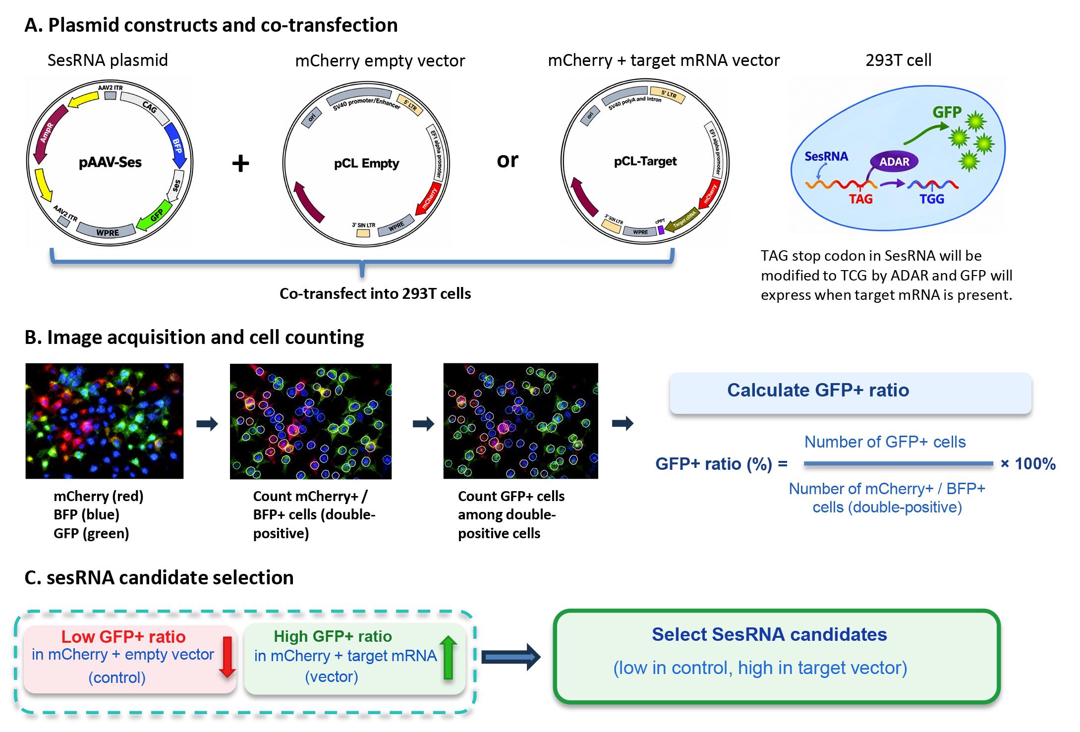
Figure 1. Schematic of target-RNA expression, HEK293T co-transfection, and sesRNA candidate screening. (A) The sesRNA reporter plasmid is co-transfected with either an mCherry-T2A empty/control plasmid or an mCherry-T2A-target mRNA expression plasmid. (B) Random fields are imaged using the same settings; mCherry/BFP double-positive cells define the co-transfected population, and EGFP-positive cells are counted within this population. The EGFP-positive ratio is calculated as EGFP-positive cells divided by mCherry/BFP double-positive cells × 100%. (C) Candidate sesRNAs are prioritized when control cultures show low reporter activity and the matching target-RNA condition shows reproducibly higher activity. ADAR, adenosine deaminase acting on RNA; BFP, blue fluorescent protein; EGFP/GFP, enhanced green fluorescent protein; mRNA, messenger RNA; sesRNA/SesRNA, sequence-encoded sensing RNA.
D. AAV6.2FF production and heparin-column purification
Timing: Approximately 5–7 days from producer-cell preparation through AAV purification and genome-titer determination.
1. Passage HEK293T cells at a 1:2 ratio for at least two consecutive days before transfection to maintain rapid proliferation.
2. Seed cells in 10-cm dishes at 4–8 × 10⁵ cells/mL in 10 mL of antibiotic-free DMEM per dish so that cultures reach approximately 70%–80% confluence the following day.
3. For the validated production batch, dilute 45 μg of the pAAV2-based sesRNA expression vector and 135 μg of pDGM6.2FF (1:3 plasmid mass ratio) in 7.5 mL of Opti-MEM. In a separate tube, dilute 540 μL of 1 mg/mL PEI in 7.5 mL of Opti-MEM, as used in the associated study [1]. Scale the preparation proportionally if the number of culture dishes is changed.
4. Add the diluted PEI to the DNA solution, mix gently, incubate for 10–15 min at room temperature, and distribute the complexes dropwise across the intended culture dishes.
5. After 5 h, replace the transfection mixture with complete DMEM.
6. At 24 h, assess BFP expression. Continue only when at least 80% of cells are BFP-positive.
7. At 72 h, collect cells and culture medium. Centrifuge at 500× g for 10 min, retain the medium, and freeze the cell pellet and medium at -80 °C overnight.
8. Resuspend the pooled cell pellets in 47.5 mL of PBS containing protease inhibitor and divide the suspension between two 50-mL tubes.
9. Add sodium deoxycholate to a 0.5% final concentration and benzonase to 50 U/mL. Incubate for 1 h at 37 °C.
10. Centrifuge at 3,000× g for 15 min to remove cell debris.
11. Combine the clarified cell lysate with the thawed culture medium, freeze the combined material overnight, thaw, incubate for 1 h at 37 °C, and centrifuge again at 3,000× g for 15 min.
12. Equilibrate the HiTrap heparin column with PBS and load the clarified viral solution at 1 mL/min using a peristaltic pump.
Critical: The lysate must be free of visible debris before loading. Residual debris can obstruct the column and reduce recovery.
13. Wash the column sequentially with 50 mL of PBS and 50 mL of 200 mM NaCl in PBS.
14. Elute the virus sequentially with 1.5 mL of 300 mM NaCl, 3 mL of 350 mM NaCl, and 1.5 mL of 450 mM NaCl, all prepared in PBS.
15. Filter the pooled eluate through a sterile 0.2 μm syringe filter.
16. Concentrate the eluate in an Amicon Ultra-4 100 kDa filter at 4,000× g until approximately 0.25 mL remains.
17. Bring the concentrated vector to a final volume of 500 μL with PBS, mix gently, aliquot, and store at -80 °C.
18. Quantify viral genome titers by SYBR Green quantitative real-time PCR (qPCR), as used for AAV6.2FF vectors [7]. Treat an aliquot of purified AAV with nuclease to remove non-encapsidated DNA, release encapsidated vector DNA, and amplify a conserved vector sequence such as the AAV inverted terminal repeat (ITR) against a linearized plasmid standard curve of known copy number. Run standards and samples in technical triplicate, confirm that amplification efficiencies and replicate Ct values are acceptable, and calculate vector genomes per milliliter (vg/mL) after correcting for all dilution factors. Use the measured titer to prepare the validated doses for mouse and PCLS delivery.
Pause point: Store single-use AAV aliquots at -80 °C and avoid repeated freeze/thaw cycles.
E. In vivo RNA-guided labeling in mouse lung
Timing: Lineage-label induction, 5 days plus a 2-week tamoxifen washout when applicable; reporter expression, at least 6 days after AAV delivery.
1. For lineage-reporter experiments, induce CreERT2-dependent tdTomato expression with tamoxifen at 50 mg/kg by intraperitoneal injection once daily for five consecutive days. After the final injection, allow a 2-week washout period before administering the AAV vector or initiating lung injury.
2. Select the experimental condition according to the target cell population:
a. For labeling alveolar type 2 or alveolar type 1 cells with Sftpc- or Pdpn-targeting sensors, respectively, proceed directly to AAV6.2FF delivery after the tamoxifen washout period, when applicable.
b. For labeling injury-induced transitional epithelial cells with the Sprr1a-targeting sensor, induce lung injury with bleomycin. On day 0, anesthetize 12-week-old mice and administer bleomycin intratracheally at 1.5 U/kg body weight in 50 μL of sterile saline. Administer 50 μL of sterile saline to control mice.
Caution: Bleomycin is hazardous. Follow institutional chemical-safety, anesthesia, and animal-procedure requirements.
3. Deliver the appropriate AAV6.2FF sesRNA-EGFP vector intratracheally at a dose of 1 × 10¹¹ vector genomes (vg) in 50 μL per mouse.
a. For Sftpc- or Pdpn-targeting vectors, administer the virus to uninjured mice.
b. For the Sprr1a-targeting vector, administer the virus on day 8 after bleomycin treatment.
4. Include appropriate experimental controls. For Sftpc- and Pdpn-targeting experiments, include mice receiving the corresponding target sensor and, where feasible, mice receiving a non-targeting or mismatched sensor. For Sprr1a-targeting experiments, include at minimum: (a) bleomycin-treated mice receiving the Sprr1a sensor; (b) saline-treated mice receiving the same sensor; and, where feasible, (c) bleomycin-treated mice receiving a non-targeting or mismatched sensor.
5. Harvest lungs at least six days after AAV administration to allow sufficient EGFP expression.
F. Lung processing and fluorescence analysis
Timing: Approximately 2 days for tissue processing and immunofluorescence, plus imaging and analysis time.
1. Euthanize mice using the institutionally approved method and perfuse the pulmonary circulation with ice-cold PBS.
2. Excise the lungs, inflate with OCT compound, embed, freeze, and prepare 30-μm cryostat sections.
3. Fix sections in 4% PFA for 15 min at room temperature and wash with PBS.
4. Permeabilize sections with 0.2% Triton X-100 in PBS for 10 min.
5. Block with 5% normal goat serum in PBS for 1 h at room temperature.
6. Incubate sections overnight at 4 °C with the required primary antibodies diluted in antibody diluent. Use SFTPC/Sftpc and Krt8 to identify AT2 and transitional epithelial cells; use KRT17/KRT5 as appropriate for human fibrotic epithelial states.
7. Wash three times with PBS and incubate with fluorophore-conjugated secondary antibodies for 1 h at room temperature.
8. Counterstain nuclei with DAPI, mount with antifade medium, and acquire images using sequential laser scanning to minimize spectral cross-detection. For quantitative comparisons, use the same objective and keep laser power, detector gain, pinhole, scan speed, pixel resolution, and post-acquisition display settings constant across experimental and control groups. Avoid saturated pixels and acquire at least three randomly selected non-overlapping fields per biological replicate when quantification is planned.
G. Ex vivo RNA-guided labeling in human PCLS
Timing: 14 days from initiation of slice culture to harvest; allow an additional 1–2 days for fixation, sectioning, staining, imaging, and analysis.
1. Generate PCLS from deidentified nonfibrotic donor lungs declined for transplantation (“failed donor” lungs) and explanted lungs from patients with IPF, as previously described [10]. Briefly, inflate the lung lobe through the airway using a 60 mL syringe and a 10 Fr catheter with prewarmed DMEM containing 2% low-melting-point agarose. Place the agarose-filled lobe on wet ice at approximately 4 °C until the agarose solidifies. Cut the lobe longitudinally and obtain 10-mm-diameter tissue cores using a biopsy punch. Secure each core to the specimen holder with tissue adhesive and cut 500-μm-thick PCLS using a VF-300-0Z vibrating microtome.
2. Culture each slice in 1 mL of complete RPMI medium for 48 h at 37 °C and 5% CO2. Inspect slices by brightfield microscopy before AAV exposure and exclude slices with gross tearing, widespread tissue disintegration, or marked loss of alveolar architecture.
3. Remove 0.5 mL of medium and replace it with 0.5 mL of fresh complete RPMI medium.
4. Apply 2.5 μL of AAV6.2FF-Sprr1a sesRNA-EGFP at a dose of 1 × 10¹⁰ vg per slice and incubate for 72 h.
5. Include untreated slices and a non-targeting sensor control.
6. Change medium every two days by replacing 0.5 mL with fresh complete RPMI medium. At each medium change, inspect slice morphology and document any deterioration before continuing the experiment.
7. Harvest human lung PCLS on day 14.
8. Rinse slices twice in PBS and fix in 4% PFA for 2 h at room temperature. Wash three times in PBS, cryoprotect in 30% sucrose at 4 °C overnight, embed in OCT, freeze, and cut 10–20 μm cryosections. Perform immunofluorescence using the blocking, permeabilization, primary-antibody, secondary-antibody, DAPI, and mounting steps described in section F, using KRT17 and KRT5 to identify KRT17-high/KRT5-low aberrant basaloid cells. Acquire comparative images with identical microscope settings and avoid saturated pixels (Figure 2).
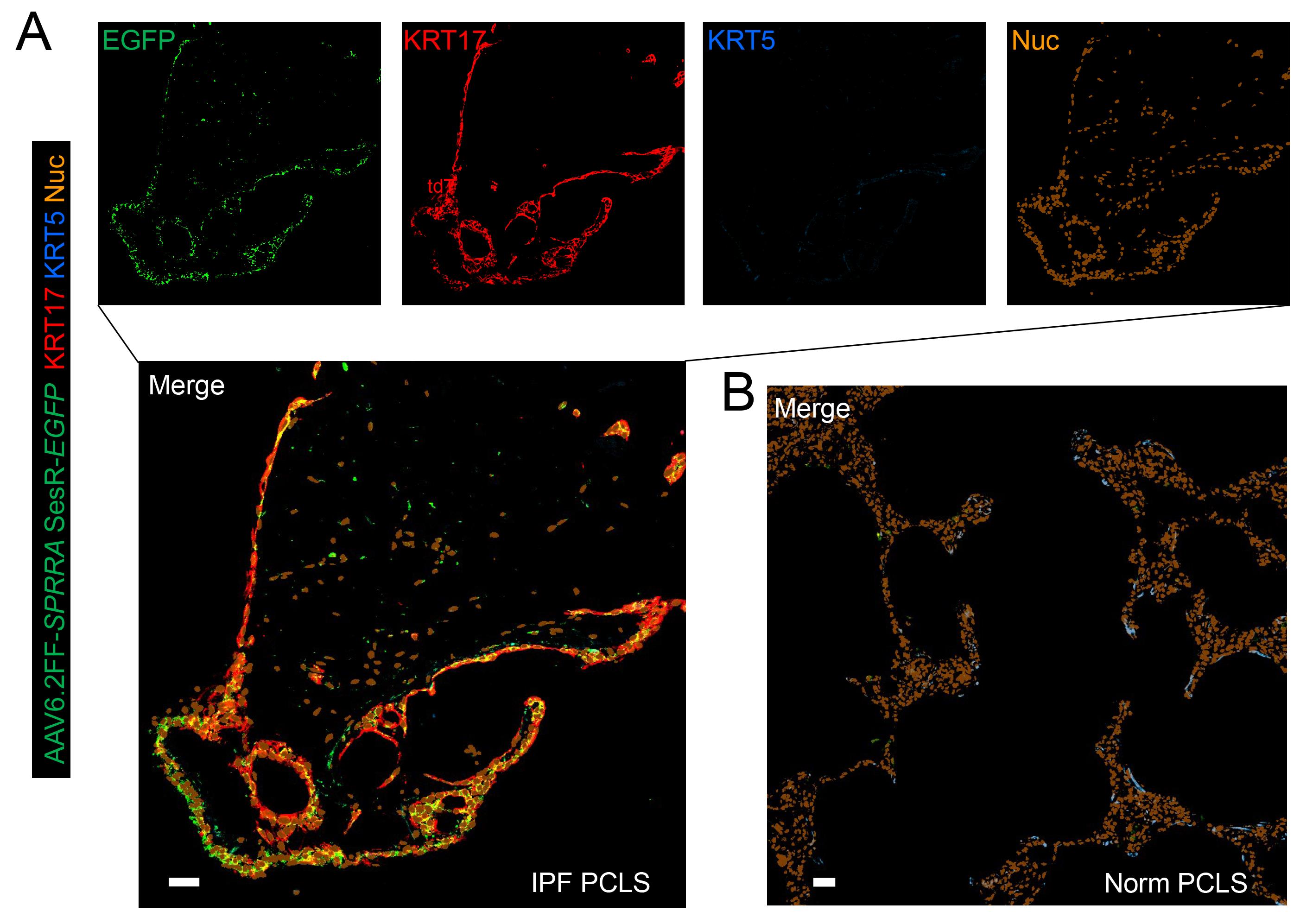
Figure 2. SPRR1A-sesRNA-guided EGFP expression in human idiopathic pulmonary fibrosis precision-cut lung slices. Representative fluorescence images show enrichment of EGFP in KRT17-high/KRT5-low aberrant basaloid cells in human IPF PCLS (A). SPRR1A-targeted EGFP expression is largely absent in normal human control PCLS (B). Scale bars, 50 μm. AAV, adeno-associated virus; EGFP, enhanced green fluorescent protein; IPF, idiopathic pulmonary fibrosis; KRT, keratin; Norm, normal; Nuc, nucleus/nuclei; PCLS, precision-cut lung slice; sesRNA/SesR, sequence-encoded sensing RNA; SPRR1A/SPRRA, small proline-rich protein 1A.
Data analysis
1. In vitro sesRNA screening. For each candidate, analyze at least three randomly selected, non-overlapping fields per condition using identical imaging settings. Identify cells positive for both mCherry and BFP as successfully co-transfected cells, then count EGFP-positive cells within this double-positive population. Calculate the EGFP-positive ratio as: EGFP-positive cells ÷ mCherry/BFP double-positive cells × 100%. Compare the ratio obtained with the target-RNA expression plasmid against the matched mCherry-only control. Promising sensors should show low control activity and a reproducible increase in the target RNA condition. Because target abundance and sesRNA efficiency vary, no universal absolute cutoff is imposed; instead, candidates are ranked by target-dependent enrichment, and promising candidates should be confirmed in at least two independent transfections before AAV production.
2. AAV quality control. Proceed with AAV harvest only when producer cultures show ≥80% BFP-positive cells at 24 h. Before animal or PCLS experiments, confirm the qPCR titer and calculate the volume needed to deliver 1 × 10¹¹ vg per mouse or 1 × 10¹⁰ vg per PCLS. Do not use preparations with visible particulate material after clarification or with titers too low to deliver the validated dose within the specified administration volume.
3. Mouse-lung fluorescence analysis. Use the mouse as the biological replicate and microscopic fields as technical sampling units. For quantitative experiments, analyze at least three non-overlapping fields per lung and include at least three biological replicates per group when feasible. Reporter specificity can be expressed as the percentage of EGFP-positive cells that co-express the expected lineage label or cell-state marker. Labeling efficiency can be expressed as the percentage of marker-positive cells that are EGFP-positive. Report transduction coverage separately from reporter specificity when patchy AAV delivery is observed.
4. Human PCLS fluorescence analysis. Treat the donor as the biological replicate and individual slices/fields as technical replicates. When tissue availability permits, analyze at least two slices per donor per condition and at least three non-overlapping fields per slice. Reporter specificity can be calculated as EGFP-positive/KRT17-high/KRT5-low cells divided by total EGFP-positive cells × 100%; labeling efficiency can be calculated as EGFP-positive/KRT17-high/KRT5-low cells divided by total KRT17-high/KRT5-low cells × 100%. Analyze donor-matched control and sensor-treated slices using identical acquisition and analysis settings. Exclude slices that failed the predefined morphology/viability criteria before treatment.
5. Statistics and replicate handling. Summarize field-level measurements within each biological replicate before statistical testing to avoid pseudoreplication. For two independent groups, use a two-tailed Student's t test when assumptions are met; use a paired test for donor-matched PCLS comparisons. For more than two groups, use one-way ANOVA with an appropriate multiple-comparison procedure. The associated study used GraphPad Prism 8 and considered p < 0.05 statistically significant [1]. Report the number of biological replicates and technical fields/slices in each analysis.
Validation of protocol
The core components of this protocol were used and validated in the following research article:
• Peng et al. [1]. Transcriptomic signature-guided depletion of intermediate alveolar epithelial cells ameliorates pulmonary fibrosis in mice. Nature Communications.
Target-dependent in vitro and in vivo labeling is shown in Figures 1B–C and 3A–C; transcriptional validation of Sprr1a-guided EGFP-positive cells is shown in Figure 4 and Supplementary Figure 5.
In that study, Sftpc- and Pdpn-targeting sensors activated EGFP preferentially in cells expressing the matching target cDNA and labeled morphologically and in genetically defined AT2 or AT1 cells in the mouse lung. The Sprr1a-targeting sensor labeled Krt8-positive transitional epithelial cells after bleomycin injury, and single-cell RNA sequencing demonstrated convergence between EGFP-positive cells and the Krt8-positive alveolar differentiation intermediate state.
To directly validate the human component of this protocol, PCLS from donors with IPF were treated with AAV6.2FF carrying the SPRR1A-targeting sesRNA-EGFP reporter, and PCLS from normal donor lungs were included to assess disease-associated reporter activation. After at least seven days to permit reporter expression, the slices were analyzed by immunofluorescence for EGFP, KRT17, and KRT5.
General notes and troubleshooting
General notes
1. Screen multiple sesRNA candidates. Not all portal-generated sensors produce efficient or specific output, and candidate performance should be established in vitro before AAV production.
2. Sensor output depends on target RNA abundance, target accessibility, endogenous ADAR activity, and the absence of cryptic translation-initiation or termination signals [6].
3. AAV transduction can be spatially incomplete. Interpret marker-positive/EGFP-negative regions together with an independent measure of viral delivery or BFP expression.
4. Use single-use AAV aliquots and minimize freeze/thaw cycles.
5. The AAV6.2FF production workflow and intratracheal delivery require appropriate biosafety approval and trained personnel.
6. The timing, dose, medium, and analytical workflow for human PCLS should be optimized independently from the mouse protocol. Begin with donor-matched control slices, preserve the validated slice thickness when possible, and change only one experimental variable at a time during optimization.
7. When adapting the protocol to another tissue, species, or disease model, first confirm that the target RNA is sufficiently enriched in the intended cell population and redesign the sesRNA against the species-specific target sequence. Screen multiple candidates in vitro before viral production.
8. AAV capsid tropism and delivery efficiency can differ substantially among tissues and species. Validate transduction independently of sesRNA activity using BFP or another constitutive vector marker, and optimize capsid, dose, route, and exposure time before interpreting EGFP-negative cells as target-negative.
9. For new experimental models, establish target-positive and target-negative controls, define imaging settings and acceptance/exclusion criteria before data collection, and keep acquisition parameters unchanged across comparison groups.
10. If the effector is changed from EGFP to another reporter or functional protein, confirm that the full sensor-effector open reading frame remains intact, that AAV packaging capacity is not exceeded, and that the new effector does not alter cell viability independently of target recognition.
Troubleshooting
| Problem | Possible cause | Solution |
|---|---|---|
| Low BFP expression after HEK293T transfection | Cells are not actively proliferating; confluence is outside 70%–80%; PEI or DNA quality is poor. | Use rapidly dividing cells passaged for at least two consecutive days, confirm DNA purity, and use freshly prepared transfection complexes. |
| EGFP signal in the target-negative control | Cryptic downstream ATG, unintended open reading frame, sensor-independent readthrough, or off-target hybridization. | Reinspect the sensor sequence, remove downstream ATGs and unintended stop/readthrough contexts, and screen a redesigned candidate. |
| Low AAV yield or low reporter signal | Poor transfection efficiency, column obstruction, loss during concentration, low viral genome dose, or inefficient delivery. | Require at least 80% BFP-positive producer cells, clarify lysate completely, monitor column flow, verify titer, and optimize the validated delivery dose. |
| Patchy in vivo EGFP signal | Incomplete AAV distribution or variable airway delivery. | Standardize the instillation technique, report transduction coverage separately from specificity, and include internal untransduced regions as delivery controls. |
| Weak or variable PCLS EGFP signal | Reduced slice viability, insufficient AAV penetration, or suboptimal exposure time. | Apply predefined viability criteria and optimize slice thickness, dose, and incubation duration using donor-matched controls. |
Acknowledgments
This work was supported by National Institutes of Health grants HL139584, HL156973, and HL174994 to Y.Z. The protocol was developed from the CellREADR method described by Qian et al. [6] and was applied and validated in Peng et al. [1].
Author contributions
Conceptualization, Z.Z., C.-s.J., and Y.Z.; Methodology and investigation, Z.Z., C.-s.J., D.S., and C.H.; Data interpretation, Z.Z., C.-s.J., C.H., I.O.R., J.A.L., V.J.T., and Y.Z.; Writing—original draft, Z.Z. and Y.Z.; Writing—review and editing, all authors.
Competing interests
The authors declare no conflicts of interest.
Ethical considerations
Animal studies were conducted in accordance with National Institutes of Health guidelines for the care and use of laboratory animals and were approved by the Institutional Animal Care and Use Committee of Tulane University (protocol 2108) and the University of Alabama at Birmingham (protocol IACUC-22215). De-identified human lung tissues used in the associated research article were reviewed by the University of Alabama at Birmingham Institutional Review Board (IRB-300000356), which determined that the project was not human-subjects research. Human lung tissue was stored at Baylor College of Medicine Chronic Lung Disease Tissue Repository under protocol H-46823. AAV transfection of PCLS was conducted under approved protocol H-55949 by the Baylor College of Medicine Institutional Review Board.
References
- Peng, F., Jiang, C. s., Zheng, Z., Aliyari, S., Shan, D., Sabharwal, A., Yin, Q., Saito, S., He, C., Rosas, I. O., et al. (2026). Transcriptomic signature-guided depletion of intermediate alveolar epithelial cells ameliorates pulmonary fibrosis in mice. Nat Commun. 17(1): 1636. https://doi.org/10.1038/s41467-026-68354-y
- Strunz, M., Simon, L. M., Ansari, M., Kathiriya, J. J., Angelidis, I., Mayr, C. H., Tsidiridis, G., Lange, M., Mattner, L. F., Yee, M., et al. (2020). Alveolar regeneration through a Krt8+ transitional stem cell state that persists in human lung fibrosis. Nat Commun. 11(1): 3559. https://doi.org/10.1038/s41467-020-17358-3
- Choi, J., Park, J. E., Tsagkogeorga, G., Yanagita, M., Koo, B. K., Han, N. and Lee, J. H. (2020). Inflammatory Signals Induce AT2 Cell-Derived Damage-Associated Transient Progenitors that Mediate Alveolar Regeneration. Cell Stem Cell. 27(3): 366–382.e7. https://doi.org/10.1016/j.stem.2020.06.020
- Kobayashi, Y., Tata, A., Konkimalla, A., Katsura, H., Lee, R. F., Ou, J., Banovich, N. E., Kropski, J. A. and Tata, P. R. (2020). Persistence of a regeneration-associated, transitional alveolar epithelial cell state in pulmonary fibrosis. Nat Cell Biol. 22(8): 934–946. https://doi.org/10.1038/s41556-020-0542-8
- Kretzschmar, K. and Watt, F. M. (2012). Lineage Tracing. Cell. 148: 33–45. https://doi.org/10.1016/j.cell.2012.01.002
- Qian, Y., Li, J., Zhao, S., Matthews, E. A., Adoff, M., Zhong, W., An, X., Yeo, M., Park, C., Yang, X., et al. (2022). Programmable RNA sensing for cell monitoring and manipulation. Nature. 610(7933): 713–721. https://doi.org/10.1038/s41586-022-05280-1
- van Lieshout, L. P., Domm, J. M., Rindler, T. N., Frost, K. L., Sorensen, D. L., Medina, S. J., Booth, S. A., Bridges, J. P. and Wootton, S. K. (2018). A Novel Triple-Mutant AAV6 Capsid Induces Rapid and Potent Transgene Expression in the Muscle and Respiratory Tract of Mice. Mol Ther Methods Clin Dev. 9: 323–329. https://doi.org/10.1016/j.omtm.2018.04.005
- Strobel, B., Duechs, M. J., Schmid, R., Stierstorfer, B. E., Bucher, H., Quast, K., Stiller, D., Hildebrandt, T., Mennerich, D., Gantner, F., et al. (2015). Modeling Pulmonary Disease Pathways Using Recombinant Adeno-Associated Virus 6.2. Am J Respir Cell Mol Biol. 53(3): 291–302. https://doi.org/10.1165/rcmb.2014-0338ma
- Limberis, M. P., Vandenberghe, L. H., Zhang, L., Pickles, R. J. and Wilson, J. M. (2009). Transduction Efficiencies of Novel AAV Vectors in Mouse Airway Epithelium In Vivo and Human Ciliated Airway Epithelium In Vitro. Mol Ther. 17(2): 294–301. https://doi.org/10.1038/mt.2008.261
- Lehmann, M., Krishnan, R., Sucre, J., Kulkarni, H. S., Pineda, R. H., Anderson, C., Banovich, N. E., Behrsing, H. P., Dean, C. H., Haak, A., et al. (2025). Precision-Cut Lung Slices: Emerging Tools for Preclinical and Translational Lung Research: An Official American Thoracic Society Workshop Report. Am J Respir Cell Mol Biol. 72(1): 16–31. https://doi.org/10.1165/rcmb.2024-0479st
Article Information
Publication history
Received: Aug 10, 2026
Accepted: Oct 2, 2026
Available online: Oct 21, 2026
Published: Nov 5, 2026
Copyright
© 2026 The Author(s); This is an open access article under the CC BY-NC license (https://creativecommons.org/licenses/by-nc/4.0/).
How to cite
Zheng, Z., Jiang, C., Shan, D., He, C., Rosas, I. O., Lasky, J. A., Thannickal, V. J. and Zhou, Y. (2026). Transcriptomic Signature-Guided EGFP Labeling of Alveolar Epithelial Cells via RNA Sensing in Mouse Lungs In Vivo and Human Idiopathic Pulmonary Fibrosis Lung Slices Ex Vivo. Bio-protocol 16(21): e5863. DOI: 10.21769/BioProtoc.5863.

 Download Citation in RIS Format
Do you have any questions about this protocol?
Post your question to gather feedback from the community. We will also invite the authors of this article to respond.
 Post a Question


