


- Protocols

- Articles and Issues
- For Authors
- About
- Become a Reviewer


Human iPSC Line SCTi003-A Differentiation Into Kidney Organoids

Published: Vol 16, Iss 21, Nov 5, 2026 DOI: 10.21769/BioProtoc.5860 Views: 43
Reviewed by: Jessica DavisNeha SaxenaAnonymous reviewer(s)
 PDF
PDF  Ask a question

Protocol Collections
Comprehensive collections of detailed, peer-reviewed protocols focusing on specific topics




Abstract
Kidney organoids derived from human induced pluripotent stem cells (hiPSCs) have emerged as valuable tools for studying human kidney development, disease mechanisms, and therapeutic responses. However, widespread application of these models has been limited by variability in differentiation efficiency, organoid composition, reproducibility, and scalability across existing protocols. Robust and standardised methods are therefore needed to improve experimental consistency and facilitate wider adoption of kidney organoid systems in basic and translational research. Here, we describe an optimised and reproducible protocol for differentiating a commercially available hiPSC SCTi003-A line into kidney organoids (KiO). The protocol builds upon earlier kidney differentiation frameworks and involves directed induction of the posterior primitive streak, intermediate mesoderm specification, and subsequent three-dimensional organoid assembly. Through systematic optimisation of differentiation media, CHIR99021 concentration and timing, growth factor supplementation, and culture format, we established a standardised workflow that yields reproducible nephron and collecting duct structures. The protocol is compatible with air–liquid interface (ALI) culture and scalable suspension-based “mini-organoid” (mKiO) and monolayer culture formats that support high-content and high-throughput applications. This validated protocol provides a reliable and accessible platform for generating human KiO, addressing key limitations of reproducibility, scalability, and assay compatibility in existing approaches, and supporting future studies of kidney development, inherited kidney diseases, and preclinical drug testing.
Key features
• An optimised kidney organoid (KiO) differentiation workflow for the commercially available normal control hiPSC line (SCTi003-A, STEMCELL Technologies), minimising line-specific re-optimisation.
• Defined control of anterior (AIM) and posterior (PIM) intermediate mesoderm specification through CHIR99021 duration and pulse timing.
• Scalable for air–liquid interface (ALI) culture, suspension-based “mini-organoid” (mKiO), and monolayer culture formats.
• Compatibility with cost-effective, high-throughput drug testing using 96-well plate formats, high content imaging, and automated image segregation.
Keywords: Kidney organoids SCTi003-A hiPSC line
Induced pluripotent stem cells
Disease models
SCTi003-A hiPSC line
Induced pluripotent stem cells
Disease models
Background
Kidney organoids (KiO) derived from induced pluripotent stem cells (hiPSCs) have emerged as powerful in vitro systems for modelling renal development, disease mechanisms, and therapeutic responses. These three-dimensional structures recapitulate key aspects of human nephrogenesis, including segmentation of nephrons into podocyte, proximal tubule, distal tubule, and collecting duct-like domains, providing a physiologically relevant alternative to conventional two-dimensional culture systems [1,2]. KiO are particularly valuable for modelling early onset genetic forms of renal disorders that are poorly recapitulated in animal models due to species-specific differences in kidney development, signalling pathways, and drug responses.
Several protocols for differentiating hiPSCs into KiO have been established, most notably those described by Takasato et al. [2,3], which use the activation of Wnt signalling by the agonist CHIR99021 to induce posterior primitive streak and intermediate mesoderm, followed by nephron patterning and three-dimensional self-organisation [1–5]. Anterior–posterior patterning of the intermediate mesoderm is regulated by the length of CHIR99021 treatment. Exposure for up to two days biases differentiation toward anterior intermediate mesoderm (AIM), while treatment for five days or longer promotes posterior intermediate mesoderm (PIM) identity. Treatment durations of 3–4 days produce a mixed anterior/posterior intermediate mesoderm phenotype [2,6]. A transient pulse of CHIR99021 is subsequently used to stimulate Wnt signalling and drive nephrogenesis, promoting the mesenchymal-to-epithelial transition and enhancing the formation of nephron structures within the developing organoids [7]. More recent refinements by Uchimura et al. [8] include the parallel induction of anterior (AIM) and posterior (PIM) intermediate mesoderm populations to better recapitulate ureteric epithelium and nephron lineages [8]. While these methods have advanced the field, challenges remain in achieving reproducible differentiation across different hiPSC lines, minimising batch-to-batch variability, and adapting protocols for scalable and cost-effective applications such as drug screening.
Here, we present an optimised and standardised protocol for differentiating a commercially available normal control hiPSC line into KiO. We describe the key parameters that influence differentiation efficiency and reproducibility, including basal medium composition, CHIR99021 concentration and exposure duration, growth factor supplementation, ALI culture conditions, cell seeding density, and total differentiation time. We identify advanced RPMI as a robust and cost-effective alternative to the previously described APEL2 medium for differentiation and define CHIR99021 timing parameters that reliably support both AIM and PIM specification.
This protocol supports KiO generation in three complementary formats: ALI cultures that facilitate structural maturation and tissue organisation, suspension-based “mini KiO” that are compatible with 96-well plate formats for high-throughput applications [9], and monolayer culture, which allows spontaneous 3D differentiation and could be applied in fluidic devices. The protocol is suitable for nephrotoxicity assessment, genetic disease modelling, and developmental studies. Its modular design allows adaptation to additional hiPSC lines, whilst the use of widely accessible reagents and defined parameters enables standardisation and reproducibility across laboratories.
Materials and reagents
Biological materials
1. Healthy control human iPSC line, female, SCTi003-A (STEMCELL Technologies, catalog number: 200-0511); this line was derived from peripheral blood mononuclear cells (PBMCs) of a 48-year-old Caucasian healthy female (https://www.stemcell.com/products/healthy-control-human-ipsc-line-female-scti003-a.html)
Reagents
1. Advanced RPMI 1640 Medium (Gibco, catalog number: 12633020)
2. Antibiotic-antimycotic (100×) (Gibco, catalog number: 15240-096)
3. Bovine albumin fraction V (7.5% solution) (Gibco, catalog number: 15260-037)
4. CHIR99021 ≥98% (HPLC-grade purity) (6 mM) (Sigma-Aldrich, catalog number: SML1046)
5. CryoStor® CS10 (STEMCELL Technologies, catalog number: 07930)
6. DMEM/F-12, HEPES (Gibco, catalog number: 31330-038)
7. Dimethyl sulfoxide (DMSO) sterile-filtered, ≥99.7% (Sigma-Aldrich, catalog number: D2650)
8. Dulbecco’s phosphate-buffered saline (DPBS) (Sigma-Aldrich, catalog number: D8537)
9. Ethanol (Supelco, catalog number: 1.00983.2511)
10. GlutaMAXTM supplement (100×) (Gibco, catalog number: 35050061)
11. Heparin solution 0.2% (2 mg/mL) (STEMCELL Technologies, catalog number: 07980)
12. Human FGF-9 (fibroblast growth factor 9) recombinant protein (200 μg/mL) (PeproTech, catalog number: 100-23)
13. Lipidure-CM5206 powder (Amsbio, catalog number: AMS.52000032GB)
14. Matrigel hESC (human embryonic stem cells)-qualified matrix (Corning, catalog number: 354277)
15. mTeSRTM Plus (STEMCELL Technologies, catalog number: 100-0276)
16. Recombinant human/murine/rat activin A (10 μg/mL) (PeproTech, catalog number: 120-14)
17. ROCK inhibitor (Y-27632) (20 mM) (CHEMDEA, catalog number: CD0141)
18. StemProTM AccutaseTM cell dissociation reagent (Gibco, catalog number: A1110501)
19. Trypan Blue 0.4% (NanoEntek, catalog number: EBT-001)
Solutions
1. 6 mM CHIR99021 (see Recipes)
2. 20 mM Y-27632 (see Recipes)
3. 10 μg/mL activin A (see Recipes)
4. 200 μg/mL FGF9 (see Recipes)
5. Advanced RPMI 1640 Medium (ARPMI) with GlutaMAX (ARPMIG) (see Recipes)
6. ARPMIG with 6 μM CHIR99021 (see Recipes)
7. ARPMIG with 200 ng/mL FGF9, 1 μg/mL heparin, 10 ng/mL activin A (see Recipes)
8. ARPMIG with antibiotic/antimycotic (ARPMIG-ABAM) (see Recipes)
9. ARPMIG-ABAM with 5 μM CHIR99021 (see Recipes)
10. ARPMIG-ABAM with 200 ng/mL FGF9, 1 μg/mL heparin (see Recipes)
11. 0.5% w/v Lipidure (see Recipes)
Recipes
1. 6 mM CHIR99021
| Reagent | Final concentration | Quantity or volume |
|---|---|---|
| CHIR99021 | 6 mM | 5 mg |
| DMSO | n/a | 1.791 mL |
Aliquot and store at -20 °C. Stock solutions are stable for up to 6 months at -20 °C. Avoid repeated freeze/thaw cycles and protect from light.
2. 20 mM Y-27632
| Reagent | Final concentration | Quantity or volume |
|---|---|---|
| Y-27632 | 20 mM | 5 mg |
| dH2O | n/a | 740 μL |
Aliquot and store at -20 °C. Avoid repeated freeze/thaw cycles and protect from light.
3. 10 μg/mL activin A
| Reagent | Final concentration | Quantity or volume |
|---|---|---|
| Activin A | 10 μg/mL | 10 μg |
| dH2O | n/a | 10 μL |
DPBS BSA 7.5% | n/a 0.1% | 976.8 μL 13.2 μL |
Working aliquots stored with a carrier protein are stable for at least 7 months at -20 to -80 °C. Avoid repeated freeze/thaw cycles. It can be stored at 4 °C for 2–7 days, or up to 1 month under sterile conditions after reconstitution.
4. 200 μg/mL FGF9
| Reagent | Final concentration | Quantity or volume |
|---|---|---|
| FGF9 | 200 μg/mL | 250 μg |
| dH2O | n/a | 250 μL |
| DPBS | n/a | 986.7 μL |
| BSA 7.5% | 0.1% | 13.3 μL |
Store for up to 1 week at 4 °C or up to 12 months at -20 to -80 °C.
5. ARPMIG
| Reagent | Final concentration | Quantity or volume |
|---|---|---|
| ARPMI | n/a | 495 mL |
| GlutaMAX 100× | 1× | 5 mL |
Store at 4 °C.
6. ARPMIG with 6 μM CHIR99021 (medium for days 0–4)
| Reagent | Final concentration | Quantity or volume |
|---|---|---|
| ARPMIG | n/a | 2 mL |
| 6 mM CHIR99021 | 6 μM | 2 μL |
Prepare fresh on the day.
7. ARPMIG with 200 ng/mL FGF9, 1 μg/mL heparin, 10 ng/mL activin A (medium for days 4–7)
| Reagent | Final concentration | Quantity or volume |
|---|---|---|
| ARPMIG | n/a | 4 mL |
| 200 μg/mL FGF9 | 200 ng/mL | 4 μL |
| 2 mg/mL heparin | 1 μg/mL | 2 μL |
| 10 μg/mL activin A | 10 ng/mL | 4 μL |
Prepare fresh on the day.
8. ARPMIG-ABAM
| Reagent | Final concentration | Quantity or volume |
|---|---|---|
| ARPMI | n/a | 490 mL |
| GlutaMAX 100× | 1× | 5 mL |
| Antibiotic/Antimycotic 100× | 1× | 5 mL |
Store at 4°C.
9. ARPMIG-ABAM with 5 μM CHIR99021 (pulse medium day 7)
| Reagent | Final concentration | Quantity or volume |
|---|---|---|
| ARPMIG-ABAM | n/a | 1.2 mL |
| 6 mM CHIR99021 | 5 μM | 1 μL |
Prepare fresh on the day.
10. ARPMIG-ABAM with 200 ng/mL FGF9, 1 μg/mL heparin (medium for days 7–12)
| Reagent | Final concentration | Quantity or volume |
|---|---|---|
| ARPMIG-ABAM | n/a | 1.2 mL |
| 200 μg/mL FGF9 | 200 ng/mL | 1.2 μL |
| 2 mg/mL heparin | 1 μg/mL | 0.6 μL |
Prepare fresh on the day.
11. 0.5% w/v Lipidure
| Reagent | Final concentration | Quantity or volume |
|---|---|---|
| Lipidure | 5 mg/mL | 1 g |
| Ethanol | 100% | 200 mL |
Store at room temperature (RT) in the dark for up to 3 months.
Laboratory supplies
1. 15 mL polypropylene centrifuge tubes (Corning, catalog number: BC030)
2. 50 mL polypropylene centrifuge tubes (Corning, catalog number: BC034)
3. 6-well plate (TPP, catalog number: 92406T)
4. 96-well plate (TPP, catalog number: 92097T)
5. Corning® Transwell® 6-well plates (Corning, catalog number: CLS3450)
6. C-slide disposable cell counting chamber slide (NanoEntek, catalog number: CS-050)
7. 1.5 mL Eppendorf tubes (Sarstedt, catalog number: 72.692)
8. Pipetboy (Integra, catalog number: 155017)
9. Pipette tips RT UNV (METTLER TOLEDO, catalog numbers: 30389172, 30389186, 30389165)
10. Thermo ScientificTM ARTTM wide bore filtered pipette tips P1000 (Thermo Fisher Scientific, catalog number: 2079G)
11. FinnpipetteTM F1 variable volume pipettes P2, P10, P200, P1000 (Thermo Fisher Scientific, catalog numbers: 4641010N, 4641030N, 4641080N, 4641100N)
12. FinnpipetteTM F1 multichannel pipette (Thermo Fisher Scientific, catalog number: 4661030N)
13. ThincertTM cell culture insert for 6-well plates, translucent (Greiner Bio-One, catalog number: 657640)
14. ThincertTM cell culture insert for 6-well plates, transparent (Greiner Bio-One, catalog number: 657641)
15. FisherbrandTM disposable pipette basins (Thermo Fisher Scientific, catalog number: 11988485)
Equipment
1. CountessTM3 (Thermo Fisher Scientific, catalog number: AMQAX2000)
2. EVOS FL Colour Imaging System (Life Technologies, catalog number: AMF4300)
3. Confocal laser scanning microscope or equivalent (Nikon A1R or Zeiss CellDiscoverer 7)
4. Humidified CO2 incubator (Sanyo, catalog number: MCO-20AIC)
5. CellGard Class II, Type A2 vertical laminar flow biological safety cabinet (NuAire, catalog number: NU-480-600E)
6. Sigma 1-14 Compact Microcentrifuge (Sigma, catalog number: 10014)
7. MultifugeTM X3 (ventilated) centrifuge (Thermo Fisher Scientific, catalog number: 75004500)
Software and datasets
1. Fiji (ImageJ)
2. Image-Pro (11.1.2, 2025) with the AI Deep Learning module
Procedure
Note: This protocol represents the final optimised version following systematic evaluation of basal media, growth factor combinations, CHIR99021 concentration and timing, and culture configuration. It is specifically validated for the SCTi003-A hiPSC line (Figure 1). SCTi003-A should be thawed, cultured, and frozen following the manufacturer’s instructions.
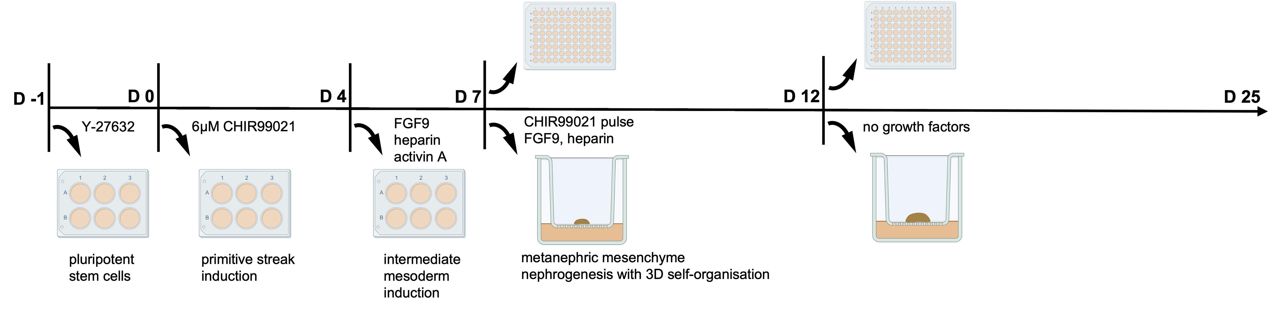
Figure 1. Schematic overview of SCTi003-A differentiation into KiO following a protocol adapted from Takasato et al. [2,3]. Created in https://BioRender.com.
A. Preparation and seeding of SCTi003-A cells (day -1)
1. Prepare reagents and materials. Prepare the following in advance:
a. DPBS.
b. Accutase equilibrated to room temperature (20–22 °C).
c. 15 mL conical tubes.
d. mTeSRTM Plus spent medium equilibrated to room temperature (20–22 °C).
Note: Collect spent mTeSRTM Plus medium from actively growing hiPSCs, filter-sterilise (0.2 μm), and store at 4 °C for up to 1 week. Spent medium improves post-dissociation cell recovery compared to fresh medium. It should only be used for spinning cells down.
e. Fresh mTeSRTM Plus medium equilibrated to room temperature (20–22 °C).
f. ROCK inhibitor (Y-27632) (see Recipe 2).
g. C-slides and 1.5 mL tubes containing 10 μL of Trypan Blue.
h. Matrigel-coated 6-well plates equilibrated to room temperature (20–22 °C) (if prepared in advance) or Matrigel aliquot.
2. Coat plates with Matrigel
a. Dilute Matrigel according to the manufacturer’s lot-specific dilution factor. Prepare aliquots sufficient for 25 mL of total media volume and store at -20 °C.
Critical: Perform all Matrigel handling on ice using pre-chilled tips and serological pipettes.
b. Thaw one Matrigel aliquot on ice (1 h) or overnight at 4 °C at the back of the fridge.
c. Prepare 25 mL of ice-cold DMEM/F-12.
d. Add Matrigel to DMEM/F-12 and mix gently by swirling; avoid bubble formation.
e. Add 1 mL of diluted Matrigel per well of a 6-well plate. Incubate plates at room temperature for ≥1 h, followed by 30 min at 37 °C before cell seeding.
Notes:
1. Diluted Matrigel can be stored at 4 °C for up to 1 week if kept undisturbed. Prepared plates, if not used immediately, can be sealed with parafilm and stored at 4 °C for up to 1 week.
2. Although growth factor–reduced Matrigel was initially effective, batch-to-batch variability required repeated validation. To improve reproducibility, we switched to hESC-qualified Matrigel, which consistently supported hiPSC maintenance and differentiation without the need for batch pre-screening.
3. Dissociate SCTi003-A cells
Note: SCTi003-A cells require approximately eight days to recover after thawing before differentiation. Once expanded, cryopreserve cells in CryoStor® CS10 (following the manufacturer’s instructions) and thaw fresh vials for each experiment.
a. Aspirate culture medium and gently wash cells once with 4 mL of DPBS.
b. Add 1 mL of Accutase per well.
Note: Accutase is recommended for cells intended for differentiation, as it improves viability and differentiation consistency.
c. Incubate at 37 °C for 5–7 min.
d. Gently tap the plate and use P1000 to pipette up and down to obtain a single-cell suspension.
e. Transfer cells into 3 mL of spent mTeSRTM Plus containing 10 μM ROCK inhibitor in a 15 mL tube.
f. Centrifuge at 180× g for 5 min.
g. Remove supernatant, gently tap the pellet, and resuspend cells in 2 mL of fresh mTeSRTM Plus with 10 μM ROCK inhibitor.
h. Mix 10 μL of cell suspension with 10 μL of Trypan Blue and add to C-slide. Count cells using a CountessTM.
i. Seed 4 × 10⁵ viable cells per well of a 6-well plate in 2 mL of mTeSRTM Plus with 10 μM ROCK inhibitor.
j. Distribute cells evenly by gentle rocking (up and down, side-to-side) and incubate overnight at 37 °C, 5% CO2.
B. Induction of primitive streak and intermediate mesoderm
1. Day 0: Initiation of differentiation
a. Confirm that cells are 40%–60% confluent the following day after plating by light microscopy.
Critical: Starting confluency strongly determines differentiation outcome. Low confluency causes early detachment; excessive confluency impairs growth factor diffusion.
b. Replace medium with 2 mL of ARPMIG (see Recipe 5) containing 6 μM CHIR99021 (see Recipe 6).
Critical: Warm ARPMIG to 37 °C throughout the protocol. Prewarming media to 37 °C prevents transient cold shock and preserves the precise kinetics of temperature-dependent signalling pathways (especially Wnt), which are critical for synchronised differentiation and proper kidney organoid patterning.
2. Day 1: Replace medium with ARPMIG + 6 μM CHIR99021 (2 mL/well).
3. Day 2: Replace medium with ARPMIG + 6 μM CHIR99021 (4 mL/well).
4. Day 3: No media change.
5. Day 4: Replace medium with ARPMIG (4 mL/well) supplemented with (see Recipe 7): 200 ng/mL FGF9 (this concentration of FGF9 is used throughout the protocol), 1 μg/mL heparin, and 10 ng/mL activin A.
Critical: Cells exhibit reduced adherence at this stage. Perform medium changes slowly by trickling medium along the well wall to minimise sheet detachment.
6. Day 5: No media change.
7. Day 6: Repeat medium change as on day 4.
C. Organoid generation and CHIR99021 pulse (day 7)
1. Prepare the following:
a. Accutase equilibrated to room temperature (20–22 °C).
b. ARPMIG equilibrated to 37 °C.
c. ARPMIG-ABAM (see Recipe 8).
d. Pulse medium: ARPMIG-ABAM + 5 μM CHIR99021 (see Recipe 9).
e. Growth medium: ARPMIG-ABAM + 200 ng/mL FGF9 + 1 μg/mL heparin (see Recipe 10).
f. Greiner translucent ThinCertTM inserts (6-well format).
g. C-slides and 1.5 mL tubes containing 10 μL of Trypan Blue.
h. Lipidure-coated 96-well round-bottom plates (prepared ≥1 day in advance). To coat round-bottom 96-well plates with Lipidure in a sterile fume hood, add 50 μL of 0.5% w/v Lipidure (see Recipe 11) to each well and allow complete evaporation of the ethanol solvent (overnight). Plates can then be sealed with parafilm and stored for up to 3 years in the dark [10].
Note: We consistently observed degradation of organoids in the outer wells of Corning ultra-low attachment 96-well plates, with this effect progressively extending to the adjacent inner wells. This phenomenon was reproducible across two different lot numbers. To address this issue, we switched to TPP round-bottom 96-well plates that were coated in-house with Lipidure.
2. Dissociation and cell counting
a. Aspirate medium and gently wash cells with 1 mL of DPBS; then, remove.
b. Add 1 mL of Accutase and incubate for 5 min at 37 °C.
c. Triturate gently to obtain a single-cell suspension.
d. Add to 3 mL of ARPMIG, centrifuge at 180× g for 5 min, and resuspend in 2 mL of ARPMIG.
e. Mix 10 μL of cell suspension with 10 μL of Trypan Blue and add to C-slide. Count cells using a CountessTM.
3. Organoid culture formats
Note: It is essential to choose at this point between the type of culture (ALI, suspension mini KiO, or monolayer).
a. Air–liquid interface (ALI)
i. Aliquot 2 × 10⁵ cells into 1.5 mL tubes.
Note: We assessed 5 × 10⁵ cells on different brands of plasticware for ALI culture. We observed minor differences in organoid size or differentiation efficiency between different starting numbers of cells (2 vs. 5 × 10⁵ cells), and we now use 2 × 10⁵ cells as a standard procedure (Figure S2).
ii. Pellet cells by centrifugation at 400× g for 2 min, three times, rotating tubes by 180° between spins.
iii. Add 1.2 mL of pulse medium to the outer well of ThinCertTM inserts.
iv. Transfer pellets carefully onto membranes with minimal medium transfer (see Figure S3).
v. Incubate for 1 h at 37 °C.
vi. Change medium to growth medium and incubate at 37 °C.
Critical: Organoids must grow at the air–medium interface. The medium must remain below the membrane (although still touching it, as shown in Figure 1) and is drawn up through the pores by capillary forces, continuously supplying medium to the organoid.
Note: Up to 10 pellets per membrane can be fitted into 6-well plate ALI plasticware when using 2 × 10⁵ cells, or 6 pellets when using 5 × 10⁵ cells. Keep the medium level in the culture below the polyester membrane and prevent overflow.
b. Mini-organoids (mini KiO) in suspension culture in 96-well plates
i. Seed 10,000 cells per well in pulse medium: ARPMIG-ABAM + 5 μM CHIR99021 (100 μL) in a Lipidure-coated 96-well round-bottom plate.
Note: If the whole 96-well plate is being set up, count the number of wells × 10,000 cells (plus 10% spare), aliquot into pipette basins containing pulse medium, and dispense with a multichannel pipette (100 μL/well).
ii. Centrifuge plate at 450× g for 30 s.
iii. Incubate for 1 h, remove 70 μL, and replace with 70 μL of growth medium.
Note: Aspirate 70 μL of media with CHIR99021 and leave 30 μL in the well. This is to avoid aspiration of cells. Calculate the growth factor’s concentrations for a total volume of 100 μL, but only add 70 μL per well.
iv. Centrifuge again at 450× g for 30 s and incubate at 37 °C.
c. Monolayer culture
Although the traditional protocol requires cell detachment, counting, and growth on ALI or suspension, we also allowed cells to remain in a monolayer culture, following standard differentiation, including CHIR99021 pulse treatment for 1 h, followed by culture in growth medium. We also observed the formation of tubular structures after this culture (Figure S4), but at a much lower differentiation efficiency.
D. Organoid maturation
Note: The subsequent days and media changes are the same across all culture formats, with the only difference being media volume.
1. Days 9–11: Change medium to ARPMIG-ABAM + FGF9 + heparin (growth medium; see Recipe 10) every other day. For ALI culture, use 1.2 mL per well; for a 96-well plate, use 70 μL per well.
2. Day 12 onward: Replace medium with ARPMIG-ABAM without growth factors. Change the medium every other day. For ALI culture, use 1.2 mL per well; for a 96-well plate, use 70 μL per well; for monolayer culture, use 2 mL per well in a 6-well plate (or scale down appropriately to culture dish size).
Harvest window: Days 19–28 (optimal: days 25–28).
Data analysis
Data processing and image analyses were performed using Fiji (ImageJ) and Image-Pro software. For Fiji-based quantification, organoid structures were segmented and their areas measured using standard thresholding analysis workflows. Image-Pro analysis was conducted using a deep learning model based on the Cellpose architecture and trained with 60 labelled images of KiOs for a total of 5,100 epochs. A bespoke analysis protocol was used to apply the deep learning model to 96-well datasets. Any KiOs that were not automatically labelled during the execution of the protocol (likely because of a large deviation from the expected KiO diameter) were labelled using the Image-Pro “Hover” tool, an AI-based zero-shot generalization labelling tool that labelled all KiOs in a manner consistent with the trained deep learning model.
All high-content mKiO experiments were performed using eight biological replicates per condition. Where applicable, at least three technical replicates were included within each biological replicate to ensure measurement consistency. Data inclusion criteria required successful organoid formation and image acquisition with sufficient contrast and focus: samples with imaging artefacts, incomplete organoids, or segmentation failure were excluded from analysis.
Validation of protocol
The robustness and reproducibility of this differentiation protocol were validated through systematic optimisation of cellular, technical, and biological parameters influencing kidney organoid formation. Validation experiments were performed using multiple independent differentiation rounds, with outcomes assessed by morphology, immunofluorescence staining for nephron and collecting duct markers, and consistency across culture formats. Representative data are shown in Figures 2–14 and Figures S1–S4.
1. Cell line specificity and transferability
This protocol was optimised for the commercially available SCTi003-A hiPSC line. Using this protocol, we consistently achieved reproducible kidney organoid differentiation across independent experimental replicates, with robust formation of nephron and collecting duct structures, after confirming these stem cells express pluripotency markers (Figure S1) and are mycoplasma-free. We have also confirmed that the hiPSC line did not acquire additional mutations or genomic rearrangements by whole genome sequencing following culture in our lab. To minimise variability and genetic drift, a structured cell banking strategy was implemented. Early-passage hiPSCs (≤ P10–P15) were expanded and cryopreserved to establish a master cell bank. Experimental cultures were derived from these low-passage aliquots and maintained only for a limited number of passages before being replaced. Cells were routinely refreshed by returning to new aliquots to avoid prolonged culture. Replacement criteria included reaching defined passage limits (e.g., ≤ P25–P30 overall or 10–15 passages post-thaw), changes in growth rate or colony morphology, loss of pluripotency marker expression, and evidence of spontaneous differentiation. Where applicable, genomic stability checks were also used. This approach ensured consistent use of low-passage cells, reducing culture-associated variability and improving reproducibility.
In addition, to assess transferability, we tested the protocol for other patient-derived hiPSC lines, achieving efficient kidney organoid differentiation [11]. In line with published observations, optimal differentiation required line-specific tuning of the protocol, primarily involving CHIR99021 concentration and exposure duration. For SCTi003-A, we determined that seeding 4 × 10⁵ cells per well in a 6-well plate reliably produced 40%–60% confluency on day 0, which was critical for downstream differentiation success. Cell counts were performed using an automated cell counter; we noted that outdated software versions underestimated cell numbers, necessitating higher seeding densities to achieve equivalent confluency.
2. Plasticware and ALI validation
a. Two-dimensional culture (days -1 to 7): We tested multiple plasticware formats (TPP, Greiner, and Ibidi 8-chamber slides) during the initial differentiation phase during monolayer culture. No detectable differences in differentiation efficiency or early morphology were observed across these platforms (note based on the authors' experiences), indicating the choice of plasticware was unimportant during the early stages of differentiation. An optimised concentration of Matrigel, specific for each manufacturing lot of this reagent, was a more critical factor.
b. ALI membranes: To identify suitable alternatives to standard Transwell® inserts, we compared Corning Transwell® membranes with Greiner ThinCertTM transparent and translucent membranes (Table 1).
Table 1. Comparison of Corning Transwell® and Greiner ThinCertTM membranes properties for ALI.
| Transwell® | ThinCertTM transparent | ThinCertTM translucent | |
|---|---|---|---|
| Company | Corning | Greiner | Greiner |
| Pore size | 0.4 μm | 0.4 μm | 0.4 μm |
| Optical membrane properties | transparent | transparent | translucent |
| Growth surface area | 4.67 cm2 | 4.524 cm2 | 4.524 cm2 |
| Pore density | 4 × 10⁶ pores/cm2 | 2 × 10⁶ pores/cm2 | 1 × 10⁸ pores/cm2 |
| Total number of pores | 18.68 × 10⁶ | 9.048 × 10⁶ | 452.4 × 10⁶ |
| Polyester membrane thickness | 10 μm | 22 ± 3 μm | 22 ± 3 μm |
| Material | polyester | polyester | polyester |
| Outcome | good | incompatible | good |
Transparent ThinCertTM membranes showed poor organoid differentiation, likely due to reduced pore density and impaired nutrient diffusion. In contrast, ThinCertTM translucent membranes supported robust tubule formation and tissue organisation comparable to, and in some cases exceeding, Transwell® inserts (Figure 2).
Although ThinCertTM translucent membranes are thicker than Transwell® membranes (~22 μm vs. ~10 μm), their substantially higher pore density appeared to compensate for increased thickness, supporting adequate nutrient exchange. Both Transwell® and ThinCertTM translucent membranes, therefore, represent suitable ALI platforms for SCTi003-A kidney organoid differentiation.
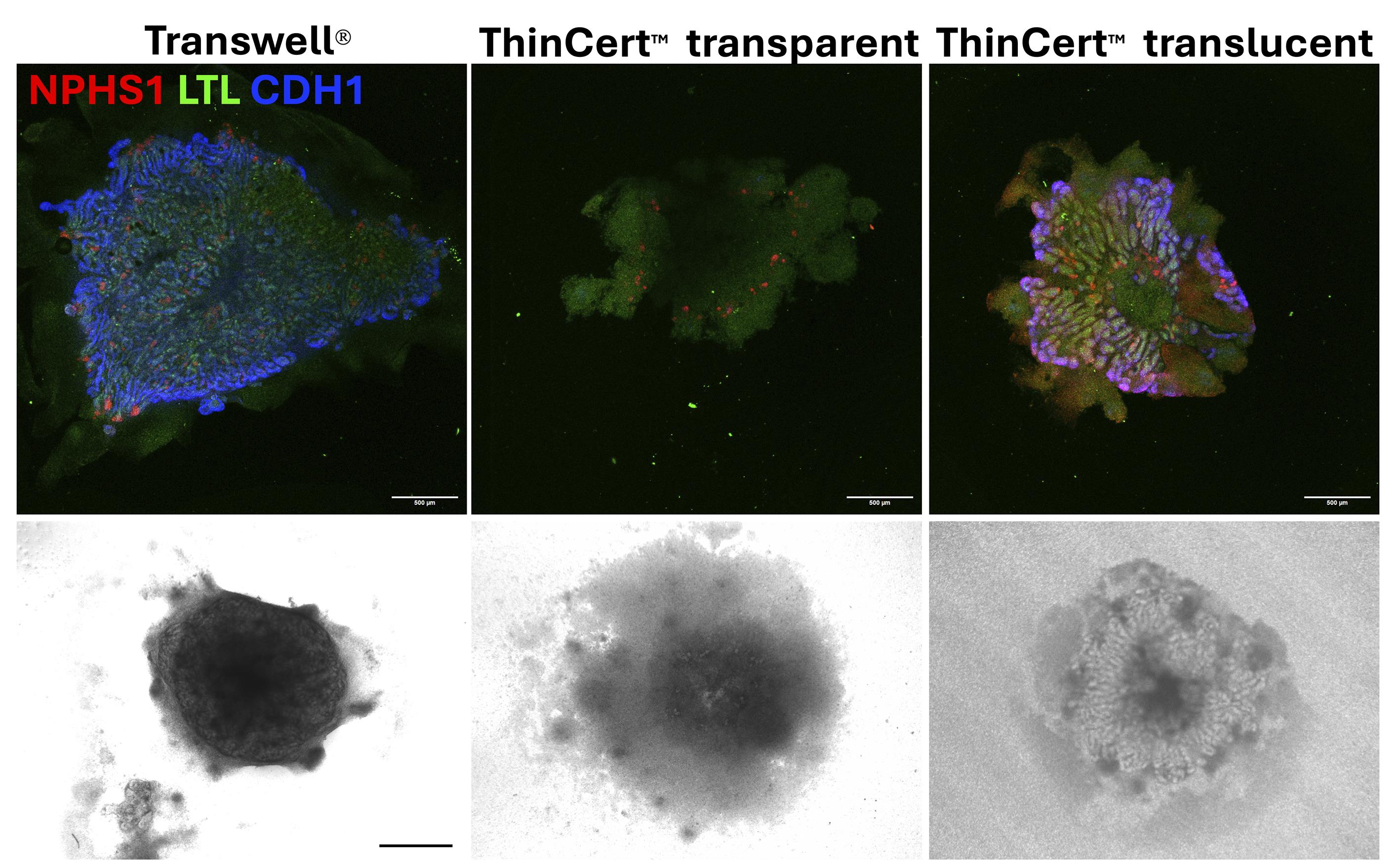
Figure 2. Comparison of differentiated kidney organoids (KiO) grown on different air–liquid interface (ALI) membranes. Top panel: Maximum intensity projection confocal images from a 4× objective: red, NPHS1 marking podocytes (sheep anti-NPHS1 1:500, R&D systems, catalog number AF4269); green, LTL marking proximal tubules (Fluorescein Lotus lectin 1:500, Vector Laboratories, catalog number FL-1321; note that this has to be vortexed before use); blue, CDH1 marking distal tubules and collecting ducts (mouse anti-E-cadherin 1:500, BD Biosciences, catalog number 610181). Scale bars = 500 μm. Bottom panel: Brightfield microscope with 2× objective; scale bar = 1,000 μm.
3. Reagent variability and optimisation
a. FGF9 source: Recombinant human FGF9 protein from two independent suppliers were tested (PeproTech vs. R&D Systems), with no detectable differences in differentiation efficiency or nephron marker expression (note based on the authors' experiences), indicating that the efficiency of the differentiation protocol is likely not dependent on any variability in this recombinant protein production.
b. CHIR99021 concentration and pulse requirement: We evaluated CHIR99021 concentrations of 6, 8, and 10 μM during primitive streak induction. For SCTi003-A, 6 μM CHIR99021 produced the most consistent differentiation outcomes with minimal cytotoxicity (Figure 3). Higher concentrations increased variability without improving organoid quality.
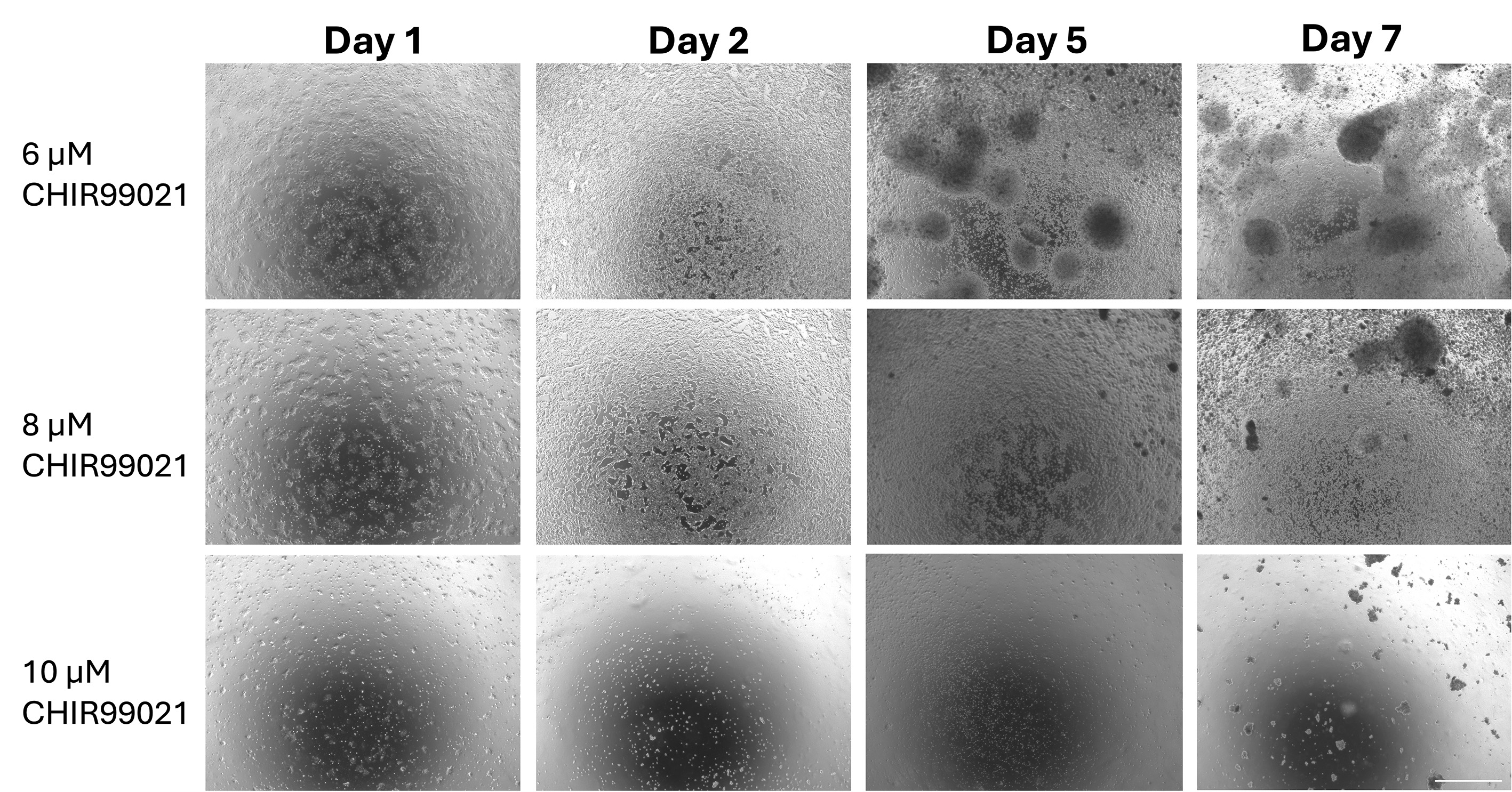
Figure 3. Optimisation of different concentrations of CHIR99021 using phase contrast images. Cells treated with 6 and 8 μM CHIR99021 show good growth and expected morphology by day 7, whereas 10 μM CHIR99021-treated cells show lack of division and cell death. This figure contains representative images for specified days of differentiation. Scale bar = 1,000 μm.
We further assessed the requirement for CHIR99021 pulse treatment at day 7. SCTi003-A organoids required a transient CHIR99021 pulse to achieve correct downstream tubular organisation [2]. Absence of this pulse resulted in severely impaired tubulogenesis across all ALI cultures (Figure 4).
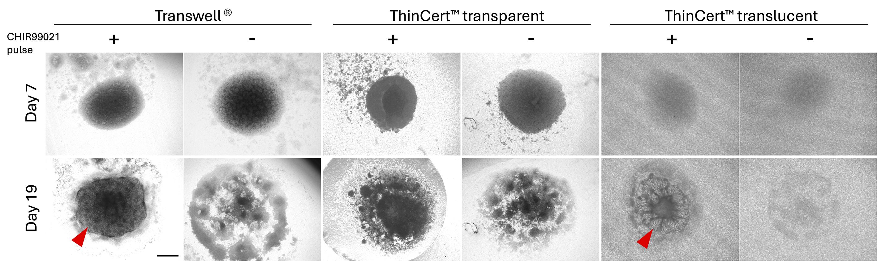
Figure 4. Checking whether CHIR99021 plus treatment is necessary on day 7 of SCTi003-A kidney organoid (KiO) differentiation, while grown on different air–liquid interface (ALI) membranes. KiO showed tubular structures only when CHIR99021-pulse-treated and grown on either Transwell® or ThinCertTM translucent membranes. Tubules are indicated by red arrowheads. Scale bar = 1,000 μm.
4. Basal medium selection and protocol benchmarking
We compared the Takasato-based protocol using either APEL2 (protocol 1) or advanced RPMI (ARPMI) (protocol 2) with the Uchimura-derived (protocol 3) and B-27 supplement (protocol 4) (Figures 5–8).
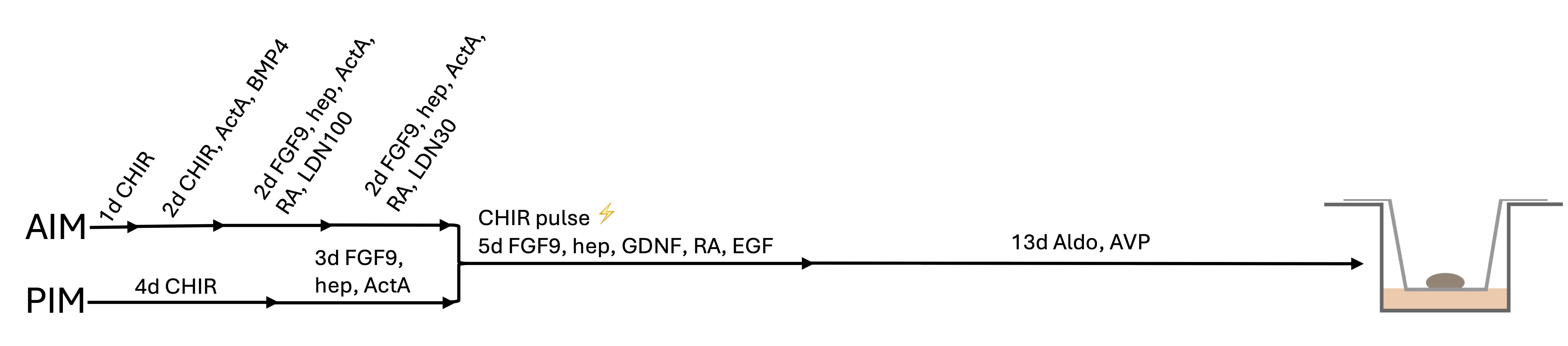
Figure 5. Schematic representation of an optimised protocol based on Uchimura et al. [8] Abbreviations: CHIR: CHIR99021 (6 and 5 μM for pulse treatment on day 7); ActA: Activin A (AIM days 1–2 and PIM days 4–6 10 ng/mL, AIM days 3–6 1 ng/mL); BMP4: BMP-4 (1 ng/mL); FGF9: FGF-9 (200 ng/mL); hep: heparin (1 μg/mL); RA: retinoic acid (100 nM); LDN: LDN193189 (days 3–4 100 nM, days 5–6 30 nM); GDNF (10 ng/mL): EGF (10 ng/mL); Aldo: aldosterone (10 nM); AVP: vasopressin (10 nM). This protocol is referred to as “protocol 3.”
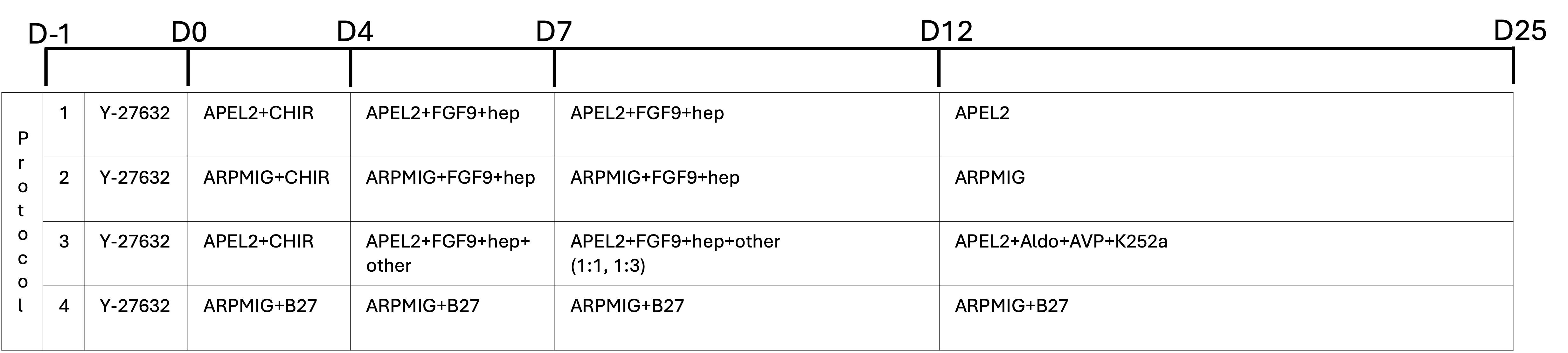
Figure 6. Experimental design illustrating four combinations of reagent and protocol tested for SCTi003-A induced pluripotent stem cell (iPSC) differentiation into kidney organoids (KiOs). Numbers on the left-hand side indicate the protocol number (1–4). Y-27632: ROCK inhibitor (10 μM); APEL2 (STEMdiffTM APELTM2 medium prepared by addition of PFHM-II protein hybridoma medium to a 5% final concentration); FGF9: FGF-9 (200 ng/mL); hep: heparin (1 μg/mL); ARPMIG (see Recipes); B27: B-27 Supplement (1×); K252a: K-252a (100 nM). Protocol 3 is presented here in simplified form; details can be found in Figure 5.
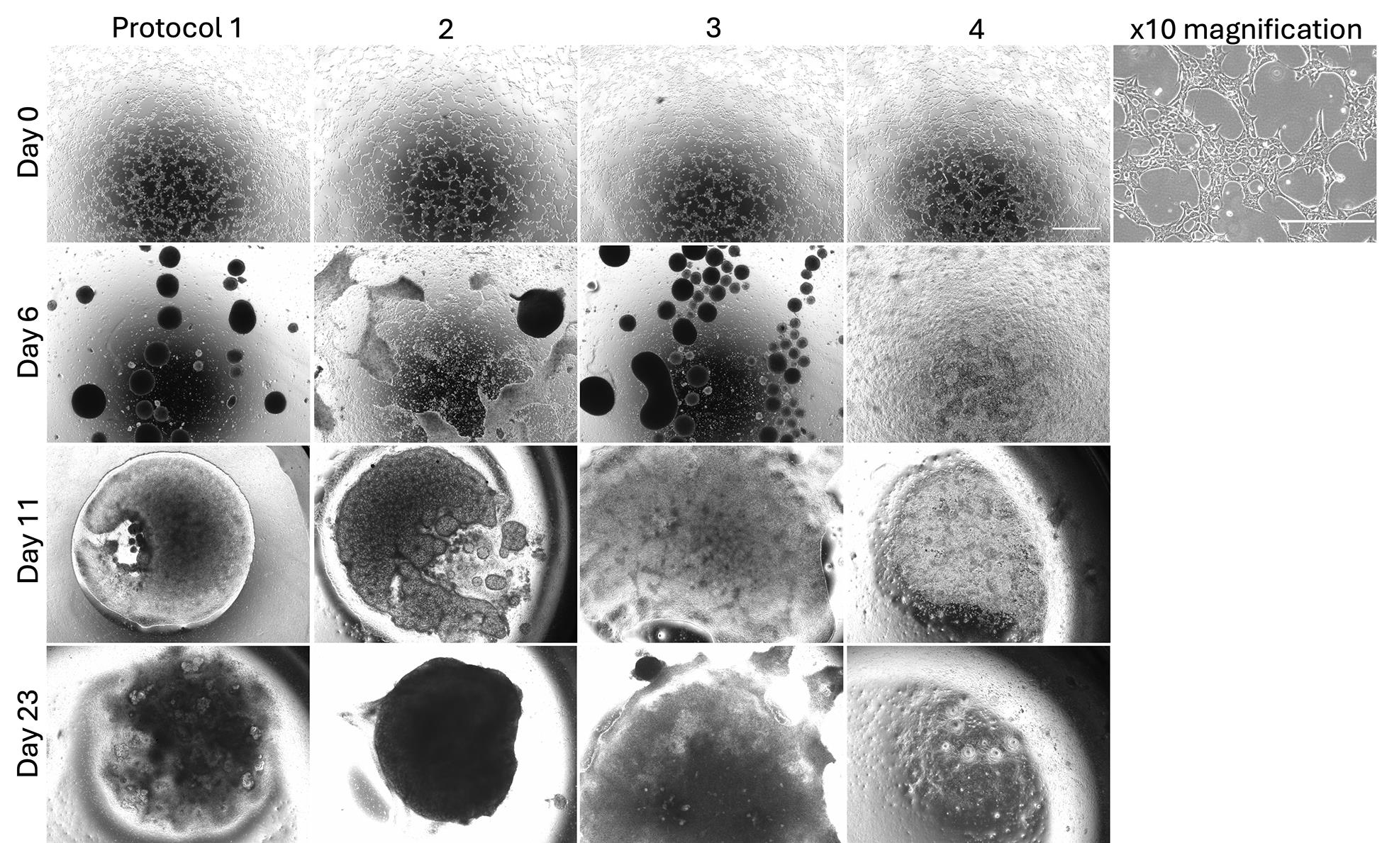
Figure 7. Brightfield microscope images of SCTi003-A differentiated into kidney organoids (KiO) following four protocols. Differentiating cells were imaged on days 0, 6, 11 and 23. All cells showed similar density at day 0 and were differentiated according to the four protocols shown in Figure 6. No tubule structures were formed in protocol 4. This figure contains representative images for specified days of differentiation. Scale bar = 1,000 μm; scale bar for 10× magnification = 400 μm.
We observed the most robust and consistent differentiation when ARPMI was used as the basal medium throughout protocol 2 [12,13], with well-defined nephron and collecting duct structures distributed across the organoid (Figures 8 and 9). Exclusive reliance on APEL2 media [3,8] reduced differentiation efficiency in SCTi003-A cultures (Figures 8 and 9).
Differentiation following the Uchimura et al.’s [8] protocol (protocol 3), implemented at this stage of protocol optimisation using APEL2 medium, was inefficient for SCTi003-A cells regardless of anterior-to-posterior intermediate mesoderm (AIM:PIM) mixing ratios (1:1 or 1:3; named protocol 3-11 and protocol 3-13, respectively, in Figures 8 and 9), resulting in poor tubular organisation and reduced marker expression. In addition, inclusion of the BDNF-NTRK2 neuronal differentiation inhibitor K252a did not appear to reduce off-target neuronal populations [14] and instead increased cell death and dedifferentiation (note based on the authors' experiences); this condition was therefore excluded from subsequent experiments (Figure 5). Protocol 4 [15–17], incorporating B27 supplementation, failed to generate stable tubular structures, and organoids cultured at an ALI did not form cohesive pellets suitable for downstream immunofluorescence analysis (Figure 7).
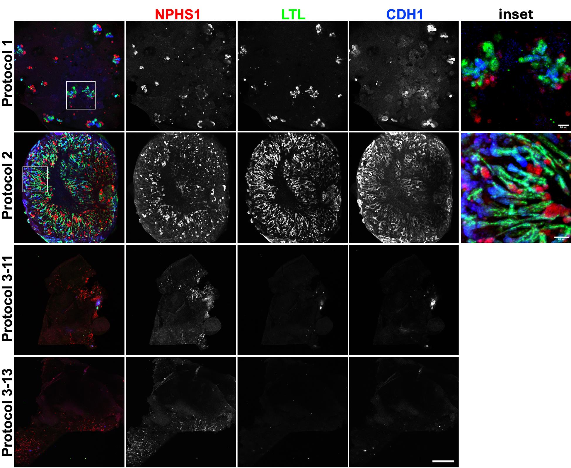
Figure 8. Immunofluorescence staining of whole kidney organoids (KiOs). 1: protocol 1 using APEL2 media. 2: protocol 2 using ARPMI; this protocol allowed successful differentiation of linear nephron and collecting duct structures. 3: protocol 3, following the Uchimura et al.’s [8] protocol using APEL2 media (AIM and PIM cells were mixed at 1:1 [protocol 3-11] or 1:3 [protocol 3-13] ratios). Differentiation with a 1:3 ratio was worse than a 1:1 ratio. Protocol 4 did not support the differentiation of any tubular structures (Figure 7), and no compact cell pellet was formed, which could be lifted for IF staining. NPHS1 (red): podocytes; LTL (green): proximal tubules; CDH1 (blue): distal tubules and collecting ducts. Scale bar = 500 μm.
To evaluate the suitability of the protocol for high-throughput applications, we assessed differentiation efficiency in suspension-based mKiO generated in Lipidure-coated 96-well plates. Mini-organoids exhibited nephron and collecting duct marker expression comparable to ALI cultures, including NPHS1, LTL, and CDH1 (Figure 9). These findings validate mKiO as a scalable and experimentally tractable format for downstream drug screening assays, whilst retaining key structural features observed in ALI-grown organoids.
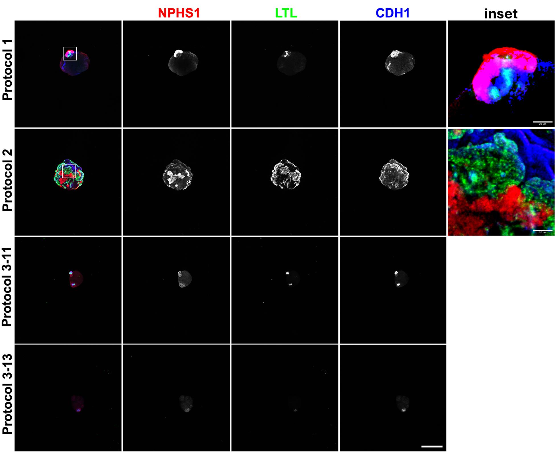
Figure 9. Immunofluorescence staining of differentiated mini-kidney organoids (mKiOs). 1: protocol 1 using APEL2 media. 2: protocol 2 using ARPMI. This protocol allowed successful differentiation of linear nephron and collecting duct structures. 3: protocol 3, following the Uchimura et al.’s [8] protocol using APEL2 media (AIM and PIM cells were mixed at 1:1 [protocol 3-11] or 1:3 [protocol 3-13] ratios). NPHS1 (red): podocytes; LTL (green): proximal tubules; CDH1 (blue): distal tubules and collecting ducts. Scale bar = 500 μm.
Based on the results from this assay, we proceeded with ARPMI culture in future differentiations.
5. Optimisation of a differentiation protocol for efficient nephron and collecting duct formation
The overarching aim of generating KiOs from the SCTi003-A hiPSC line was to establish a disease-relevant model for cystic kidney disorders. In autosomal dominant polycystic kidney disease (ADPKD), cysts arise from both nephron and collecting duct epithelia, whereas in autosomal recessive polycystic kidney disease (ARPKD), cyst formation is largely restricted to collecting ducts. Therefore, a key requirement of the differentiation protocol was the reliable generation of both nephron and collecting duct structures within the same organoid.
The canonical Takasato et al. [2,3] protocol was selected as a starting point because of its reported ability to generate both nephron and collecting duct lineages. However, multiple independent studies have noted that this approach generates collecting duct structures that remain immature. We therefore sought to determine whether parallel differentiation of posterior intermediate mesoderm (PIM; nephron lineage) and anterior intermediate mesoderm (AIM; ureteric epithelium lineage) would enhance representation of both compartments and improve suitability for in vitro modelling of cystic disease (Figure 10).
To address this, we directly compared three differentiation strategies: (i) the canonical Takasato protocol; (ii) a modified Takasato protocol with temporally separated induction of AIM (3 days) and PIM (4 days); and (iii) the Uchimura et al. [8] protocol, in which PIM is induced using a Takasato-based approach, and AIM is generated using a defined growth-factor regimen prior to the combination of both cultures at day 7. The Uchimura et al. [8] protocol employs a defined cocktail of developmental and maturation factors, including EGF, BMP-4, activin A, retinoic acid, LDN193189, GDNF, aldosterone and vasopressin, to promote AIM specification and subsequent nephron development.
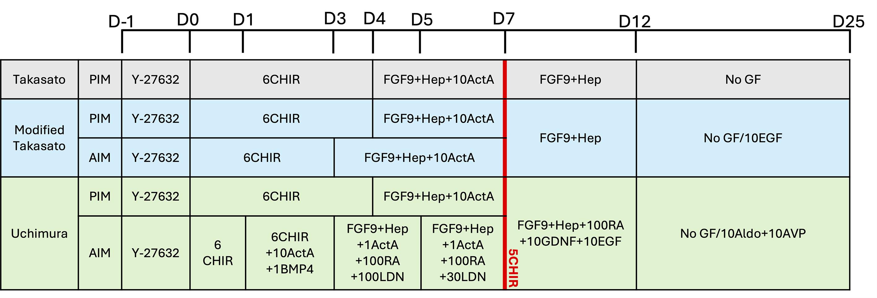
Figure 10. Schematic representation of tested protocols. Three protocols were compared for the differentiation efficiency of AIM and PIM structures. All PIM differentiation protocols here are based on Takasato et al. [2,3]. 6CHIR: 6 μM CHIR99021; FGF9: FGF-9 (200 ng/mL); Hep: heparin (1 μg/mL); 10ActA: 10 ng/mL activin A; 10EGF: 10 ng/mL EGF; 1BMP4: 1 ng/mL BMP-4; 1ActA: 1 ng/mL activin A; 100RA: 100 nM retinoic acid; 100/30LDN: 100/30 nM LDN193189; 10GDNF: 10 ng/mL GDNF; 10Aldo: 10 nM aldosterone; 10AVP: 10 nM vasopressin; GF: growth factors. The red line indicates the CHIR99021 pulse treatment on day (D) 7.
All three protocols produced organoids containing structures positive for established nephron and collecting duct markers, including NPHS1 (podocytes), LTL (proximal tubules), CDH1 (distal tubules and collecting ducts), and GATA3 (collecting ducts) (Figure 11). These findings indicate that, at the level of immunofluorescence detection, each protocol can initiate renal lineage differentiation in SCTi003-A cells.
Notably, however, organoids generated using the Uchimura-based protocol displayed extensive spatial overlap between GATA3 and NPHS1 staining. Such co-localisation is not expected in mature human kidney tissue and likely reflects populations of partially specified or under-differentiated epithelial progenitors rather than fully committed collecting duct and podocyte lineages. In contrast, both the canonical and modified Takasato protocols more consistently produced spatially segregated nephron and collecting duct domains, supporting clearer lineage resolution within individual organoids.
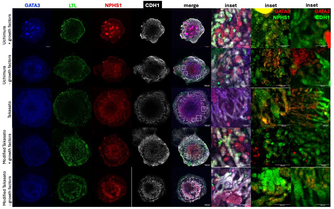
Figure 11. Immunofluorescence staining of SCTi003-A differentiated to kidney organoids (KiOs) following three protocols. Based on publications by Uchimura [8] (with/out growth factors past day 12), Takasato [3], and modified Takasato (with different incubation times of CHIR99021 to induce both PIM and AIM) with/out growth factors past day 12. Blue: GATA3 (rabbit anti-GATA3 1:500, Cell Signalling, catalog number 5852), collecting ducts; green: LTL, proximal tubules; red: NPHS1, podocytes; white: CDH1, distal tubules and collecting ducts. Scale bar on whole organoids = 500 μm, zoomed in = 100 μm.
We further evaluated the performance of each protocol in suspension-based mKiO, which are more suitable for high-throughput applications such as drug screening. All protocols generated mKiO expressing nephron and collecting duct markers with patterns broadly comparable to those observed in ALI cultures (Figure 12), indicating that suspension culture did not inherently compromise lineage specification.
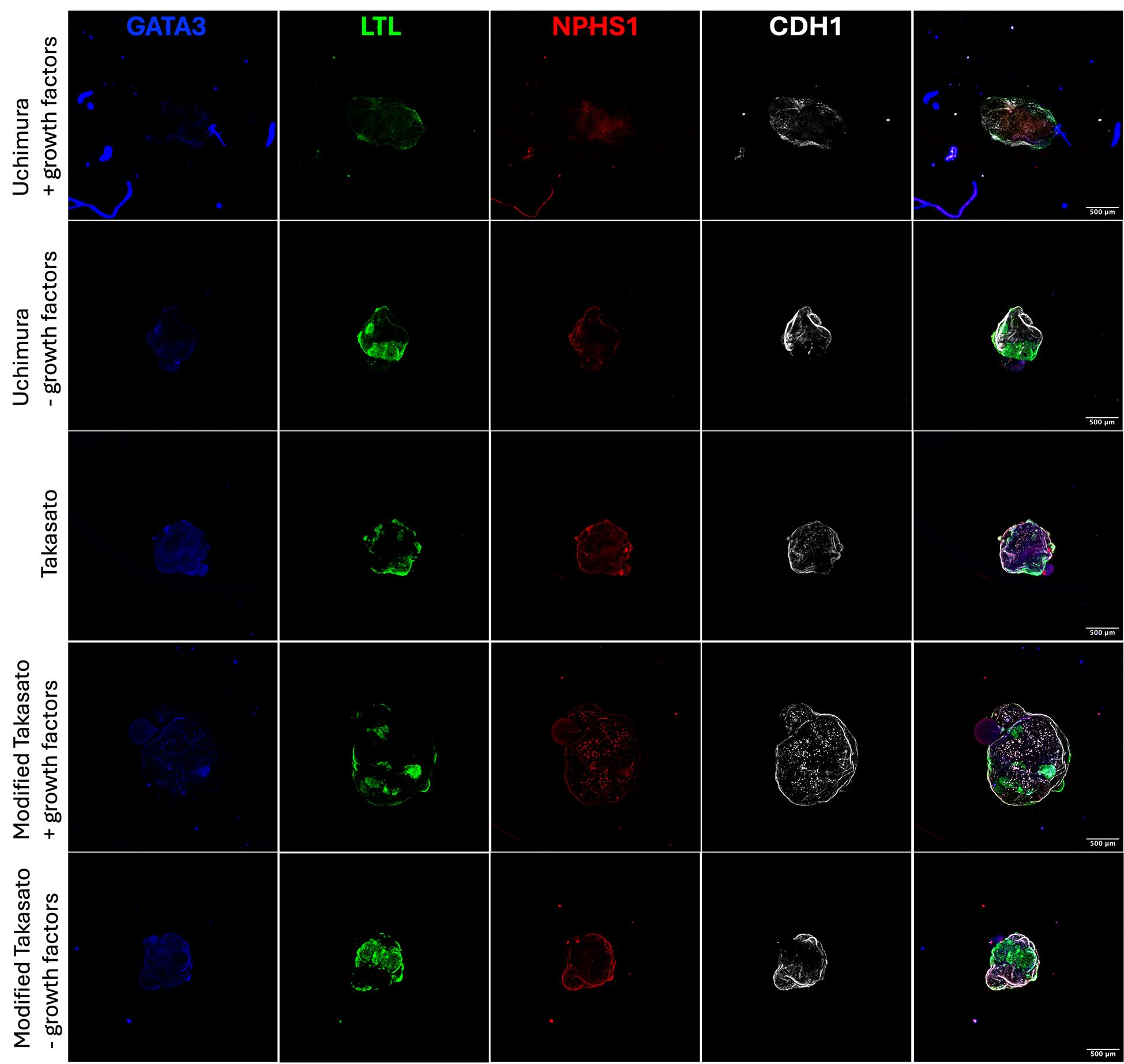
Figure 12. Immunofluorescence staining of SCTi003-A differentiated into mini-kidney organoids (mKiOs) following three protocols. Based on publications by Uchimura [8] (with/out growth factors past day 12), Takasato [3], and modified Takasato (with different incubation times of CHIR99021 to induce both PIM and AIM) with/out growth factors past day 12. Blue: GATA3, collecting ducts; green: LTL, proximal tubules; red: NPHS1, podocytes; white: CDH1, distal tubules and collecting ducts. Scale bar = 500 μm.
To support scalable phenotypic analysis of mKiO cultures, we implemented automated organoid recognition and area quantification using a trained Cellpose model within Image-Pro software (Media Cybernetics). Traditional threshold-based segmentation tools struggle to detect transparent regions within mKiO, necessitating extensive manual annotation. The Cellpose-based workflow reliably identified organoid boundaries, dramatically reducing analysis time and user bias. This approach transformed image analysis from a rate-limiting step requiring manual tracing of hundreds of organoids into a rapid, reproducible pipeline suitable for multi-plate, multi-timepoint studies, and represents an under-appreciated but critical component of scalable organoid-based drug screening (Figure 13).
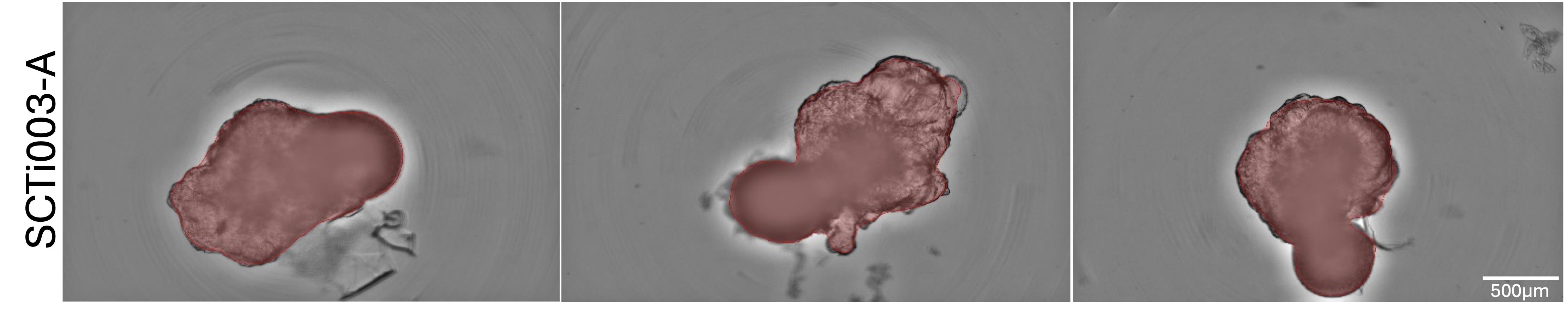
Figure 13. Automated segmentation and area quantification of mini kidney organoids (KiOs) using a trained Cellpose model. Representative brightfield images of suspension-based mKiO grown in Lipidure-coated 96-well plates and imaged on a high-content CellDiscoverer7 system. Organoid boundaries were automatically identified using a trained Cellpose model implemented within Image-Pro software (highlighted in red). Scale bar = 500 μm.
To assess collecting duct differentiation in greater detail, we examined expression of the principal cell marker aquaporin 2 (AQP2), co-stained with the collecting duct markers, GATA3 and DBA (Dolichos biflorus agglutinin, a lectin that is a specific marker of collecting tubules). All protocols generated DBA+/GATA3+ collecting duct-like structures with low-level AQP2 expression (Figure 14), consistent with the limited maturation typically observed in current KiO systems. Among the tested conditions, the modified Takasato protocol, supplemented with EGF, produced the lowest level of principal cells marked by AQP2, and this protocol was excluded from subsequent experiments.
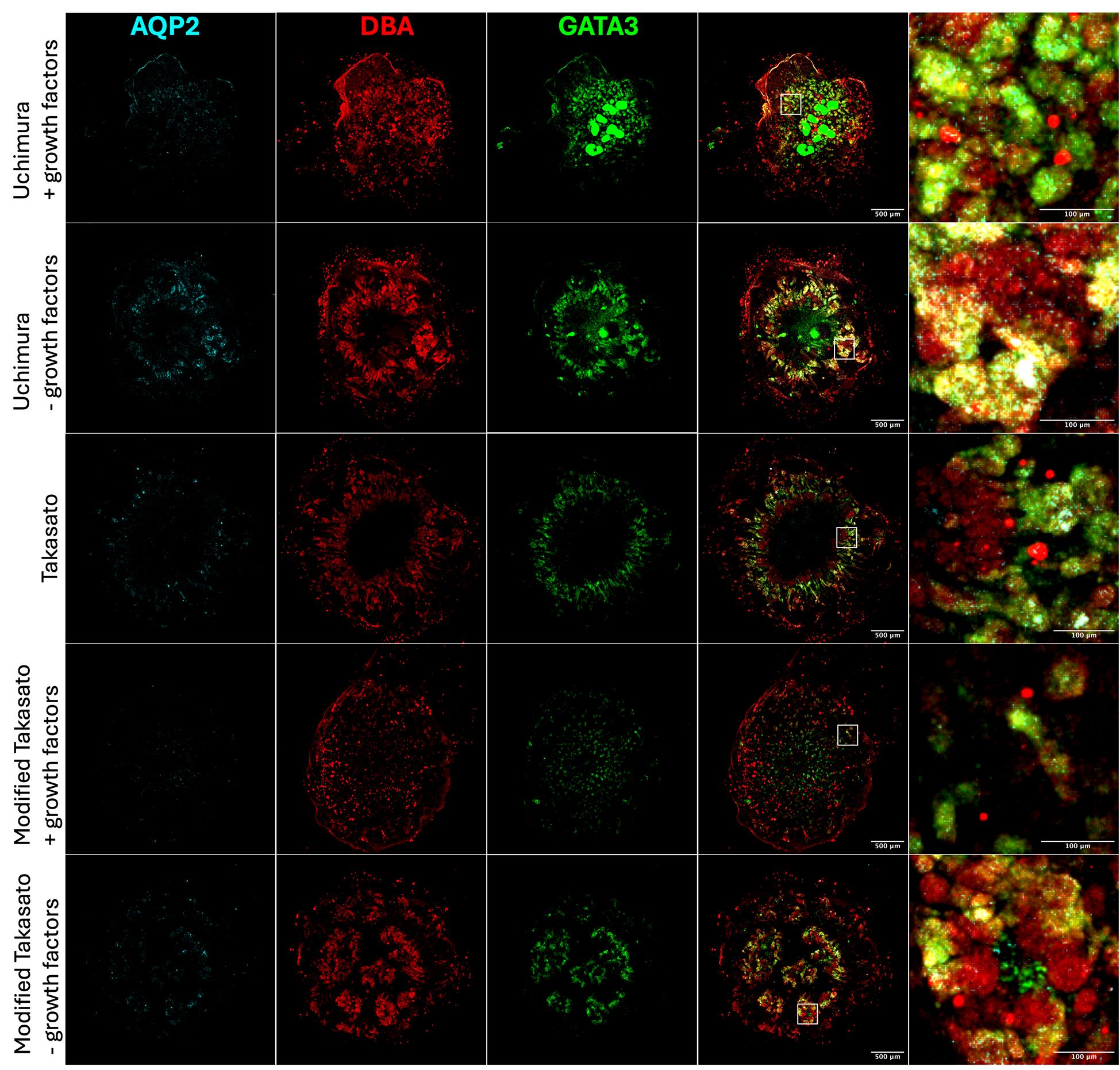
Figure 14. Immunofluorescence staining of collecting duct structures of SCTi003-A differentiated to kidney organoids (KiOs) following three protocols. Based on publications by Uchimura [8] (with/out growth factors past day 12), Takasato [3], and modified Takasato (with different incubation times of CHIR99021 to induce both PIM and AIM) with/out growth factors past day 12. Cyan: AQP2 (mouse anti-aquaporin 2 1:50, Santa Cruz Biotechnology, catalog number: sc-515770), principal cells; red: DBA (Dolichos biflorus agglutinin, Rhodamine 1:200, Vector labs, catalog number: RL-1032), collecting ducts; green: GATA3, collecting ducts. Scale bar on whole organoids = 500 μm, zoomed in = 100 μm.
Taken together, these comparisons indicate that while multiple differentiation strategies can initiate nephron and collecting duct specification in SCTi003-A cells, Takasato-based protocols provide superior spatial segregation of renal lineages, a key criterion for modelling cystic kidney disease. Nevertheless, we recognise that immunofluorescence alone cannot fully resolve differences in lineage maturity or differentiation efficiency. Future quantitative analyses, including qRT-PCR or single-cell RNAseq profiling of nephron, collecting duct, and progenitor markers, will be required to more conclusively assess relative maturation states across differentiation strategies.
6. Biological rationale for differentiation ending on day 25
Beyond day 25:
• SIX2+ nephron progenitors become depleted [18]
• No new nephrons form [14]
• Organoids enlarge without vascular maturation [19]
• Central hypoxia, apoptosis, and necrotic cores emerge [20]
• Mesenchymal drift and structural disorganisation increase [14]
Functional assays (e.g., dextran uptake, cisplatin toxicity) remain reliable until approximately day 30, after which the tubular architecture collapses.
7. Summary of validation
Together, these analyses demonstrate that the protocol robustly generates KiO from SCTi003-A hiPSCs, tolerates moderate variation in reagents and plasticware, and performs reproducibly across differentiation formats. Systematic optimisation of CHIR99021 timing, basal media, and ALI configuration was essential for achieving consistent nephron and collecting duct patterning. Suspension-based mKiOs generated using this protocol differentiate reproducibly and exhibit nephron and collecting duct marker expression comparable to ALI cultures, supporting their use in high-throughput drug screening. Coupling this format with automated organoid area recognition enables rapid, unbiased, and scalable phenotypic analysis across multiple plates and timepoints. This protocol therefore provides a reproducible workflow for disease modelling and high-throughput applications.
General notes and troubleshooting
Troubleshooting
Problem 1: Increased spontaneous differentiation before day 0.
Possible cause: hiPSC cultures over-passaged or stressed.
Solution: Use low-passage cultures, routinely return to master cell-bank stocks, and monitor pluripotency markers.
Problem 2: Poor cell survival after day -1 seeding.
Possible causes: Excessive dissociation stress; omission of ROCK inhibitor.
Solutions: Use Accutase for no longer than 7 min; include 10 μM Y-27632 during recovery.
Problem 3: Failure to reach 40%–60% confluency on day 0.
Possible causes: Incorrect cell counting; low viability after thawing; suboptimal seeding density.
Solutions: Seed 4 × 10⁵ viable cells/well. Confirm automated cell counter calibration. Use freshly thawed low-passage SCTi003-A cultures.
Problem 4: Extensive cell detachment between days 4 and 7.
Possible causes: Medium changes performed too rapidly; cells becoming fragile during IM induction.
Solution: Aspirate and add media slowly along the wall of the well. Avoid direct dispensing onto the cell layer.
Problem 5: Heterogeneous differentiation across wells.
Possible causes: Uneven Matrigel coating; uneven cell distribution during seeding.
Solutions: Prepare lot-specific Matrigel dilutions, maintain reagents on ice during coating, and gently rock plates after seeding to ensure uniform cell distribution.
Problem 6: Excessive cell death during primitive streak induction.
Possible cause: CHIR99021 concentration too high.
Solution: Use 6 μM CHIR99021. Avoid concentrations of 8–10 μM unless re-optimising for a different iPSC line.
Problem 7: Poor nephron and tubule formation.
Possible cause: Incorrect CHIR99021 treatment duration or concentration.
Solutions: Confirm 6 μM CHIR99021 from days 0 to 4 and a 5 μM pulse on day 7. Use freshly prepared CHIR-containing media.
Problem 8: No tubular structures after day 20.
Possible cause: Omission of day 7 CHIR pulse.
Solution: Include the transient day 7 CHIR pulse. Omission severely impairs tubulogenesis.
Problem 9: Small or poorly organised organoids on ALI membranes.
Possible causes: Insufficient cell number transferred; disrupted pellet during transfer.
Solution: Pellet 2 × 10⁵ cells and transfer with minimal handling. Use wide-bore pipette tips when lifting pellets.
Problem 10: Organoids submerged during ALI culture.
Possible cause: Medium level above the membrane surface.
Solutions: Ensure the medium remains below the membrane. Maintain a true air–liquid interface throughout culture.
Problem 11: Successful cell pellets but poor differentiation on ALI.
Possible cause: Use of an inappropriate insert membrane.
Solution: Use Corning Transwell® or Greiner ThinCertTM Translucent inserts. Avoid ThinCertTM Transparent inserts, which showed poor differentiation.
Problem 12: Poor organoid recovery after transfer to ALI.
Possible cause: Pellet disruption during handling.
Solution: Perform the triple centrifugation step (400× g, 2 min, three times) and minimise medium carryover during transfer.
Problem 13: High variability between organoids in 96-well format.
Possible cause: Inconsistent aggregation or attachment of spheroids.
Solutions: Use Lipidure-coated TPP round-bottom plates. Centrifuge immediately after seeding to promote uniform aggregation.
Problem 14: Degradation of outer-well organoids in 96-well plates.
Possible causes: Plate surface incompatibility or edge effects.
Solution: Avoid Corning ULA plates and use Lipidure-coated TPP plates, which showed uniform differentiation across the plate.
Problem 15: Reduced nephron marker expression.
Possible cause: Use of APEL2 as the sole differentiation medium.
Solution: Use ARPMI throughout the differentiation protocol, as this resulted in superior nephron and collecting duct formation.
Problem 16: Poor collecting duct development.
Possible causes: Inefficient AIM/PIM specification or protocol deviation.
Solution: Verify CHIR timing and growth factor additions. Canonical Takasato-based differentiation gave the best lineage segregation.
Problem 17: Variable differentiation between experiments.
Possible causes: Cell line drift or loss of pluripotency.
Solution: Replace cultures after 10–15 passages post-thaw and routinely confirm morphology, OCT4, and SSEA4 expression.
Problem 18: Organoid necrosis at late time points (day >30).
Possible cause: Overgrowth and diffusion limitations.
Solution: Harvest between days 25 and 28 whenever possible. Avoid prolonged culture unless perfusion or vascularisation strategies are used.
Problem 19: Weak immunofluorescence staining.
Possible causes: Poor antibody penetration or over-fixation.
Solutions: Optimise fixation time, extend antibody incubation, and ensure adequate permeabilisation of whole organoids.
Acknowledgments
This work was supported by Kidney Research UK (project grant PKD_RP_006_20211124). The authors also acknowledge financial support from the Medical Research Council (project grant MR/T017503/1), the MRC "Discovery Medicine North" Doctoral Training Partnership (MR/R015902/1), and the University of Leeds (alumnus funding from Sue and Peter Cheney). The protocol was developed as part of the research funded by these organisations; no separate prior publication describing its validation is available.
Author contributions
Protocol Development, K.S.; Protocol Optimization or Validation, K.S., M.K., G.P., E.L.H., A.L.B.; Writing—Original Draft, K.S.; Writing—Review & Editing, K.S., M.K., C.A.J.; Funding acquisition, C.A.J.; Supervision, C.A.J.
Competing interests
The corresponding author declares, on behalf of all authors, that there are no financial or non-financial competing interests.
Ethical considerations
No ethical approval was required for this study, as it exclusively utilised the commercially available SCTi003-A cell line and did not involve human or animal subjects.
Supplementary information
The following supporting information can be downloaded here:
1. Figure S1. SCTi003-A KiO labelled with pluripotency markers and brightfield images of cell colonies at different magnifications.
2. Figure S2. SCTi003-A KiO differentiated on ALI using different cell numbers.
3. Figure S3. Schematic illustration demonstrating how to efficiently transfer a pellet of IM cells with minimal disruption to ALI culture.
4. Figure S4. Brightfield (left) and IF images (right) of stem cells differentiated into kidney structures in 2D culture.
References
- Freedman, B. S., Brooks, C. R., Lam, A. Q., Fu, H., Morizane, R., Agrawal, V., Saad, A. F., Li, M. K., Hughes, M. R., Werff, R. V., et al. (2015). Modelling kidney disease with CRISPR-mutant kidney organoids derived from human pluripotent epiblast spheroids. Nat Commun. 6(1): 8715. https://doi.org/10.1038/ncomms9715
- Takasato, M., Er, P. X., Chiu, H. S., Maier, B., Baillie, G. J., Ferguson, C., Parton, R. G., Wolvetang, E. J., Roost, M. S., Chuva de Sousa Lopes, S. M., et al. (2015). Kidney organoids from human iPS cells contain multiple lineages and model human nephrogenesis. Nature. 526(7574): 564–568. https://doi.org/10.1038/nature15695
- Takasato, M., Er, P. X., Chiu, H. S. and Little, M. H. (2016). Generation of kidney organoids from human pluripotent stem cells. Nat Protoc. 11(9): 1681–1692. https://doi.org/10.1038/nprot.2016.098
- Morizane, R., Lam, A. Q., Freedman, B. S., Kishi, S., Valerius, M. T. and Bonventre, J. V. (2015). Nephron organoids derived from human pluripotent stem cells model kidney development and injury. Nat Biotechnol. 33(11): 1193–1200. https://doi.org/10.1038/nbt.3392
- Garreta, E., Prado, P., Tarantino, C., Oria, R., Fanlo, L., Martí, E., Zalvidea, D., Trepat, X., Roca-Cusachs, P., Gavaldà-Navarro, A., et al. (2019). Fine tuning the extracellular environment accelerates the derivation of kidney organoids from human pluripotent stem cells. Nat Mater. 18(4): 397–405. https://doi.org/10.1038/s41563-019-0287-6
- Taguchi, A. and Nishinakamura, R. (2017). Higher-Order Kidney Organogenesis from Pluripotent Stem Cells. Cell Stem Cell. 21(6): 730–746.e6. https://doi.org/10.1016/j.stem.2017.10.011
- Gupta, N. and Morizane, R. (2022). Kidney development to kidney organoids and back again. Semin Cell Dev Biol. 127: 68–76. https://doi.org/10.1016/j.semcdb.2021.09.017
- Uchimura, K., Wu, H., Yoshimura, Y. and Humphreys, B. D. (2020). Human Pluripotent Stem Cell-Derived Kidney Organoids with Improved Collecting Duct Maturation and Injury Modeling. Cell Rep. 33(11): 108514. https://doi.org/10.1016/j.celrep.2020.108514
- Boreström, C., Jonebring, A., Guo, J., Palmgren, H., Cederblad, L., Forslöw, A., Svensson, A., Söderberg, M., Reznichenko, A., Nyström, J., et al. (2018). A CRISP(e)R view on kidney organoids allows generation of an induced pluripotent stem cell–derived kidney model for drug discovery. Kidney Int. 94(6): 1099–1110. https://doi.org/10.1016/j.kint.2018.05.003
- Chichagova, V., Dorgau, B., Felemban, M., Georgiou, M., Armstrong, L. and Lako, M. (2019). Differentiation of Retinal Organoids from Human Pluripotent Stem Cells. Curr Protoc Stem Cell Biol. 50(1): e95. https://doi.org/10.1002/cpsc.95
- Atkinson, R., Georgiou, M., Yang, C., Szymanska, K., Lahat, A., Vasconcelos, E. J. R., Ji, Y., Moya Molina, M., Collin, J., Queen, R., et al. (2024). PRPF8-mediated dysregulation of hBrr2 helicase disrupts human spliceosome kinetics and 5’-splice-site selection causing tissue-specific defects. Nat Commun. 15(1): 3138. https://doi.org/10.1038/s41467-024-47253-0
- Bas-Cristóbal Menéndez, A., Du, Z., van den Bosch, T. P. P., Othman, A., Gaio, N., Silvestri, C., Quirós, W., Lin, H., Korevaar, S., Merino, A., et al. (2022). Creating a kidney organoid-vasculature interaction model using a novel organ-on-chip system. Sci Rep. 12(1): 20699. https://doi.org/10.1038/s41598-022-24945-5
- Monteil, V., Kwon, H., Prado, P., Hagelkrüys, A., Wimmer, R. A., Stahl, M., Leopoldi, A., Garreta, E., Hurtado del Pozo, C., Prosper, F., et al. (2020). Inhibition of SARS-CoV-2 Infections in Engineered Human Tissues Using Clinical-Grade Soluble Human ACE2. Cell. 181(4): 905–913.e7. https://doi.org/10.1016/j.cell.2020.04.004
- Wu, H., Uchimura, K., Donnelly, E. L., Kirita, Y., Morris, S. A. and Humphreys, B. D. (2018). Comparative Analysis and Refinement of Human PSC-Derived Kidney Organoid Differentiation with Single-Cell Transcriptomics. Cell Stem Cell. 23(6): 869–881.e8. https://doi.org/10.1016/j.stem.2018.10.010
- Czerniecki, S. M., Cruz, N. M., Harder, J. L., Menon, R., Annis, J., Otto, E. A., Gulieva, R. E., Islas, L. V., Kim, Y. K., Tran, L. M., et al. (2018). High-Throughput Screening Enhances Kidney Organoid Differentiation from Human Pluripotent Stem Cells and Enables Automated Multidimensional Phenotyping. Cell Stem Cell. 22(6): 929–940.e4. https://doi.org/10.1016/j.stem.2018.04.022
- Facioli, R., Lojudice, F. H., Anauate, A. C., Maquigussa, E., Nishiura, J. L., Heilberg, I. P., Sogayar, M. C. and Boim, M. A. (2021). Kidney organoids generated from erythroid progenitors cells of patients with autosomal dominant polycystic kidney disease. PLoS One. 16(8): e0252156. https://doi.org/10.1371/journal.pone.0252156
- Suhito, I. R., Kim, J. W., Koo, K., Nam, S. A., Kim, Y. K. and Kim, T. (2022). In Situ Detection of Kidney Organoid Generation From Stem Cells Using a Simple Electrochemical Method. Adv Sci. 9(20): e202200074. https://doi.org/10.1002/advs.202200074
- Howden, S. E., Vanslambrouck, J. M., Wilson, S. B., Tan, K. S. and Little, M. H. (2019). Reporter‐based fate mapping in human kidney organoids confirms nephron lineage relationships and reveals synchronous nephron formation. EMBO Rep. 20(4): e201847483. https://doi.org/10.15252/embr.201847483
- Homan, K. A., Gupta, N., Kroll, K. T., Kolesky, D. B., Skylar-Scott, M., Miyoshi, T., Mau, D., Valerius, M. T., Ferrante, T., Bonventre, J. V., et al. (2019). Flow-enhanced vascularization and maturation of kidney organoids in vitro. Nat Methods. 16(3): 255–262. https://doi.org/10.1038/s41592-019-0325-y
- Przepiorski, A., Sander, V., Tran, T., Hollywood, J. A., Sorrenson, B., Shih, J. H., Wolvetang, E. J., McMahon, A. P., Holm, T. M., Davidson, A. J., et al. (2018). A Simple Bioreactor-Based Method to Generate Kidney Organoids from Pluripotent Stem Cells. Stem Cell Rep. 11(2): 470–484. https://doi.org/10.1016/j.stemcr.2018.06.018
Article Information
Publication history
Received: Jul 20, 2026
Accepted: Sep 23, 2026
Available online: Oct 12, 2026
Published: Nov 5, 2026
Copyright
© 2026 The Author(s); This is an open access article under the CC BY license (https://creativecommons.org/licenses/by/4.0/).
How to cite
Szymanska, K., Karwatka, M., Parpas, G., Harris, E. L., Barlow, A. L. and Johnson, C. A. (2026). Human iPSC Line SCTi003-A Differentiation Into Kidney Organoids. Bio-protocol 16(21): e5860. DOI: 10.21769/BioProtoc.5860.

 Download Citation in RIS Format
Do you have any questions about this protocol?
Post your question to gather feedback from the community. We will also invite the authors of this article to respond.
 Post a Question


