


- Protocols

- Articles and Issues
- For Authors
- About
- Become a Reviewer


A Simplified Density Gradient–Free Method for Isolating Functional Symbiosomes From Legume Nodules

(*contributed equally to this work) Published: Vol 16, Iss 19, Oct 5, 2026 DOI: 10.21769/BioProtoc.5854 Views: 26
Reviewed by: Anonymous reviewer(s)
 PDF
PDF  Ask a question

Protocol Collections
Comprehensive collections of detailed, peer-reviewed protocols focusing on specific topics




Abstract
Symbiosomes are plant membrane–bound, organelle-like structures that enclose single or multiple bacteroids (differentiated rhizobia) within legume cells, serving specifically as the site for N2 fixation. Although isolated symbiosomes have been used to study nitrogen fixation, ion transport, and metabolite translocation, conventional density gradient–based isolation methods for their isolation are labor-intensive. Here, we describe a simplified, density gradient–free protocol for rapid enrichment of symbiosome-containing fractions from mature legume nodules using the Medicago truncatula–Sinorhizobium meliloti symbiosis as an example. The method combines razor blade homogenization, filtration through a 40 μm cell strainer, and sequential low-speed centrifugation to remove debris and enrich symbiosome-associated fractions. The resulting material is compatible with downstream mass spectrometry–based proteomics and mutant phenotypic analyses. Compared with conventional approaches, this protocol is rapid, simple, and suitable for higher-throughput biochemical and proteomic studies of symbiosomes.
Key features
• Density gradient–free enrichment of symbiosome-containing fractions from mature legume nodules.
• Rapid workflow using homogenization, filtration, and low-speed centrifugation with minimal specialized equipment.
• Compatible with downstream mass spectrometry–based proteomic analysis of symbiosome-associated proteins.
• Suitable for comparative analysis of wildtype and mutant nodules in symbiotic nitrogen fixation studies.
Keywords: Symbiosome enrichment Functional symbiosome
Legume nodules
Symbiotic nitrogen fixation
Medicago truncatula
Functional symbiosome
Legume nodules
Symbiotic nitrogen fixation
Medicago truncatula
Background
Symbiotic nitrogen fixation in legumes depends on the formation of symbiosomes, specialized membrane-bound compartments that house differentiated rhizobia within infected nodule cells [1,2]. Symbiosomes provide the biochemical environment required for N2 fixation and mediate extensive metabolite and ion exchange between the host plant and bacterial symbiont. Because of their central role in symbiosis, isolated symbiosomes have been used to investigate nitrogen fixation activity, membrane transport, and metabolite exchange [3,4]. Despite the establishment of early symbiosome isolation methods, relatively few studies have subsequently employed isolated symbiosomes for functional analyses, largely due to the fact that conventional density gradient–based procedures are technically demanding, low-throughput, and often require substantial nodule material. Several modified approaches have attempted to simplify the procedure through homogenization and low-speed centrifugation to obtain crude symbiosome or infected-cell fractions [5,6]. Although these methods improve experimental accessibility, the resulting preparations are often heterogeneous and may exhibit reduced structural integrity, and their suitability for downstream physiological analyses has not been extensively evaluated. A recent proteomic study identified 111 symbiosome membrane-associated proteins through purification of intact symbiosomes followed by symbiosome membrane enrichment [7,8]. However, this procedure relied on multiple purification steps and Tris-HCl-based buffers, which may not be optimal for preserving the physiological activity of symbiosomes during downstream in vitro functional assays. Here, we describe a simplified density gradient-free protocol for the enrichment of symbiosome-containing fractions from mature legume nodules using the Medicago truncatula–Sinorhizobium meliloti symbiosis as an example. This protocol was adapted from [3,4] and optimized based on [5]. The method combines mechanical homogenization, filtration, and sequential low-speed centrifugation to reduce cellular debris while enriching symbiosome-associated particles. Although the resulting fractions are less highly purified than those obtained by density gradient centrifugation, they are suitable for downstream mass spectrometry–based proteomics and comparative analyses of wildtype and mutant nodules. In addition, the protocol may facilitate studies of symbiosome-associated protein localization, mutant phenotyping, and regulatory mechanisms involved in symbiotic nitrogen fixation.
Materials and reagents
Biological materials
1. Mature nodules from legume plants (Medicago truncatula nodulated with Sinorhizobium meliloti, harvested 21 days post-inoculation)
Reagents
1. Mannitol (Sigma-Aldrich, CAS: 69-65-8)
2. Ethylene glycol bis (2-aminoethylether)-N,N,N′,N′-tetraacetic acid (EGTA) (Sigma-Aldrich, CAS: 67-42-5)
3. MgSO4·7H2O (Sigma-Aldrich, CAS: 10034-99-8)
4. Dithiothreitol (DTT) (Sigma-Aldrich, CAS: 3483-12-3)
5. Bovine serum albumin (BSA), fatty acid-free (Sigma-Aldrich, CAS: 9048-46-8)
6. Ascorbic acid (Sigma-Aldrich, CAS: 50-81-7)
7. MES (Sigma-Aldrich, CAS: 4432-31-9)
8. KOH (Sigma-Aldrich, CAS: 1310-58-3); 1 M KOH solution was used to adjust MES to pH 7.0
9. KNO3 (Sigma-Aldrich, CAS: 7757-79-1)
10. (Optional) Protease inhibitor cocktail (Sigma-Aldrich, catalog number: P8340 or P9599; recommended for protein/proteomic analyses)
11. Sodium dodecyl sulfate (SDS) (Sigma-Aldrich, CAS: 151-21-3); used for protein lysis
12. Ammonium bicarbonate (ABC) (Sigma-Aldrich, CAS: 1066-33-7); 50 mM working solution prepared in water for protein digestion
13. Methanol (Sigma-Aldrich, CAS: 67-56-1); ice-cold, used for chloroform/methanol precipitation
14. Chloroform (Sigma-Aldrich, CAS: 67-66-3); used for protein precipitation
15. Acetone (Sigma-Aldrich, CAS: 67-64-1); 90% (v/v) in water, used for washing protein pellets
16. Urea (Sigma-Aldrich, CAS: 57-13-6); 8 M solution prepared in water for protein solubilization
17. Bicinchoninic acid (BCA) assay kit (Sigma-Aldrich, catalog number: BCA1 or equivalent); used for protein quantification
18. Tris(2-carboxyethyl)phosphine (TCEP) (Sigma-Aldrich, CAS: 51805-45-9); used as reducing agent
19. Chloroacetamide (CAA) (Sigma-Aldrich, CAS: 79-07-2); used for alkylation
20. Trypsin (Sigma-Aldrich, catalog number: T6567 or equivalent); used for protein digestion
21. Trifluoroacetic acid (TFA) (Sigma-Aldrich, CAS: 76-05-1); 20% and 0.1% (v/v) solutions prepared in water for acidification and peptide loading
22. Acetonitrile (ACN), HPLC grade (Sigma-Aldrich, CAS: 75-05-8); used for peptide elution and as mobile phase B for LC-MS/MS
23. Formic acid (FA), HPLC grade (Sigma-Aldrich, CAS: 64-18-6); 0.1% (v/v) solution in water used as mobile phase A and peptide reconstitution buffer
24. Colorimetric peptide assay kit (ThermoFisher Scientific, catalog number: 23275 or equivalent); used for peptide quantification after desalting
Solutions
1. Isolation buffer (see Recipes)
2. Wash buffer (see Recipes)
3. Testing buffer (see Recipes)
Recipes
1. Isolation buffer (100 mL, pH 7.0)
| Reagent | Final concentration | Quantity or volume |
|---|---|---|
| Mannitol | 350 mM | 6.36 g |
| EGTA | 10 mM | 0.38 g |
| MgSO4·7H2O | 10 mM | 0.246 g |
| MES | 25 mM | 0.488 g |
| BSA | 1% | 1 g |
| Ascorbic acid | 20 mM | 0.352 g |
| DTT | 5 mM | 77 mg |
2. Wash buffer (100 mL, pH 7.0)
| Reagent | Final concentration | Quantity or volume |
|---|---|---|
| Mannitol | 350 mM | 6.36 g |
| MES (pH 7.0) | 25 mM | 0.488 g |
| MgSO4·7H2O | 3 mM | 82 mg |
3. Testing buffer
Prepare by supplementing wash buffer (see Recipe 2) with the substrate of interest, depending on the functional assay to be performed. For example, for nitrate uptake assays, the testing buffer consists of wash buffer containing 2 mM KNO3 as the substrate. The buffer should be prepared fresh on the day of the experiment and kept on ice until use.
Laboratory supplies
1. 1 mL pipette tip (Maisinuo, catalog number: HZX005-1)
2. Double-edged razor blade (Shanghai Feiying Brand, catalog number: FEIYING-DE)
3. 10 × 10 cm Petri dish (LABSTAR, catalog number: BX051)
4. 40 μm cell strainer (WHB, catalog number: WHB-40UM-F)
5. 15 mL sterile centrifuge tube (LABSELECT, catalog number: CT-002-15A)
6. Forceps (Maisinuo, catalog number: HZX186-1)
7. 1.5 mL microcentrifuge tube (Maisinuo, catalog number: HZX018-1)
Equipment
1. Refrigerated centrifuge (Eppendorf, model: 5424 R)
2. Microscope (Olympus, model: BX51)
3. Balance (Mettler Toledo, model: ML203T)
4. 1 mL single-channel pipette (Eppendorf)
Procedure
A. Enrichment of symbiosome fractions
1. Prepare buffers: Prepare all buffers as described in the Recipes section and maintain them at 4 °C throughout the procedure.
2. Harvest nodules: Wash mature plants thoroughly with tap water. Using forceps, detach mature nodules (21-day post-inoculation M. truncatula nodules; Figure 1A, B) and collect them into a Petri dish containing ice-cold isolation buffer on ice. A total fresh nodule weight greater than 200 mg is recommended.
3. Homogenization and filtration: Place the nodules in a Petri dish on ice with pre-cooled isolation buffer to just cover them (approximately 1 mL per 100 mg nodules). Finely mince the nodules using a razor blade on ice until the homogenate can be readily pipetted (Figure 1C). Add five volumes of isolation buffer relative to the homogenate volume and mix thoroughly by pipetting. Transfer the homogenate directly to a 40 μm cell strainer placed on a 15 mL centrifuge tube; the filtrate, which represents the crude symbiosome-containing extract, is collected in the tube below (Figure 1C, D).
4. Removal of large debris: Centrifuge the filtrate at 200× g for 2 min at 4 °C (approximately 1,500 rpm in an Eppendorf 5424 R centrifuge; Figure 1E) (centrifuge speed adapted from [5]). The pellet contains low-speed nuclei and large cellular debris (Figure 1F). Carefully transfer the supernatant to a new tube. Before use, weigh an empty 1.5-mL microcentrifuge tube and record its weight (W1). Transfer the entire supernatant into the pre-weighed tube.
5. Enrichment of symbiosome fractions: Centrifuge the supernatant at 2,500× g for 15 min at 4 °C (approximately 5,000 rpm in the Eppendorf 5424 R centrifuge; centrifuge speed adapted from [5] (Figure 1G). The resulting pellet is enriched in symbiosome-associated particles (Figure 1H). Carefully discard the supernatant and immediately weigh the tube with the pellet (W2). The fresh weight of the symbiosome pellet is calculated as W2−W1. Then, gently resuspend the pellet in pre-cooled wash buffer (1 mL). The preparation may then be used for downstream analyses.
B. Validation of symbiosome integrity using mCherry-labeled Sinorhizobium meliloti
Note: To confirm that the symbiosomes isolated by this protocol are viable and retain their functional integrity, we performed an additional validation experiment using the S. meliloti 2011 strain constitutively expressing the mCherry fluorescent protein. The fluorescent signal of mCherry is stably maintained inside viable bacteroids and can be used as a marker for intact symbiosomes.
1. Plant inoculation: Grow Medicago truncatula plants in a sterilized mixture of vermiculite and perlite (1:1). At 7 days post-germination, the plants were inoculated with an overnight culture of S. meliloti 2011 strain carrying a constitutive mCherry expression plasmid. The rhizobia culture was diluted to an optical density (OD600) of 0.02 in sterile water, and 2 mL was applied per plant.
2. Nodule harvest: At 21 days post-inoculation, pink, healthy nodules were harvested from the root system and immediately placed in ice-cold isolation buffer.
3. Symbiosome isolation: The nodules were processed exactly as described in Section A, using the same isolation and wash buffers, without any modification.
4. Microscopic observation: An aliquot of the final symbiosome suspension (resuspended in wash buffer) was placed on a glass slide and covered with a coverslip. An aliquot of the final symbiosome suspension (resuspended in wash buffer) was placed on a glass slide and covered with a coverslip. The sample was examined using a Zeiss LSM 880 confocal laser scanning microscope (excitation:561 nm, emission: 580 nm ~ 610nm). Brightfield images were acquired in parallel to visualize the overall morphology.
5. Signal interpretation: Symbiosomes isolated from nodules harboring mCherry-labeled S. meliloti 2011 exhibited bright-red fluorescence, which was clearly visible within the symbiosome compartments (see representative images in Figure 1I). The presence of strong, intact fluorescent signals within the isolated symbiosomes confirms that the bacteroids remain viable and that the symbiosome membranes are sufficiently intact to retain the soluble mCherry protein during the isolation procedure. This observation validates that the protocol yields functional symbiosomes suitable for downstream assays.
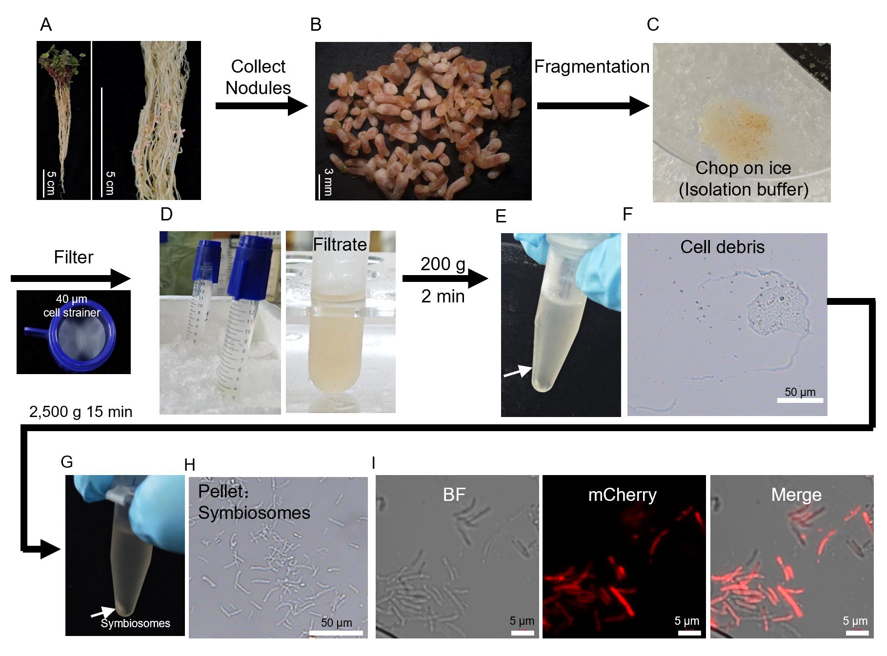
Figure 1. Workflow for the enrichment of symbiosome-containing fractions from Medicago truncatula nodules. (A) Nodulated Medicago truncatula plant. (B) Collected nodules. (C) Chopping nodules on ice with a razor blade. (D) Filtration through a 40 μm cell strainer. (E) Eppendorf tube after low-speed centrifugation (200× g); the arrow shows a small pellet (large debris and nuclei). (F) Cell debris fraction. (G) Eppendorf tube after medium-speed centrifugation (2,500× g); the arrow indicates the pellet enriched with symbiosomes. (H) Representative microscopic image of the pellet from (G), showing intact symbiosomes. (I) Representative views from fluorescent symbiosomes isolated from nodules inoculated with mCherrylabeled S. meliloti 2011.
C. Functional assay example: Nitrate uptake
1. Normalize sample amounts: After determining the fresh weight of the symbiosome pellet by differential weighing (as described in Section A), resuspend the pellet in an appropriate volume of wash buffer to achieve the desired concentration. For initial trials, we recommend starting with a relatively high concentration (e.g., 2 mg of fresh weight per milliliter). In the representative example shown in this protocol (nitrate uptake assay), the final symbiosome suspension was adjusted to 1.5 mg of fresh weight per milliliter using wash buffer.
2. Initiate the uptake assay: Add KNO3 to a final concentration of 2 mM. Incubate the symbiosome suspension statically in the dark at room temperature or at 4 °C, depending on the experimental design (Figure 2A).
3. Sampling and nitrate measurement: Collect aliquots at designated time points. Immediately pellet symbiosome fractions by centrifugation at 2,500× g for 2 min and collect the supernatant. Determine residual nitrate concentrations in the supernatant using the nitrate quantification assay [9].
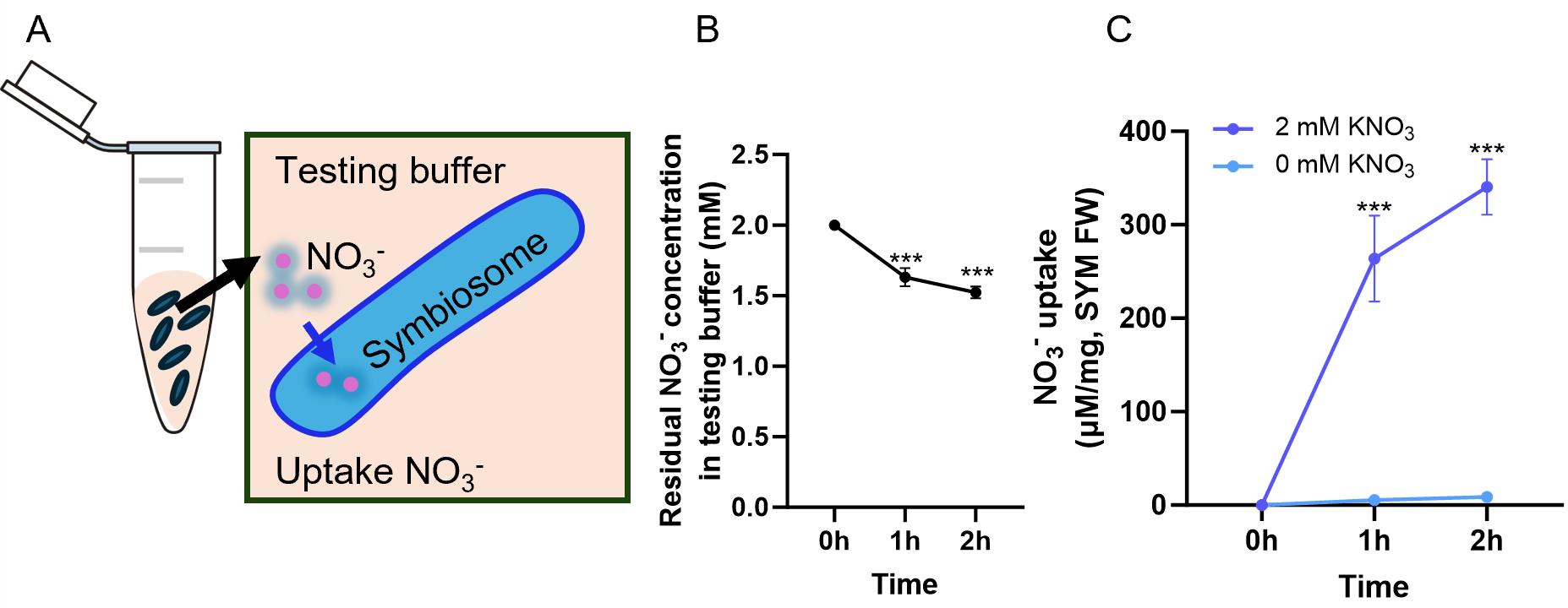
Figure 2. Example nitrate uptake assay using enriched symbiosome fractions. (A) Schematic representation of the nitrate uptake assay. Purified symbiosome pellets were resuspended in wash buffer containing 2 mM KNO3 as the substrate, adjusted to a final concentration of 1.5 mg of fresh weight per milliliter, and incubated in the dark without shaking to allow nitrate uptake. Aliquots were withdrawn at 0, 1, and 2 h post-incubation and immediately centrifuged (2,500× g, 2 min). The resulting supernatants were collected for nitrate quantification. (B) Residual nitrate concentration in the testing buffer over time. (C) NO3- uptake calculated from the data in (B), normalized to symbiosome fresh weight. Data in (B–C) are presented as mean ± SD (n = 3). Symbiosomes incubated in testing buffer without KNO3 (0 mM) served as a negative control, and no detectable nitrate was observed throughout the time course. Asterisks indicate statistically significant differences compared to the 0-h time point within each treatment group (two-tailed Student’s t-test; ***p < 0.001)
D. Proteomic analysis of symbiosome fractions
Note: This section does not aim to exhaustively describe the proteomic method, nor is it essential for symbiosome isolation. Rather, it offers a step-by-step operational outline that users may reference to validate the protocol.
1. Lysis and protein precipitation: Resuspend symbiosome pellets (approximately 50 mg of fresh weight from section A) in 200 μL of lysis buffer containing 1% SDS and 50 mM ABC. Sonicate briefly and centrifuge at 13,000× g at 4 °C for 10 min to remove insoluble debris. Transfer 150 μL of supernatant to a new tube and perform chloroform/methanol precipitation by sequentially adding 600 μL of ice-cold methanol, 150 μL of chloroform, and 450 μL of ice-cold water, vortexing after each addition. Centrifuge (13,000× g, 4 °C, 10 min), wash the protein disc twice with methanol and once with 90% acetone, air-dry the pellet, and dissolve it in 8 M urea to a final concentration of 5–10 mg/mL. Determine the protein concentration using a bicinchoninic acid (BCA) assay.
2. Trypsin digestion: Take 100 μg of protein (in 20 μL of 8 M urea) and dilute with 20 μL of 100 mM ABC. Add 10 μL of a solution containing 50 mM TCEP and 250 mM CAA in 100 mM ABC (final 10 mM TCEP and 50 mM CAA) and incubate at 37 °C for 30 min for reduction and alkylation. Add 150 μL of 100 mM ABC (urea <1 M, pH ~8), then add 2 μg of trypsin and incubate at 37 °C for 2 h. Add another 2 μg of trypsin and incubate overnight. Acidify the digest with 20% TFA (1/20 volume) to pH <3 and dilute to 1 mL with 0.1% TFA.
3. Peptide desalting: Desalt the peptides using Oasis HLB 1 cc (50 mg) cartridges. Activate the cartridge with methanol, equilibrated with 0.1% TFA, load the sample twice, wash with 0.1% TFA, and elute with 70% ACN/0.1% TFA. Dry the eluate in a vacuum concentrator, reconstitute in 0.1% FA, quantify using a colorimetric peptide assay, and adjust the concentration to 250 ng/μL.
4. LC-MS/MS analysis: Perform mass spectrometry analysis on a timsTOF Pro2 (Bruker) equipped with a CaptiveSpray source. Inject 200 ng peptides and separate them on a 25 cm × 75 μm C18 column at 300 nL/min using a 60-min gradient (2%–22% B for 45 min, 22%–37% B for 5 min, 37%–80% B for 5 min; mobile phase A: 0.1% FA in water; B: 0.1% FA in ACN) in DIA-PASEF mode. Set the MS parameters as follows: MS1 m/z 100–1700, MS2 m/z 319.5–1159.5; ion mobility 0.71–1.33 V·s/cm2; 84 isolation windows; collision energy ramped 20–59 eV.
5. Data processing: Process the raw data with Spectronaut 19 (DirectDIA+) against the Medicago truncatula or Sinorhizobium meliloti 1021 (UniProt) database. Specify trypsin as the enzyme with a maximum of two missed cleavages. Set carbamidomethylation (Cys) as a fixed modification, and oxidation (Met) and N-terminal acetylation as variable modifications. Apply a false discovery rate (FDR) of 1% at both peptide and protein levels. For label-free quantification, use MaxLFQ with LOESS cross-run normalization.
Data analysis
As shown in Figure 2B, the residual NO3- concentration in the testing buffer decreased progressively over time in the presence of isolated symbiosomes, indicating depletion of nitrate from the external medium. This decrease was used to calculate the amount of NO3- taken up by the symbiosomes, which is presented in Figure 2C as NO3- uptake normalized to symbiosome fresh weight. The uptake increased over time, consistent with the observed decrease in residual nitrate concentration (Figure 2B).
To rule out potential background effects, a negative control was included in which symbiosomes were incubated in testing buffer without KNO3 (0 mM). Under this control condition, no detectable nitrate was observed over the entire time course, confirming that the decrease in nitrate concentration shown in Figure 2B and the corresponding uptake shown in Figure 2C are specifically attributable to symbiosome-associated nitrate transport activity, rather than to nonspecific adsorption or buffer artifacts.
Protein abundances between whole nodules and symbiosome fractions were compared directly based on the normalized intensity values (Tables 1 and 2). For each protein, fold change was calculated as the abundance in whole nodules divided by that in isolated symbiosomes.
Validation of protocol
The protocol described here has been experimentally validated using three independent experiments. First, symbiosomes were isolated using this procedure from plants nodulated with mCherry-tagged rhizobia and observed under a confocal microscope. They were found to exhibit clear mCherry fluorescence (Figure 1G), confirming that the isolation procedure yields viable rhizobia. Second, the symbiosomes were assayed using an in vitro nitrate uptake assay (Figure 2B). This showed that wildtype (WT) symbiosomes can deplete nitrate from the medium over 2 h. No nitrate depletion was observed in control samples lacking exogenous KNO3 (Figure 2C). This demonstrates that the isolated symbiosomes remain metabolically active after purification. Third, proteomic analysis was carried out on enriched symbiosomes and compared to whole-nodule samples (Table 1). The most abundant protein detected in whole nodules was the host-derived sucrose synthase enzyme, which showed a 160-fold lower abundance in the symbiosome-enriched sample. Similar depletion patterns were observed for several other highly abundant nodule proteins, including two leghemoglobins. This substantial reduction of abundant host proteins indicates that the isolation procedure effectively removes most host cytosolic contaminants while enriching for the symbiosome fraction. Moreover, analysis of the rhizobial proteins showed high abundance of nitrogenase subunits and TCA cycle enzymes, as expected (Table 2).
Table 1. Comparison of the abundances of the 10 most abundant host proteins in whole nodules and isolated symbiosomes
| Protein ID | Gene ID | Description | Abundances | ||
| Whole nodules | Symbiosomes | FoldChange | |||
| Q9T0M6 | Medtr4g124660 | Sucrose synthase | 107604.4 | 672.0 | 160.1 |
| A0A072VPF0 | Medtr1g095703 | Elongation factor 1-alpha | 65282.1 | ND | ND |
| G7K9E9 | Medtr5g018370 | PPR containing plant-like protein | 62576.1 | ND | ND |
| A0A072V8Q4 | Medtr2g046710 | S-adenosylmethionine synthase | 61822.6 | 416.3 | 148.5 |
| A0A072UPF9 | Medtr4g103920 | Glyceraldehyde-3-phosphate dehydrogenase | 58887.4 | 147.6 | 399.0 |
| A0A072VAQ7 | Medtr3g085850 | Glyceraldehyde-3-phosphate dehydrogenase | 57322.1 | 128.4 | 446.4 |
| G7JZK0 | Medtr5g071360 | Asparagine synthetase | 54902.2 | 395.3 | 138.9 |
| I3SU63 | Medtr5g069055 | Fructose-bisphosphate aldolase | 53508.0 | 188.8 | 283.4 |
| P27992 | Medtr5g066070 | Leghemoglobin 6 | 50419.7 | 373.2 | 135.1 |
| G7K1Z7 | Medtr5g081000 | Leghemoglobin Lb120-1 | 46932.4 | 131.2 | 357.7 |
Table 2. Comparison of the abundances of the 15 most abundant rhizobia proteins in whole nodules and isolated symbiosomes
| Protein ID | Protein name | Description | Abundances | ||
| Whole nodules | Symbiosomes | FoldChange | |||
| P00460 | nifH | Nitrogenase iron protein | 155,903.6 | 110,016.4 | 1.4 |
| P35469 | groEL1 | Chaperonin GroEL 1 | 59,133.8 | 55,823.3 | 1.1 |
| O33915 | gltA | Citrate synthase | 97,120.4 | 52,003.6 | 1.9 |
| Q92ZL4 | nifD | Nitrogenase protein alpha chain | 67,367.2 | 44,312.0 | 1.5 |
| Q92LK8 | atpD | ATP synthase subunit beta | 55,611.8 | 38,702.0 | 1.4 |
| P09818 | fixA | Protein FixA | 48,455.6 | 30,325.2 | 1.6 |
| P35471 | groEL5 | Chaperonin GroEL 5 | 3,217.8 | 29,729.9 | 0.1 |
| Q92LK6 | atpA | ATP synthase subunit alpha | 31,462.0 | 28,853.4 | 1.1 |
| Q92QH8 | rplL | Large ribosomal subunit protein bL12 | 39,609.5 | 27,122.9 | 1.5 |
| P27865 | recA | Protein RecA | 16,858.9 | 25,289.6 | 0.7 |
| Q9EYG9 | sucC | Succinate-CoA ligase | 48,822.4 | 24,829.8 | 2.0 |
| Q925Y6 | tufA | Elongation factor Tu | 50,387.0 | 24,178.4 | 2.1 |
| Q92ZL3 | nifK | Nitrogenase molybdenum-iron protein β-chain | 48,839.9 | 22,050.3 | 2.2 |
| Q92PL0 | SMc00531 | ABC transporter ATP-binding protein | 30,166.1 | 21,914.2 | 1.4 |
| Q92Q12 | tig | Trigger factor | 31,573.0 | 21,758.5 | 1.5 |
Note: Proteomic comparison between whole nodules and isolated symbiosomes. The 15 proteins with the highest abundance of rhizobia proteins in whole nodules and their corresponding abundances in isolated symbiosomes are listed. Fold change was calculated as abundance in whole nodules divided by that in isolated symbiosomes (whole nodules/symbiosomes).
General notes and troubleshooting
General notes
1. Applicability to other legumes: This protocol is optimized for isolating symbiosomes from Medicago truncatula nodules, but it can be adapted to other legume species as well, including soybean (Glycine max) and pea (Pisum sativum). Minor adjustments to centrifugation speed may be needed.
2. Substrate flexibility for transport assays: The nitrate uptake assay described here can be replaced with other transport assays by substituting the corresponding substrate. For instance, this protocol can be used to measure the activity of symbiosome membrane-localized transporters such as sulfate/bicarbonate/oxalate exchangers or vacuolar iron transporters, making it a versatile tool for studying diverse ion transport processes across the symbiosome membrane.
3. Sources of variability: The yield and activity of isolated symbiosomes depend on plant age and harvesting time. Use plants of the same developmental stage and harvest nodules at a consistent time of day.
4. High-precision measurement of substrate uptake: For more accurate quantification of substrate uptake by isolated symbiosomes, radiolabeled substrates (e.g., 15NO3-, 14C-labeled organic acids, or 35S-sulfate) can be used. This approach allows detection of low transport activities and provides higher sensitivity than measuring ion depletion from the buffer.
5. Handling nodule homogenization: When chopping nodules with a razor blade or scalpel to prepare homogenates, patience is required. It is recommended to chop the nodules gently and thoroughly rather than grinding them directly, as excessive grinding or crushing can damage symbiosome integrity.
6. Optional validation: The mCherry labeling experiment described above is not required for routine symbiosome isolation. However, we recommend performing this validation step as a quality-control check when setting up the protocol for the first time, or when testing a new legume–rhizobia symbiosis system, to confirm that the isolated symbiosomes remain intact and functional.
7. Nuclei contamination: For proteomic analysis, trace contamination by nuclei may be observed. This does not affect functional assays; however, for high-sensitivity applications, we recommend referencing whole-nodule proteomic profiles to differentiate symbiosome-specific proteins from nuclear contaminants.
8. Limitations of the protocol: The current procedure does not distinguish between nitrate uptake across the host-derived peribacteroid membrane (PBM) and uptake by the bacteroids themselves. For mechanistic studies, additional permeabilization (e.g., with Triton X-100) or separation of bacteroids from PBM vesicles may be required, for example, using 25 mM N-tris(hydroxymethyl)-methyl-2-amino-ethanesulfonic acid (TES) buffer, as reported in the literature.
Troubleshooting
Problem 1: Low yield of isolated symbiosomes.
Possible causes: Small sample size; insufficient homogenization.
Solutions: Increase the amount of the nodule tissue. Thoroughly chop the nodules into a fine homogenate. For larger nodules (e.g., soybean), pre-chop the tissue into small pieces before homogenization.
Problem 2: High variability between replicates.
Possible causes: Plant material not uniform (different age or harvest time); inconsistent substrate concentration in assay buffer; pipetting errors.
Solutions: Use plants of the same developmental stage (e.g., 3 weeks post-inoculation). Harvest nodules at the same time of day. Prepare fresh assay buffer and verify substrate concentration via a standard curve. Additionally, measure substrate concentration using a 96-well microplate reader, which minimizes pipetting errors and improves consistency across replicates.
Acknowledgments
This work was supported by the Ministry of Science and Technology of the People’s Republic of China (2024YFA0918203), Strategic Priority Research Program of Chinese Academy of Sciences (XDB0630000), and the National Natural Science Foundation of China (grant 32150710527). We thank the Mass Spectrometry Platform at the Center for Excellence in Molecular Plant Sciences (CEMPS), Chinese Academy of Sciences, for performing the mass spectrometry analysis.
Author contributions
Conceptualization, F.L.; Investigation, F.L. and Y.H.; Formal analysis, F.L.; Writing—Original Draft, F.L.; Writing—Review & Editing, J.M.; Funding acquisition, J.M.; Supervision, J.M.
Competing interests
The authors declare that they have no competing interests.
References
- Clarke, V. C., Loughlin, P. C., Day, D. A. and Smith, P. M. C. (2014). Transport processes of the legume symbiosome membrane. Front Plant Sci. 5: e00699. https://doi.org/10.3389/fpls.2014.00699
- Gao, Y., Chen, L., Yang, W., Yue, T., Li, Q., Chen, K., Yuan, J., Li, X., Ott, T., Su, C., et al. (2025). Symbiosome functionality in Medicago truncatula nodules requires continuous clearing of pectins from the symbiosome space. Nat Commun. 16(1): e1038/s41467–025–67404–1. https://doi.org/10.1038/s41467-025-67404-1
- Day, D., Price, G. and Udvardi, M. (1989). Membrane Interface of the Bradyrhizobium japonicum–Glycine max Symbiosis: Peribacteroid Units From Soyabean Nodules. Aust J Plant Physiol. 16(1): 69–84. https://doi.org/10.1071/pp9890069
- Price, G. D., Day, D. A. and Gresshoff, P. M. (1987). Rapid Isolation of Intact Peribacteroid Envelopes from Soybean Nodules and Demonstration of Selective Permeability to Metabolites. J Plant Physiol. 130: 157–164. https://doi.org/10.1016/s0176-1617(87)80219-5
- D’Agostino, L. W., Yong-Villalobos, L., Herrera-Estrella, L. and Patil, G. B. (2023). Development of High-Quality Nuclei Isolation to Study Plant Root–Microbe Interaction for Single-Nuclei Transcriptomic Sequencing in Soybean. Plants. 12(13): 2466. https://doi.org/10.3390/plants12132466
- Yang, J., Zhai, N., Chen, Y., Wang, L., Chen, R. and Pan, H. (2023). A signal peptide peptidase is required for ER-symbiosome proximal association and protein secretion. Nat Commun. 14(1): e1038/s41467–023–40008–3. https://doi.org/10.1038/s41467-023-40008-3
- Catalano, C. M., Lane, W. S. and Sherrier, D. J. (2004). Biochemical characterization of symbiosome membrane proteins from Medicago truncatula root nodules. Electrophoresis. 25(3): 519–531. https://doi.org/10.1002/elps.200305711
- Wu, J., Zhang, X., Fan, Z., Huang, Y., Cao, Y., Ren, J., Yang, L., Tian, J., Yu, Y., Kong, Z., et al. (2026). Symbiosome membrane-localized cationic amino acid transporters support symbiotic nitrogen fixation in Medicago truncatula. Plant Commun. 7(2): 101636. https://doi.org/10.1016/j.xplc.2025.101636
- Hachiya, T. and Okamoto, Y. (2017). Simple Spectroscopic Determination of Nitrate, Nitrite, and Ammonium in Arabidopsis thaliana. Bio Protoc. 7(10): e2280. https://doi.org/10.21769/bioprotoc.2280
Article Information
Publication history
Received: Jun 30, 2026
Accepted: Aug 19, 2026
Available online: Sep 30, 2026
Published: Oct 5, 2026
Copyright
© 2026 The Author(s); This is an open access article under the CC BY-NC license (https://creativecommons.org/licenses/by-nc/4.0/).
How to cite
Li, F., Hou, Y. and Murray, J. D. (2026). A Simplified Density Gradient–Free Method for Isolating Functional Symbiosomes From Legume Nodules. Bio-protocol 16(19): e5854. DOI: 10.21769/BioProtoc.5854.

 Download Citation in RIS Format
Do you have any questions about this protocol?
Post your question to gather feedback from the community. We will also invite the authors of this article to respond.
 Post a Question


