


- Protocols

- Articles and Issues
- For Authors
- About
- Become a Reviewer


An Improved Method for Rapid Unmarked Gene Deletion in Bacteria: Pseudomonas aeruginosa as an Example

Published: Vol 16, Iss 20, Oct 20, 2026 DOI: 10.21769/BioProtoc.5847 Views: 34
Reviewed by: Alba BlesaVandana SinghAnonymous reviewer(s)
 PDF
PDF  Ask a question
Original research article
The authors used this protocol in:
Sep 2012
Advertisement


Protocol Collections
Comprehensive collections of detailed, peer-reviewed protocols focusing on specific topics




Related protocols
Abstract
The release and updating of whole-genome sequences of several representatives of the human opportunistic pathogen Pseudomonas aeruginosa have laid the ground for investigating the mechanisms of antibiotic resistance, biofilm formation, and virulence, while also offering opportunities for researchers to find new therapeutic targets to control this bacterium using a functional genomics approach. However, there is still a lack of detailed protocols describing gene inactivation methods in P. aeruginosa, resulting in failures and extra time spent designing in-house protocols. Here, we introduce a rapid, efficient, and unmarked deletion mutagenesis method combining overlap extension PCR, efficient conjugation, and the traditionally used sacB-based counter-selection procedure. Efficient generation of deletion mutants using this detailed protocol can be easily completed in one week using standard lab reagents. Importantly, this method may be adaptable to other bacteria where the sacB-based counter-selection system works.
Key features
• Using modified overlap extension PCR (MOE-PCR), the deletion construct can be generated rapidly in a single fusion PCR without intermediate gel purification.
• Only one transformation step is involved in the whole protocol, thus saving time compared to marked mutagenesis involving Flp (flippase) recombinase recognition target (FRT)-mediated resolution.
• Pseudomonas agar P medium (PM) and casamino acid medium (CAA) reduce reactive oxygen species (ROS)-mediated toxicity, facilitating deletion of oxidative stress–sensitive genes.
• The sacB-based counter-selection works in many Gram-negative bacteria and even some Gram-positive actinobacteria, so the protocol may be adaptable to such species.
Keywords: P. aeruginosa Gene deletion
Unmarked mutagenesis
Overlap extension PCR
sacB counter-selection
Allelic exchange
MOE-PCR
Gene deletion
Unmarked mutagenesis
Overlap extension PCR
sacB counter-selection
Allelic exchange
MOE-PCR
Graphical overview
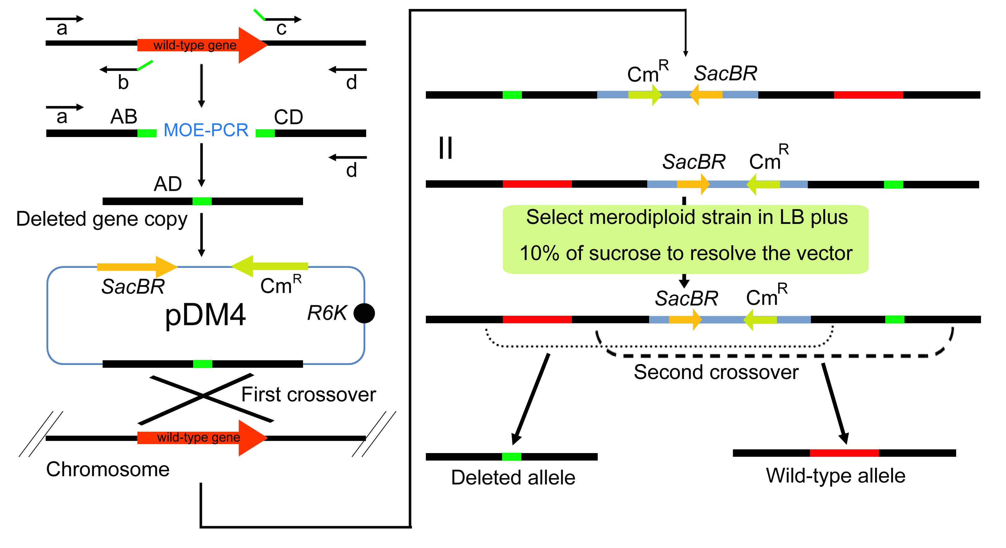
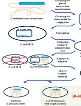
Workflow of deletion mutagenesis in P. aeruginosa. (A) The upstream (AB) and downstream (CD) homologous arms are amplified from the wild-type chromosome with primer pairs a/b and c/d. AB and CD share complementary sequences at their internal junction and are fused by MOE-PCR to generate the deletion cassette AD, in which the target gene is removed. The AD cassette is cloned into the sacB-containing suicide vector pDM4, and the recombinant plasmid is established in E. coli before its transfer into P. aeruginosa by conjugation. A single crossover between the cassette and the chromosomal target integrates pDM4 and creates a merodiploid carrying both the wild-type allele and the deleted allele. (B) The merodiploid is plated on Luria-Bertani (LB) agar without NaCl, containing 10% sucrose. Cells that still carry the integrated pDM4 vector express sacB and die from the accumulation of levan, so only cells in which a second crossover has excised the vector together with one duplicated allele survive. The chromosome of such cells retains either the wild-type allele or the deleted allele, and colony PCR with primers a and d identifies the markerless deletion mutants among the survivors. Note: The flanking primers (a and d) contain at least 17 nt perfectly matched to the target sequence plus a unique restriction site at their 5′ ends for cloning into pDM4. The internal primers (b and c) are 17–35 nt in length and share a 15–25 bp complementary overlap at their 5′ ends, and their Tm values are balanced against those of the flanking primers. The pDM4-AD construct is first established in E. coli strain S17-1 λpir and is then transferred into P. aeruginosa by conjugation.
Background
P. aeruginosa is a ubiquitous Gram-negative environmental bacterium that has remarkable metabolic diversity, resulting in the colonization of a multitude of ecological niches. This bacterium is noted as an important human opportunistic pathogen with the ability to cause acute and chronic infections. In 2000, Stover and his colleagues reported the whole genome sequence of P. aeruginosa type strain PAO1, providing insights into the basis of its pathogenesis, as well as multiple antibiotic resistance and versatile metabolism [1]. However, more than 2,000 genes (ca. 40% of the entire genome) of P. aeruginosa were predicted to encode (conserved) hypothetical proteins, with no clearly predicted functions. This blueprint provides potential opportunities for researchers to explore the functions of undefined genes of this bacterium, thus opening the post-genomic era for Pseudomonas.
Multiple genetic tools and methods have been generated and applied to study the biology of P. aeruginosa, including transposon (knockin) and deletion mutagenesis (knockout). Both have proved to be powerful techniques in the identification of functions of some of those uncharacterized genes, although the functions of certain essential genes (ca. 300–400 for P. aeruginosa) could not be resolved this way [2]. A variety of transposon libraries have been developed for P. aeruginosa strains based on different reporter genes, such as phoA [2–4], lacZ [2], gfp [5], and lux [6]. In addition, a plasposon and a mariner transposon library were also constructed to study the pathogenesis of P. aeruginosa [4,7,8]. While these mutant libraries have greatly facilitated high-throughput genomic studies, functional genomics involving specific non-polar gene knockouts necessitates an efficient deletion mutagenesis approach. Several methods based on allelic exchange have been developed, which combine the use of suicide vectors and counter-selectable markers [9–11]. Among these, Hmelo et al. (2015) [12] established a field-standard two-step allelic exchange protocol for P. aeruginosa, which has since served as the methodological benchmark for unmarked deletion methods in this organism. Unfortunately, although the previously described methods for rapid and efficient mutant generation have been useful in numerous studies, detailed protocols are often still lacking and require further modification to reduce the time and cost of mutant generation and improve its speed and reproducibility.
In this protocol, we describe an efficient approach to generate unmarked deletion mutants in P. aeruginosa based on the gene deletion method we used in previous studies [13–17]. Briefly, MOE-PCR was used to amplify a region containing the flanking sequences of the target gene to be deleted, which was subsequently ligated to a suicide vector (pDM4) [18–20] and introduced into P. aeruginosa by triparental mating with the mobilizable pDM4 derivative. The donor E. coli S17-1 λpir carries the recombinant suicide plasmid, the helper E. coli S17-1 λpir carrying pRK2013 supplies the RP4-derived Mpf machinery in trans, and the recipient is P. aeruginosa PAO1. Because pDM4 carries only the oriT and the TraJ relaxosome but lacks the Mpf machinery, it is mobilizable but not self-transmissible, and the helper strain is essential for plasmid transfer into PAO1. This configuration gives robust conjugation efficiencies sufficient for routine unmarked deletion mutagenesis and avoids the lower efficiencies typical of direct electroporation or chemical transformation of P. aeruginosa, which are limited by its restriction-modification barrier [21]. Second, merodiploids arising from chromosomal integration of the suicide vector through homologous recombination are resolved by the use of the counter-selection imposed by the sacB gene harbored on pDM4 [22]. Finally, gene disruption is confirmed by colony PCR (coPCR) and sequencing, if required. The entire procedure takes less than one week, is easy to perform, is not laborious, and is suitable for large-scale deletion analysis. In addition, the lethal effect of the sacB gene when cells are grown in the presence of sucrose is due to the accumulation of the levan polysaccharide in the periplasm [23,24], which means that this selection method works in most Gram-negative bacteria such as Acinetobacter baumannii, Erwinia chrysanthemi, Legionella pneumophila, Anabaena sp., Yersinia sp., Rhizobium sp., Xanthomonas sp., Pseudomonas sp., Klebsiella pneumoniae, Helicobacter pylori, Agrobacterium sp., and even in some Gram-positive actinobacteria, such as Corynebacterium glutamicum and Mycobacterium sp. [25]. Therefore, the method described here could serve as a general protocol for the genetic manipulation of those bacteria.
This proposed deletion mutagenesis approach has several advantages over other methods. First, MOE-PCR eliminates the intermediate gel-purification step required in conventional overlap extension PCR, saving approximately 4 h of hands-on time per target, reducing gel extraction reagent cost, and avoiding UV exposure of the amplicon, a known source of UV-induced mutations. Second, only one transformation step is involved, saving substantial time relative to marked mutagenesis with FRT-mediated resolution. Third, PM and CAA make it considerably easier to delete oxidative stress-response genes. Aerated LB auto-oxidizes during shaking culture and generates micromolar concentrations of H2O2 lethal to ROS-defective mutants [26]. In our hands, PM and CAA result in a higher recovery of oxidative stress–defective mutants than LB.
Materials and reagents
Biological materials
1. P. aeruginosa PAO1 (wild type; recipient strain) [1]
2. E. coli S17-1 λpir (thi pro hsdR hsdM+ recA RP4-2-Tc::Mu-Km::Tn7 λpir; cloning host and donor strain) [27]
3. E. coli S17-1 λpir (pRK2013) (helper strain for triparental conjugation) [28]
The strains and plasmids used in this protocol are listed in Table 1.
Table 1. Strains and plasmids used in this protocol
| Strain or plasmid | Relevant features | Use in this protocol | Antibiotic concentration |
|---|---|---|---|
| P. aeruginosa PAO1 | Wild type | Recipient strain for conjugation | None; transconjugants are selected on LB agar with Cm 300 μg/mL (section M) |
| E. coli S17-1 λpir | thi pro hsdR hsdM+ recA RP4-2-Tc::Mu-Km::Tn7 λpir | Cloning host. Donor strain carrying pDM-AD | Cm 50 μg/mL on LB agar (section J); Cm 25 μg/mL in LB broth for pDM-AD clones (section K) |
| E. coli S17-1 λpir (pRK2013) | Helper plasmid pRK2013 (ColE1 replicon, Tra+, KmR) | Helper strain for triparental conjugation (section L) | Km 50 μg/mL |
| pDM4 | Suicide vector. oriR6K (requires λpir), sacB, CmR, mob/oriT (RP4) | Suicide vector for allelic exchange | Cm as above |
| pDM-AD | pDM4 carrying the fused deletion construct AD | Recombinant suicide vector | Cm as above |
Reagents
1. Phusion High-Fidelity PCR kit (200 reactions) (New England BioLabs, catalog number: F553L) and Taq DNA polymerase (Qiagen, catalog number: 201203)
Critical: Use high-fidelity DNA polymerase to amplify the deleted gene copy, but use normal DNA polymerase (which is cheaper than the high-fidelity polymerase) to perform coPCRs when screening for positive clones containing the deletion. Phusion High-Fidelity DNA Polymerase is used in section B, and Taq DNA Polymerase is used in section N.
2. Gentra Puregene Yeast/Bact. kit (200 mL) (Qiagen, catalog number: 158567)
3. QIAprep Spin Miniprep kit (250) (Qiagen, catalog number: 27106)
4. Deionized H2O or Milli-Q ultrapure H2O or double-distilled H2O, autoclaved
5. 10 mM dNTP mix (10 mM each dNTP) (Invitrogen, catalog number: 18427-013)
6. Ethidium bromide (Sigma-Aldrich, catalog number: E1510)
7. DNA marker (Smart ladder 1000 lanes) (Eurogentec, catalog number: MW-1700-10)
8. Restriction enzymes: SalI (Thermo Fisher Scientific, Fermentas, catalog number: ER0641) and XbaI (Thermo Fisher Scientific, Fermentas, catalog number: ER0681)
9. Rapid DNA Ligation kit (Fermentas, catalog number: K1422)
10. GenElute Gel Extraction kit (50) (Sigma-Aldrich, catalog number: NA1111-1KT)
11. QIAquick PCR Purification kit (250) (Qiagen, catalog number: 28106)
12. UltraPure agarose (Invitrogen, catalog number: 16500500)
13. 6× DNA loading dye (Fermentas, catalog number: R0611)
14. Tris-HCl (1 M, pH 8.0) (Thermo Fisher, catalog number: 15567-027)
15. EDTA (0.5 M, pH 8.0) (Thermo Fisher, catalog number: 15575-038)
16. Sucrose (Merck, catalog number: 1.07687)
17. RbCl (Sigma-Aldrich, catalog number: R2252)
18. MnCl2·4H2O (Sigma-Aldrich, catalog number: M3634)
19. Potassium acetate (Sigma-Aldrich, catalog number: P1190)
20. CaCl2·2H2O (Sigma-Aldrich, catalog number: C5080)
21. Glycerol (Sigma-Aldrich, catalog number: G5516)
22. MOPS (Sigma-Aldrich, catalog number: M3183)
23. Tris base (Sigma-Aldrich, catalog number: T1503)
24. Glacial acetic acid (Sigma-Aldrich, catalog number: A6283)
25. Chloramphenicol (Sigma-Aldrich, catalog number: C0378)
26. Gelysate peptone (BD BBL, catalog number: 211870)
27. K2SO4 (Sigma-Aldrich, catalog number: P9458)
28. MgCl2 (Sigma-Aldrich, catalog number: M8266)
29. MgSO4·7H2O (Sigma-Aldrich, catalog number: M1880)
30. DMSO (Sigma-Aldrich, catalog number: D8418)
31. Agar (Sigma-Aldrich, catalog number: A1296)
32. Casamino acids (BD, Bacto, catalog number: 223050)
33. K2HPO4·3H2O (Sigma-Aldrich, catalog number: P9666)
34. GelRed nucleic acid gel stain (Biotium, catalog number: 41003) or equivalent safer DNA stain
35. Oligonucleotide primers a–d (17–35 nt each, designed as described in Primer design), HPLC-purified grade (Sangon Biotech, Shanghai, China; custom-synthesized)
Critical: All primers must be ordered as HPLC-purified grade to ensure high fidelity and avoid synthesis errors that could compromise the overlap extension PCR.
36. Absolute ethanol (Sinopharm Chemical Reagent Co., Ltd., Shanghai, China, catalog number: 10009218)
37. Tryptone (BD Bacto, catalog number: 211705)
38. Yeast extract (BD Bacto, catalog number: 212750)
39. Sodium chloride (NaCl) (Sigma-Aldrich, catalog number: S9888)
40. Potassium hydroxide (KOH) (Sigma-Aldrich, catalog number: 221473)
41. Sodium hydroxide (NaOH) (Sigma-Aldrich, catalog number: 221465)
42. Hydrochloric acid (HCl) (Sigma-Aldrich, catalog number: 258148)
43. Syringe filter (0.22 μm, PES) (Millipore, catalog number: SLGP033RS)
Solutions
1. Chemically competent E. coli S17-1 λpir cells (see Recipes)
a. RF1 buffer
b. RF2 buffer
2. Bacterial plates containing appropriate antibiotics (see Recipes)
a. LB broth
b. LB broth without NaCl containing 10% (w/v) sucrose
c. Cm stock solution
d. King's A medium (Pseudomonas agar P medium, PM)
3. 50× TAE stock buffer (see Recipes)
4. TE buffer (see Recipes)
5. 50% sucrose (see Recipes)
6. CAA medium (see Recipes)
Recipes
1. Chemically competent E. coli S17-1 λpir cells
For cloning of the deleted copy into the suicide vector pDM4 (Figure 1), E. coli strain S17-1 λpir (thi pro hsdR hsdM+ recA RP4-2-Tc::Mu-Km::Tn7 λpir) was used, since it produces the λpir protein to maintain the replication of pDM4 plasmid and its derivatives [27]. Chemically competent cells are used in our protocol and made according to the rubidium chloride (RbCl) method [29], which is supposed to increase competence compared to the calcium chloride (CaCl2) method. Alternatively [30], the CaCl2 method can be substituted. The relevant features and antibiotic concentrations of the strains and plasmids used in this protocol are summarized in Table 1 above.
Critical: Prepare RF1 and RF2 buffers freshly. Add metal salts (RbCl, MnCl2, CaCl2) after pH adjustment to avoid precipitation.
a. RF1 buffer (Buffer I for inducing chemical competence)
| Reagent | Final concentration | Quantity or volume |
|---|---|---|
| RbCl | 100 mM | 1.21 g |
| MnCl2·4H2O | 50 mM | 0.99 g |
| Potassium acetate | 30 mM | 0.294 g |
| CaCl2·2H2O | 10 mM | 0.148 g |
| Glycerol | 15% (w/v) | 15 g |
| Deionized H2O | n/a | Up to 100 mL |
Note: Dissolve potassium acetate and glycerol in ~80 mL of deionized H2O. Adjust pH to 5.8 with 0.2 M acetic acid. Do NOT overshoot (Mn2+ precipitates at alkaline pH). Then, dissolve metal salts. Sterilize by filtration (0.22 μm). Store at 4 °C.
b. RF2 buffer (Buffer II for inducing chemical competence)
| Reagent | Final concentration | Quantity or volume |
|---|---|---|
| 3-(N-morpholino) propane sulfonic acid (MOPS) | 10 mM | 0.209 g |
| RbCl | 10 mM | 0.121 g |
| CaCl2·2H2O | 75 mM | 1.10 g |
| Glycerol | 15% (w/v) | 15 g |
| Deionized H2O | n/a | Up to 100 mL |
Note: Dissolve MOPS and glycerol in ~80 mL of deionized H2O. Adjust pH to 6.5 with 1 M KOH. Do NOT overshoot. Then, dissolve metal salts. Sterilize by filtration (0.22 μm). Store at 4 °C.
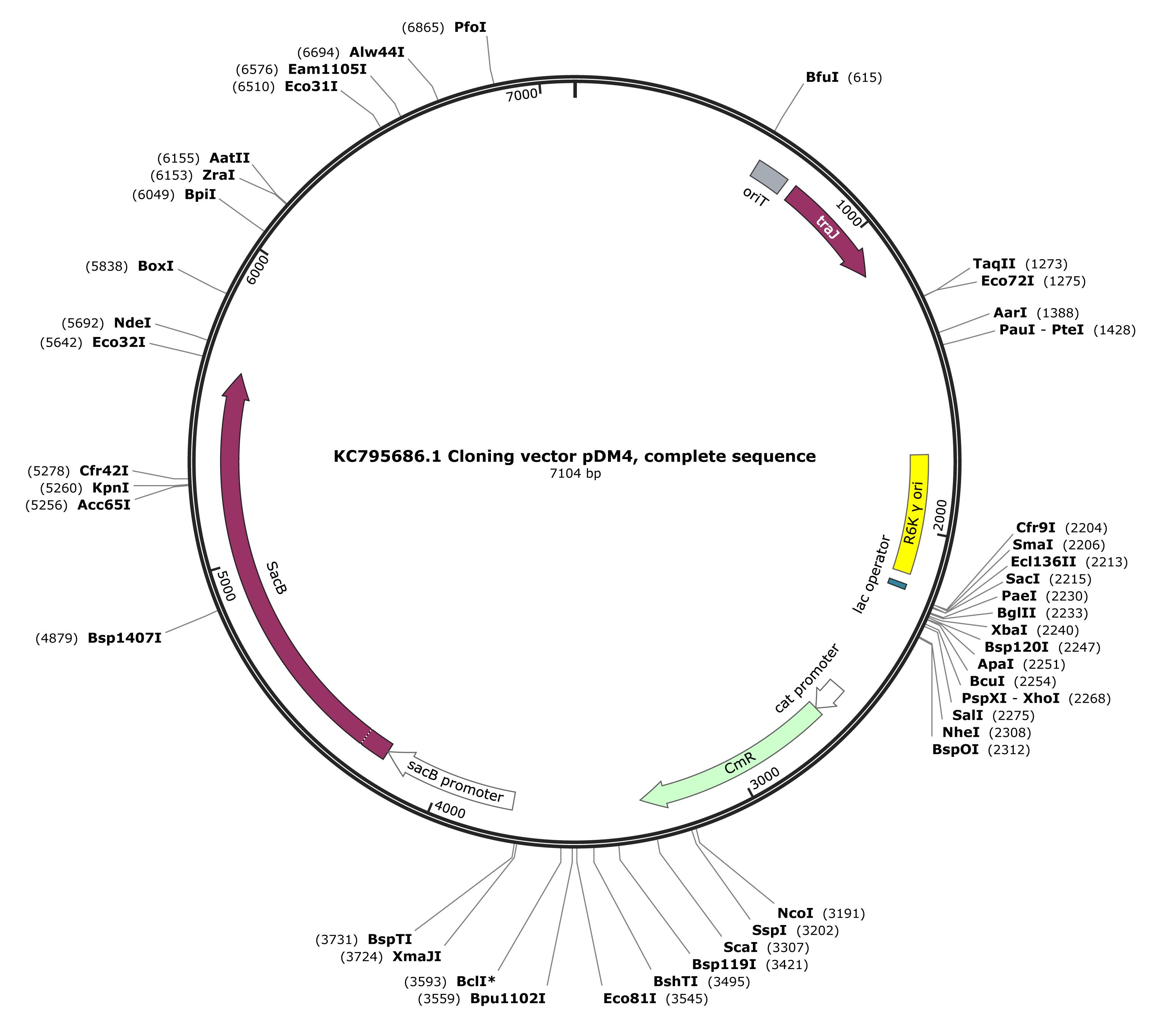
Figure 1. Map of the pDM4 suicide vector. The pDM4 vector (KC795686.1, 7104 bp) carries the R6K γ origin of replication, which requires the λpir protein and therefore restricts propagation to E. coli S17-1 λpir, the RP4 origin of transfer (oriT) and the traJ gene for conjugative mobilization, the sacB gene and its promoter for sucrose counter-selection, and the chloramphenicol resistance gene (cat) under the control of its own promoter. Restriction sites suitable for cloning the deletion cassette AD are indicated.
2. Bacterial plates containing appropriate antibiotics
For cloning of the deletion construct, LB agar plates containing 50 μg/mL chloramphenicol (Cm) are used. Cm serves as the selection marker, because the pDM4 suicide vector carries a chloramphenicol resistance gene. For the selection of P. aeruginosa transconjugants, LB agar plates containing 300 μg/mL Cm are used. For the counter-selection of double recombinants, LB medium without NaCl and containing 10% (w/v) sucrose is used. King's A medium (Pseudomonas agar P medium, PM) is suitable for the isolation of mutants, especially for strains with defects in oxidative stress defense. The recipes of these media are given below.
Critical: Sucrose must be filter-sterilized and added to the cooled autoclaved medium to avoid caramelization.
a. LB broth
| Reagent | Final concentration | Quantity or volume |
|---|---|---|
| Tryptone | 1% (w/v) | 10 g |
| Yeast extract | 0.5% (w/v) | 5 g |
| NaCl | 1% (w/v) | 10 g |
| Deionized H2O | n/a | Up to 1 L |
Note: Dissolve the components in about 900 mL of deionized water, adjust the pH to 7.0 with NaOH if necessary, and bring the volume to 1 L. Sterilize by autoclaving at 121 °C for 20 min. For LB agar plates, add 15 g of agar per liter before autoclaving. LB medium without NaCl is prepared by omitting NaCl from this recipe. Add the Cm stock solution only after the autoclaved medium has cooled to about 50 °C.
b. LB broth without NaCl containing 10% (w/v) sucrose (1 L, approx. 40 plates)
Critical: Sucrose must be filter-sterilized (0.22 μm) and added to the autoclaved medium after it has cooled to approximately 50 °C to avoid caramelization.
| Reagent | Final concentration | Quantity or volume |
|---|---|---|
| LB broth (prepare as above, but omit NaCl) | n/a | 800 mL |
| Sucrose stock [50% (w/v), filter-sterilized] | 10% (w/v) | 200 mL |
Note: The purpose of removing NaCl from the LB broth is to decrease the osmotic potential, which enhances the osmotic stress mediated by the levansucrase encoded by the sacB gene integrated into the chromosome of P. aeruginosa, leading to the rapid resolution of the plasmid from the genome. A sucrose concentration of 10% (w/v) is recommended as a starting concentration for counter-selection. The concentration can be increased up to 20% (w/v) if background growth or false-positive survivors indicate insufficient counter-selection, or decreased to 5% (w/v) if sucrose toxicity substantially impairs the recovery or growth of candidate recombinants. For LB agar plates, add 15 g of agar per liter before autoclaving.
c. Cm stock solution
| Reagent | Final concentration | Quantity or volume |
|---|---|---|
| Chloramphenicol | 30 mg/mL | 300 mg |
| Absolute ethanol | n/a | 10 mL |
Note: Filter sterilization is not required, as the stock is prepared in absolute ethanol. Store at -20 °C in the dark.
d. King's A medium (Pseudomonas agar P medium, PM)
| Reagent | Final concentration | Quantity or volume |
|---|---|---|
| Gelysate peptone | 20 g/L | 20 g |
| Glycerol | 10 mL/L | 10 mL |
| Agar | 15 g/L | 15 g |
| K2SO4 | 10 g/L | 10 g |
| MgCl2 | 1.4 g/L | 1.4 g |
| Deionized H2O | n/a | Up to 1 L |
Note: Glycerol serves as the carbon source. Agar is added before autoclaving when plates are required for the isolation of mutants. Adjust pH to 7.0 with 1 M KOH or HCl before autoclaving. Supplement with appropriate antibiotics as described above.
3. 50× TAE stock buffer
50× TAE is diluted with distilled water (1:50) to make a 1× working solution for the preparation of agarose gel.
| Reagent | Final concentration | Quantity or volume |
|---|---|---|
| Tris base (MW 121.1) | 242 g/L (2 M, 50×) | 242 g |
| Glacial acetic acid | 57.1 mL/L (50×) | 57.1 mL |
| EDTA (0.5 M, pH 8.0) | 50 mM (50×) | 100 mL |
Note: All solutions can be stored at room temperature.
4. TE buffer
| Reagent | Final concentration | Quantity or volume |
|---|---|---|
| Tris-HCl (1 M, pH 8.0) | 10 mM | 10 mL |
| EDTA (0.5 M, pH 8.0) | 1 mM | 2 mL |
| Deionized H2O | n/a | Up to 1 L |
Note: Autoclave before use. TE buffer can be stored at room temperature for up to 6 months.
5. 50% sucrose
| Reagent | Final concentration | Quantity or volume |
|---|---|---|
| Sucrose | 50% (w/v) | 50 g |
| Deionized H2O | n/a | Up to 100 mL |
Note: Filter-sterilize. The 50% sucrose solution can be stored at 4 °C for up to 4 months.
6. CAA medium
| Reagent | Final concentration | Quantity or volume |
|---|---|---|
| Casamino acids | 5 g/L | 5 g |
| K2HPO4·3H2O | 1.18 g/L | 1.18 g |
| MgSO4·7H2O | 0.25 g/L | 0.25 g |
| Agar | 15 g/L | 15 g (when plates are required) |
| Deionized H2O | n/a | up to 1 L |
Note: This is an iron-poor casamino acid medium used for growing P. aeruginosa under iron-limiting conditions, as previously described [17]. Adjust pH to 7.0 with 1 M KOH before autoclaving. Add agar 15 g/L when plates are required. Sterilize by autoclaving.
Equipment
1. Thermal cycler (Eppendorf Mastercycler Gradient, catalog number: 5331 000.011 or equivalent)
2. Agarose gel electrophoresis system (Bio-Rad, model: Sub-Cell GT, catalog number: 170-4406 or equivalent)
3. Gel imaging system (Bio-Rad, model: Gel Doc XR+, catalog number: 1708195 or equivalent)
4. PCR tubes (0.2 mL thin-walled, e.g., Axygen, catalog number: PCR-02-C or equivalent)
5. NanoDrop 2000 spectrophotometer (Thermo Scientific, catalog number: ND-2000, or equivalent)
6. Benchtop incubator (Innova 40, New Brunswick Scientific or equivalent)
7. Biological safety cabinet (Class II, or equivalent)
8. Benchtop centrifuge 5417C (Eppendorf, catalog number: 5417 000.315, rotor FA-45-30-11, or equivalent)
Procedure
The timeline of the complete protocol is summarized in Table 2.
Table 2. Timeline of the complete protocol
| Day | Sections | Work | Approximate time | Pause point |
|---|---|---|---|---|
| Day 1 | A–D | Extract genomic DNA and amplify fragments AB and CD | 4 h | PCR products can be stored at -20 °C |
| Day 1 | E–G | Generate product AD by MOE-PCR and purify it from the gel | 4 h | The excised gel slice can be stored at -20 °C |
| Day 1 (evening) | H | Digest product AD and pDM4 with restriction enzymes | Overnight, about 14 h | Digested DNA can be stored at -20 °C |
| Day 2 | I–J | Ligate AD into pDM4 and transform into E. coli S17-1 λpir | 3 h, then overnight incubation | Transformation plates can be stored at 4 °C |
| Day 3 | K–L | Verify positive clones, extract pDM-AD, and perform conjugation | 8–10 h, followed by overnight incubation | Conjugation plates are incubated overnight at 37 °C |
| Day 4 | M | Select transconjugants on LB plates with 300 μg/mL chloramphenicol | 1 h, then overnight incubation | Plates can be stored at 4 °C |
| Day 5 | N–O | Optional coPCR confirmation and start the counter-selection | 3 h, then about 8 h of growth | The sucrose culture is grown until turbid |
| Day 6–7 | O | Plate serial dilutions and screen for mutants by coPCR | 1–2.5 h per day | Verified mutants can be stored in 15% glycerol at -70 °C |
Primer design
This is the most critical step for the success of the whole protocol. The basic principles for designing primers for overlap extension PCR have been widely described in different articles [31–34], and the strategy is illustrated in the Graphical overview. Proceed as follows:
1. Analyze the DNA sequence of the gene to be deleted together with its upstream and downstream flanking regions. Careful analysis of the sequence greatly facilitates the PCR procedure and the cloning work.
2. Design the flanking primers a and d. Each should contain at least 17 nt perfectly matched to the fragment to be amplified, preceded at the 5′ end by one unique restriction site plus a few protection nucleotides, as recommended by the enzyme manufacturer, to ensure maximum digestion efficiency. Different restriction sites are used in primers a and d, so that the vector is cut at two positions and the insert is ligated in a directed manner. In the validation experiment, the AD cassette was digested with SalI and XbaI and inserted between the corresponding sites of pDM4.
3. Design the internal primers b and c. Special attention should be paid to b and c: they should share about 15–25 bp of overlapping sequence at their 5′ ends, spanning the deletion junction, and their design must not introduce a frameshift mutation after the fusion of fragments AB and CD. Their lengths range from 17 to 35 nt, depending on the balance of the melting temperatures (Tm) of the four primers (a, b, c, and d). The internal primers b and c may also contain a unique restriction site to facilitate the insertion of selective markers or other cassettes.
4. Check each primer against general primer-design guidelines, such as GC content above 50% and avoidance of secondary structures.
5. Check the sizes of the expected PCR products: AB and CD should each range from 200 to 1000 bp, resulting in a 400–2000-bp AD product. It is preferable to obtain amplicons of similar size for AB and CD, which will maximize the efficiency of homologous recombination after the sewing step and transfer to the host cells.
Note: An improved overlap extension PCR cloning method has been described very recently, whereby flanking primers containing a plasmid sequence at the 5′ ends and the insert sequence at the 3′ ends provide a straightforward and efficient way to perform the cloning step without restriction endonucleases, bypassing the ligation step [35].
A. Purification and quantification of genomic DNA
1. Inoculate 5 mL of LB broth with a single colony of P. aeruginosa from a fresh bacterial plate (less than one week old). Bacterial strains are grown overnight at 37 °C with shaking.
2. Genomic DNA is extracted with the Gentra Puregene Yeast/Bact. kit according to the manufacturer’s protocol.
Note: For genomic DNA isolation, different commercial kits such as DNAzol Reagent (GibcoBRL, catalog number: 10503) and DNeasy Blood & Tissue Kit (Qiagen, catalog number: 69504) can be used.
3. Purified genomic DNA can be dissolved in autoclaved deionized H2O or in TE buffer and quantified using a spectrophotometer (NanoDrop ND-2000).
Critical: DNA quality is assessed by the A260:A280 ratio. A value of 1.7–1.9 indicates DNA that is free of protein contamination and suitable for downstream applications. The DNA solution is then diluted to approximately 50 ng/μL for the following steps. Typically, about 20–50 μg of genomic DNA is obtained from 5 mL of an overnight culture, which is far more than needed for the entire protocol.
Pause point: Purified genomic DNA can be stored at -20 °C in TE buffer for months and at -80 °C for years.
B. Set up PCRs to amplify AB and CD DNA fragments
1. Label PCR tubes for the AB and CD amplicons and for one negative control per PCR, that is, one no-template control for the AB reaction and one for the CD reaction. For each primer pair, prepare a master mix for two tubes, one sample, and one negative control, and dispense it into the two tubes to minimize pipetting errors. Combine all reagents in individual PCR tubes on ice. Add the Phusion DNA polymerase at the end, which should be kept at -20 °C until its addition. The negative control reactions contain all components, including the corresponding primer pair, except that the template DNA is replaced by the same volume of water.
2. The reaction mix volume is usually 50 μL for each sample, which is enough for most applications, as shown in Table 3.
Table 3. Reaction mixture for the first PCR to amplify the AB and CD fragments
| Component | Volume/50 μL (product AB) | Volume/50 μL (product CD) | Final concentration$ |
| 5× Phusion HF buffer | 10 μL | 10 μL | 1× |
| 10 mM dNTPs | 1 μL | 1 μL | 200 μM for each |
| Primer a | x* μL | - | 0.5 μM |
| Primer b | x* μL | - | 0.5 μM |
| Primer c | - | x* μL | 0.5 μM |
| Primer d | - | x* μL | 0.5 μM |
| DMSO# | 1.5 μL | 1.5 μL | 3% |
| Template DNA | x* μL | x* μL | 50–125 ng |
| H2O | Up to 50 μL | Up to 50 μL | |
| Phusion DNA polymerase | 0.5 μL | 0.5 μL |
* x indicates variable since the concentration of primers and template DNA differs between users; the volume used per reaction must be calculated individually. For a 10 μM primer stock, add 2.5 μL to obtain a final concentration of 0.5 μM in a 50 μL reaction.
# Addition of DMSO is recommended for GC-rich amplicons, but not in the case of low GC content amplicons or amplicons larger than 20 kb.
$ The use of 0.5 μM as the final concentration of the primers is suitable for most PCRs and is also recommended in the Phusion High-Fidelity PCR system.
Critical: Introduction of Phusion DNA polymerase to this modified protocol has obvious and significant advantages over other systems, especially in terms of efficiency and time. Before starting, one should carefully read the manual provided by the supplier (New England BioLabs).
C. Run PCRs for products AB and CD
1. Place the PCR tubes in the thermal cycler and run the program shown in Table 4.
Table 4. Cycling conditions for the first PCR
| Cycle number | Denaturation | Annealing | Extension | Hold |
|---|---|---|---|---|
| 1 | 98 °C, 30 s | |||
| 2–30 | 98 °C, 5–10 s | 50–60 °C, 10–30 s | 72 °C, 15–30 s | |
| 31 | 72 °C, 5–10 min | |||
| 32 | 4 °C, hold |
Critical: The Phusion DNA polymerase uses 98 °C for denaturation and generates blunt ends in amplification products. For extension, 15–30 s per kilobase is recommended for Phusion polymerase. Since AB and CD fragments are designed to be 200–1,000 bp, a 15–30 s extension step is sufficient for the entire range; extend proportionally if longer amplicons are used.
Critical: The choice of annealing temperature depends on the primers used; in this protocol, we recommend using an annealing temperature higher than 50 °C, since a lower annealing temperature will cause the formation of nonspecific products.
Pause point: Hold PCR tubes at 4 °C or store at -20 °C for one month for further use.
D. Gel electrophoresis analysis of PCR products
1. Pour a 0.8% (w/v) agarose gel by melting 0.8 g of UltraPure agarose in 100 mL of 1× TAE buffer. The agarose percentage should be adjusted to the expected size of the PCR products. A 0.8% (w/v) gel (0.8 g in 100 mL) effectively resolves fragments of 800–10,000 bp, while a 1.5% (w/v) gel (1.5 g in 100 mL) resolves fragments of 200–3,000 bp and is recommended for products shorter than 800 bp. In the validation experiment (Figure 2), a 0.8% gel was used and was sufficient to resolve the three products of the PA4203 deletion (356, 539, and 895 bp), since only one fragment was expected per lane, although a higher percentage gives a better resolution of the two shorter products.
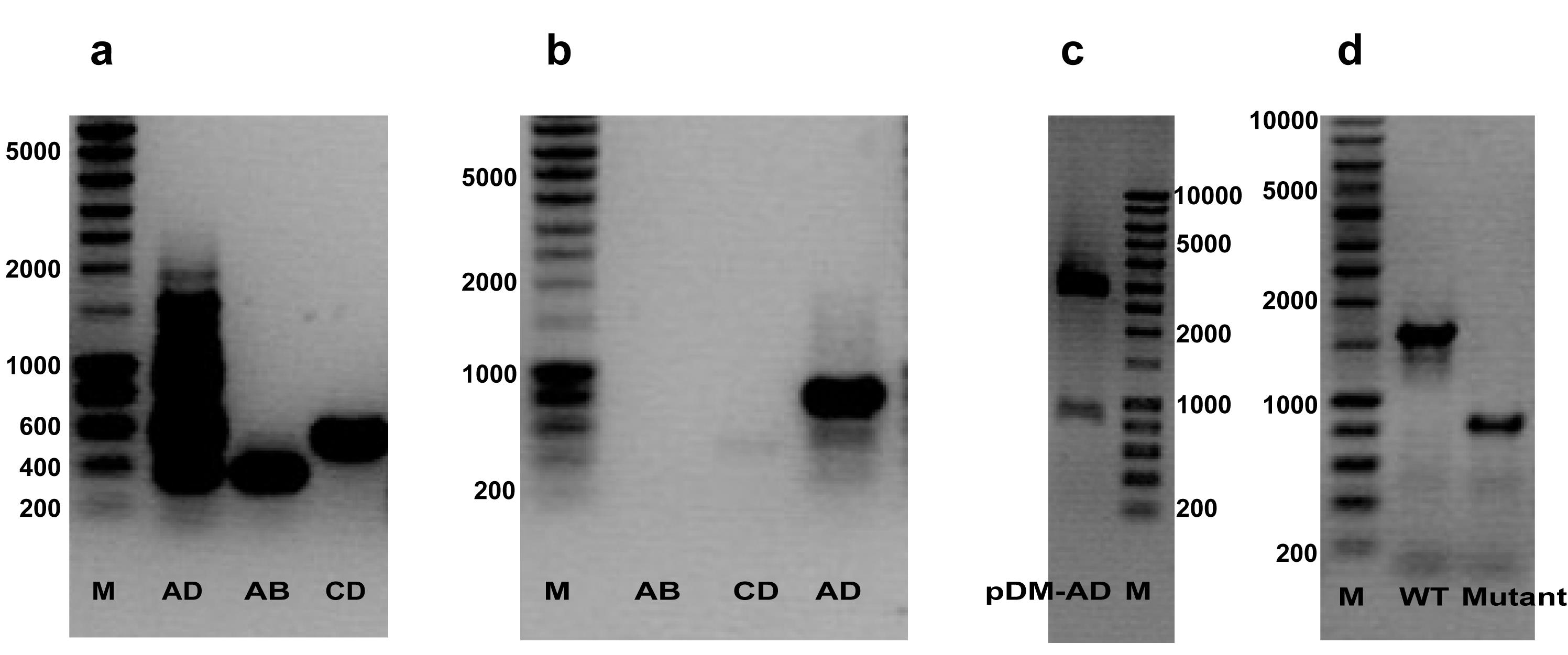
Figure 2. Deletion of the PA4203 gene in P. aeruginosa. (a) PCR product AD, AB, and CD. (b) PCR product AB, CD, and AD. In (a), the ratio of flanking primers to internal primers was 1:1, and in (b), it was 10:1 (see the second Critical point in section E, which describes the primer-ratio adjustment made in the first PCR setup). The weak or invisible AB and CD bands in (b) are expected because the low concentration of internal primers limits the yield of the first PCR, while the small amount of AB and CD is sufficient as the template for the MOE-PCR. (c) Digested recombinant suicide vector pDM-AD. (d) coPCR analysis of the deletion mutant, with wild type as a positive control. Lanes are labeled in each panel. All DNA markers used here are Smart Ladder (200–10,000 bp).
2. Place the gel into the Bio-Rad gel electrophoresis apparatus and remove the comb. Immerse the gel in 1× TAE buffer and make sure that the buffer fills each end of the electrophoresis apparatus.
3. Load 5 μL of a suitable DNA marker (Smart ladder from Eurogentec) into the first well in the gel. Then, load 10 μL of a mixture containing 2 μL of 6× DNA loading buffer and 8 μL of PCR products.
4. Run the gel at 100 V and monitor the migration of the indicator dye periodically. Stop the electrophoresis when the dye front has reached the middle of the gel (for products larger than 1 kb) or approximately 1 cm from the bottom (for products smaller than 1 kb), depending on the expected size of the products. The dye front serves as a visual guide only; the exact stopping point can be adjusted according to the gel length and the size range of the DNA fragments.
5. Check the results by staining the gel in DNA staining solution containing ethidium bromide (EB) for 10 min and visualize using a Bio-Rad Imaging System or equivalent.
Note: In this protocol, we do not purify the PCR products AB and CD from the gel, but use them directly in step E1, which is a critical change in overlap extension PCR and one of the reasons we refer to the protocol as modified overlap extension PCR (MOE-PCR).
Caution: EB is very toxic; avoid contact with skin by using gloves.
Note: The remaining PCR products (approximately 42 μL per reaction) are kept on ice for direct use in section E.
E. Set up PCR for product AD
1. Usually, a reaction volume of 50 μL is sufficient to support the subsequent experiments (restriction enzyme digestion and gel purification). One can eventually increase the volume of the reaction to 100 μL. Once the AB and CD fragments have been successfully amplified, prepare the second PCR (MOE-PCR) as shown in Table 5.
Table 5. Reaction mixture for the overlap extension PCR (MOE-PCR) to generate product AD
| Component | Volume/50 μL (product AD) | Negative control | Final concentration |
|---|---|---|---|
| 5× Phusion HF buffer | 10 μL | 10 μL | 1× |
| 10 mM dNTPs | 1 μL | 1 μL | 200 μM for each |
| Primer a$ | x μL | x μL | 0.25 μM (range 0.125–0.5) |
| Primer d$ | x μL | x μL | 0.25 μM (range 0.125–0.5) |
| DMSO | 1.5 μL | 1.5 μL | 3% |
| Product AB# | 5–15 μL | - | |
| Product CD# | 5–15 μL | - | |
| H2O | Up to 50 μL | Up to 50 μL | |
| Phusion DNA polymerase | 0.5 μL | 0.5 μL |
# Products AB and CD should always be kept on ice to avoid degradation. As a starting point, use 5 μL of each product when the bands are of normal intensity on the gel (section D). Increase the volume toward 15 μL when the yield of the first PCR is low, and decrease it when the bands are strong. Use similar volumes of AB and CD so that the two fragments are balanced and keep the combined volume at or below 30 μL to limit the carryover of dNTPs, primers, and buffer from the first PCR.
$As a starting point, use primers a and d at a final concentration of 0.25 μM (1.25 μL of a 10 μM stock in a 50 μL reaction), i.e., half of the amount used in the first PCR (0.5 μM, section B). This concentration is sufficient because the template of the MOE-PCR consists of the already amplified, high-copy AB and CD products rather than genomic DNA, and it minimizes nonspecific amplification, which increases with primer concentration. If the yield of product AD is low, the concentration can be raised up to 0.5 μM (2.5 μL of a 10 μM stock); if nonspecific bands are still visible on the gel, it can be reduced to 0.125 μM (0.625 μL of a 10 μM stock).
Critical: The direct use of PCR product AB and CD in the second PCR further simplifies the protocol and avoids using gel purification or a PCR purification kit to isolate relatively pure PCR products, which could possibly introduce mutations via exposure to UV and poor DNA recovery. Doing so saves a lot of time and money.
Critical: If desired, one can also purify the PCR fragments AB and CD from the gel according to [31]. One possible disadvantage of our MOE-PCR is a higher background in the gel due to the presence of reagents from the first PCR. This can easily be solved by either carefully and rapidly cutting the desired bands from the gel or by increasing the ratio of the flanking primers (primer a and d) to internal primers (primer b and c) from 1:1 to 10:1 in the first PCR setup. A PCR purification kit can be used instead to remove the carryover reagents, which avoids exposing the DNA to UV light. The gel intended for excision of product AD should not be stained with ethidium bromide. Instead, a safer DNA stain such as GelRed can be used, which can be visualized under long-wavelength UV (365 nm) or, preferably, on a blue-light transilluminator to avoid exposing the DNA to UV altogether. Locate the band under long-wavelength UV (365 nm) and minimize the exposure time.
F. Run PCR for product AD
1. Place the PCR tubes in a thermal cycler carefully and run the program shown in Table 6.
Table 6. Cycling conditions for the MOE-PCR
| Cycle number | Denaturation | Annealing | Extension | Hold |
|---|---|---|---|---|
| 1 | 98 °C, 30 s | |||
| 2–30 | 98 °C, 5–10 s | 50–60 °C, 10–30 s | 72 °C, 15–30 s | |
| 31 | 72 °C, 5–10 min | |||
| 32 | 4 °C, hold |
Pause point: PCR fragments can be kept in the thermal cycler at 4 °C overnight or in the freezer for up to one month. If planning to continue with step G1, one can prepare the gel for in-gel purification of product AD to further save time.
G. Isolate and in-gel purify product AD
1. Repeat steps D1–5 to verify the fusion PCR product AD.
Critical: A thicker preparative gel should be used in step D1 in order to accommodate all 50 μL product in one or two wells maximum. Use a 15 × 10 cm gel tray with a wide-tooth preparative comb. Pour the gel to a thickness of 8 mm using 120 mL of 0.8% (w/v) agarose.
Note: The gel percentage is chosen according to the expected size of product AD, as described in step D1. A 0.8% (w/v) gel is suitable for fragments of 800–10,000 bp. A 1.5% (w/v) gel improves the resolution of shorter fragments and is recommended when product AD is shorter than 800 bp. Proceed to excision only if a single, clear band of the expected size is observed; if multiple bands or smearing are present, see section E, second Critical point, for troubleshooting.
2. Locate the product AD band without exposing the excised DNA to EB. Either stain the gel with a safer DNA stain such as GelRed and visualize under 365 nm UV, or stain side strips of the gel briefly with EB for reference and excise the band from the unstained central portion.
3. Excise the product AD very carefully from the gel containing the desired bands with a razor blade and place it into a sterile 2-mL microfuge tube.
Pause point: The excised gel containing the AD product can be stored in the freezer for up to one month.
4. Purify the PCR product AD using the GenElute Gel Extraction kit according to the manufacturer’s protocol. Finally, dissolve the purified AD product in double-distilled H2O. The concentration and quality of the DNA are determined in step I2 before ligation.
Note: Other gel extraction kits or DNA extraction methods are also applicable in this step.
H. Restriction enzyme digestion of AD product and pDM4 vector
1. pDM4 is maintained in E. coli S17-1 λpir. An overnight culture is used for plasmid extraction with the QIAprep Spin Miniprep Kit before setting up the digestion. Label individual autoclaved 1.5-mL microfuge tubes for product AD and the suicide vector pDM4. Set up restriction digestion reactions by combining the reagents given in Table 7 on ice, keeping the enzyme cold and adding it at the end.
Table 7. Reaction mixture for restriction digestion of product AD and the pDM4 vector
| Component | Volume/50 μL (product AD) | Volume/50 μL (vector pDM4) | Final concentration |
|---|---|---|---|
| 10× digestion buffer | 5 μL | 5 μL | 1× |
| Restriction enzyme a | x μL | x μL | 2 U/μg DNA |
| Restriction enzyme b | x μL | x μL | 2 U/μg DNA |
| pDM4 | - | x μL | 1–3 μg |
| Product AD | x μL | - | Use all eluted AD |
| H2O | Up to 50 μL | Up to 50 μL |
Note: To obtain a relatively high concentration of insert DNA (product AD) and vector pDM4, elute with a small volume of water (10–20 μL).
2. Let the reaction run overnight (~14 h) at 37 °C (SalI and XbaI, the enzymes used here, are both active at this temperature).
Critical: Since this step is likely to be performed at the end of the first day, we suggest doing the digestion overnight and continuing the work the next day. Alternatively, one can use the FastDigest restriction enzyme (Fermentas), which only requires 0.5 h to complete the digestion of the two DNAs.
Caution: Pay attention to the star activity introduced by overtime digestion; for detailed information, please refer to the accompanying manual for the restriction enzymes. Enzymes susceptible to cause star activity include EcoRI, BamHI, EcoRV, HindIII, PstI, SalI, and XmnI.
I. Ligation of digested product AD and vector pDM4
1. Purify the digested product AD and vector pDM4 with the QIAquick PCR Purification kit and elute with double-distilled H2O.
2. Quantify the concentration and assess the quality of purified product AD and vector pDM4 using a NanoDrop ND-2000 spectrophotometer. DNA purity is evaluated by measuring the A260:A280 ratio, and a ratio of 1.7–1.9 indicates good-quality DNA.
Pause point: One can store the purified digested DNA at -20 °C for months.
3. Ligate product AD into cut vector pDM4 using the Rapid DNA Ligation kit. Prepare the reactions according to the manufacturer’s protocol, as shown in Table 8. Incubate for 10 min at 22 °C.
Table 8. Reaction mixture for ligation of product AD into the pDM4 vector
| Component | Volume/20 μL |
|---|---|
| Linear vector DNA | 20–100 ng |
| Insert DNA (product AD) | 1:1 to 10:1 molar ratio over vector |
| 10× DNA ligase buffer | 2 μL |
| T4 DNA ligase | 1 μL |
| H2O | Up to 20 μL |
Note: One should use a small volume of water (10–20 μL) to reach a relatively high concentration of insert DNA (product AD) and vector pDM4. Alternatively, other types of T4 DNA Ligation kits can also be used in this step, and ligation could be done within 1 h or even overnight if desired. A 3:1 insert-to-vector molar ratio is commonly used as a starting point for PCR product cloning, but the optimal ratio may vary with different inserts. We therefore recommend setting up parallel ligations at 1:1, 3:1, and 5:1 and selecting the condition that gives the best result. The ratio can be determined through the following equation:
where N is the ratio of insert DNA to vector DNA. Recommended control: Prepare a parallel ligation reaction containing only the linearized vector (no insert) to estimate the background of self-ligation.
Pause point: Ligation reactions can be stored at -20 °C for months.
J. Transform into E. coli S17-1 λpir
1. Heat-inactivate T4 ligase at 65 °C for 10 min or 70 °C for 5 min.
Note: This step can greatly improve transformation efficiency in some cases.
2. Thaw chemically competent E. coli S17-1 λpir cells (see Recipe 1) on ice.
3. Add 2–5 μL of the inactivated ligation mixture to 50–100 μL of competent cells. Mix gently and incubate on ice for at least 5 min, using the standard heat shock transformation protocol [29].
Note: Include a negative control (no DNA) and a positive control (uncut plasmid vector) to exclude contamination and verify transformation efficiency.
4. Heat-shock the cells at 42 °C for 90 s and immediately transfer the tube to ice for approximately 2 min.
5. Add 500 μL to 1 mL of LB broth without antibiotics and incubate at 37 °C with shaking for 45–60 min to allow recovery.
6. Spread 100 or 150 μL of the recovered culture onto LB agar containing 50 μg/mL Cm.
7. Air-dry the plates and incubate overnight at 37 °C.
Note: Keep the remaining cells at 4 °C as backup in case additional plating is needed.
K. Verify the presence of insert
1. Label 10 colonies on the transformation plate and the corresponding PCR tubes.
2. Prepare PCR solutions using primers a and d as described in section N (Table 9) and distribute equal volumes into individual PCR tubes.
Table 9. Colony PCR (coPCR) reaction mix
| Reagent | Quantity or volume | Final concentration |
|---|---|---|
| 10× Taq buffer | 5 μL | 1× |
| 10 mM dNTPs | 1 μL | 200 μM each |
| Primer a (10 μM) | 2.5 μL | 0.5 μM |
| Primer d (10 μM) | 2.5 μL | 0.5 μM |
| DMSO | 1.5 μL | 3% |
| Taq DNA polymerase | 0.5 μL | 2.5 U |
| Colony (on sterile toothpick) | 1 piece | n/a |
| Deionized H2O | Up to 50 μL | n/a |
3. Use a clean and sterile toothpick to scrape a tiny piece of a bacterial colony into the labeled PCR tube, swirl for 2–3 rounds, and remove it immediately and carefully without touching the tube wall.
4. Run the standard PCR program described in section N (Table 10).
Table 10. Colony PCR cycling conditions
| Cycle number | Denaturation | Annealing | Extension | Hold |
|---|---|---|---|---|
| 1 | 95 °C, 5 min | |||
| 2–36 | 95 °C, 45 s | 55 °C, 30 s | 72 °C, 2 min | |
| 37 | 72 °C, 10 min | |||
| 38 | 4 °C, hold |
Note: For longer amplicons, the 72 °C extension time during cycles 2–36 may be adjusted to approximately 1 min/kb.
5. Check the PCR products by running an agarose gel according to steps D1–5.
6. Streak the confirmed positive clones on an LB agar plate containing 50 μg/mL Cm and on an LB agar plate without NaCl but with 10% (w/v) sucrose. The construct renders the cells sucrose-sensitive, so no growth should be observed on the sucrose-containing plate after incubation.
7. Inoculate at least two confirmed positive clones into LB broth containing 25 μg/mL chloramphenicol and grow them at 37 °C with shaking at 220 rpm until sufficient growth is obtained for plasmid extraction later that day.
8. The next morning, extract the recombinant plasmid pDM-AD using the QIAprep Spin Miniprep kit (or equivalent) and send the plasmid for sequence analysis to confirm the deleted gene sequence.
9. After confirming the positive pDM-AD clones, inoculate the donor strain (E. coli S17-1 λpir carrying pDM-AD), the helper strain (E. coli S17-1 λpir/pRK2013), and wild-type P. aeruginosa, each from a single colony, into 3 mL of LB broth. Incubate for 3–4 h at 37 °C with shaking at 220 rpm until log phase and perform conjugation later on the same day.
Note: Hundreds of colonies should appear on the transformation plates. Screening 5–20 colonies is generally enough to obtain at least one positive clone. Picking colonies directly from the transformation plate is an easy and rapid screening approach. Colony PCR is generally fast and saves time compared with other alternatives. It is unnecessary to use a high-fidelity DNA polymerase in colony PCR. Taq polymerase is sufficient. A permanent culture can be made from each positive strain in LB broth containing 15% glycerol and stored at -70 °C.
L. Conjugate the suicide recombinant vector pDM-AD into P. aeruginosa
The suicide vector pDM4 carries the RP4-type origin of transfer (mob/oriT) but no transfer genes, so its mobilization depends on the RP4 transfer functions provided in trans. The donor strain E. coli S17-1 λpir carries an RP4 derivative (RP4-2-Tc::Mu-Km::Tn7) integrated in its chromosome and can therefore supply the basic transfer functions. In this protocol, however, we use the classical triparental mating scheme, in which a separate helper strain (E. coli S17-1 λpir/pRK2013) supplies the complete RP4 transfer region (tra and trb genes) on the multicopy plasmid pRK2013. Providing the transfer machinery from a helper strain in high abundance increases the efficiency of pDM-AD mobilization and follows the established convention for allelic exchange in P. aeruginosa.
1. Place the wild-type P. aeruginosa culture in a 42 °C incubator for 30 min.
Note: This preincubation minimizes the efficiency of the bacterium's restriction systems and production of the phenazine compound pyocyanin.
2. From each culture [donor strain (E. coli S17-1 λpir harboring pDM-AD), recipient strain (wild-type PAO1) and helper strain (E. coli S17-1 λpir harboring pRK2013)], transfer 1 mL into a microcentrifuge tube and centrifuge at 12,850× g for 1 min, wash the pellet twice with 1 mL of fresh LB medium, and then resuspend in 1 mL of fresh LB medium.
3. Mix 500 μL of the donor strain culture, 500 μL of the helper strain, and 200 μL of the recipient strain culture (P. aeruginosa PAO1) into a sterile microfuge tube and centrifuge at 12,850× g for 1 min. Wash the mixed cell pellet twice again with 1 mL of fresh LB medium to remove as much pyocyanin and other antibiotics as possible. Finally, resuspend the pellet in 250 μL of LB medium. A negative control should be set up in which the donor culture is replaced by LB medium.
Note: Electroporation can also be used to introduce the recombinant suicide vector into the recipient cells, which is rapid and easy to perform but has lower efficiency. Therefore, we recommend using the conjugation procedure described in this protocol.
4. Inoculate a plate with the mixture in several dots of approximately 5 μL or plate 0.1 mL of the bacterial cell suspension on a LB plate. Incubate the air-dried plate at 37 °C overnight.
Critical: These two steps will ensure the conjugal transfer of the suicide vector into the recipient P. aeruginosa.
Note: No antibiotic is added to the mating plate because the recipient P. aeruginosa is chloramphenicol-sensitive and must remain viable during conjugation. Selection for transconjugants is applied in section M.
M. Select P. aeruginosa transconjugants (first crossover)
1. Scrape the bacterial lawn from LB plates with 3 mL of fresh LB and dilute the cells 100 times in sterile LB or 10 mM MgSO4 in microfuge tubes. Duplicate LB-medium plates containing 300 μg/mL Cm are used for each deletion analysis. Incubate overnight at 37 °C. P. aeruginosa colonies (mucus, greenish, and not very smooth in appearance) will appear after overnight incubation.
2. Replicate approximately 50 colonies onto a new LB plate with 300 μg/mL Cm.
Note: Since we conjugated a suicide vector into P. aeruginosa, where it cannot replicate, this indicates that antibiotic resistance encoded by the plasmid results from the integration of the vector into the P. aeruginosa chromosome via homologous recombination between the cloned fragment and the corresponding region of the recipient chromosome. This process is also called single crossover.
N. Confirm P. aeruginosa conjugant
1. If confirmation of suicide-vector integration by coPCR is desired, set up colony PCR reactions using primers a and d as described below. Pick a small portion of each colony with a sterile toothpick and place it into the reaction mix. We suggest screening approximately 20 colonies. The standard 50-μL reaction mixture and cycling conditions are given in Tables 9 and 10.
2. Run the agarose gel as described in steps D1–5. If the suicide vector has integrated into the target site in the recipient chromosome, the corresponding merodiploid is expected to contain both the wild-type and the recombinant allele. However, coPCR at this stage is optional, because one or both expected products may fail to amplify or may not be equally visible.
Note: The transconjugants may be taken directly to the sucrose counter-selection in section O without performing coPCR at this stage. The appearance of dead cells or loss of turbidity in sucrose-containing cultures indicates sacB-associated sucrose sensitivity but does not by itself definitively confirm chromosomal integration. CoPCR at this stage should therefore be regarded as supportive rather than mandatory. If only one band is detected, this should not alone be interpreted as evidence that integration has failed. The final genotype should be confirmed by coPCR of sucrose-resistant candidate colonies.
O. Screen P. aeruginosa mutants (second crossover)
1. Inoculate one positive P. aeruginosa conjugant from a single colony into 4 mL of LB medium without NaCl containing 10% (w/v) sucrose, without antibiotics. Incubate at 37 °C with shaking at 220 rpm for approximately 8 h, or until the culture becomes turbid.
Critical: The turbid bacterial cultures can either be serially diluted for an additional 2–3 generations or directly plated onto the LB medium using serial dilutions to select mutants. Importantly, dead cells should appear on top of the liquid cultures because of cell lysis caused by the high osmotic pressure generated by the accumulation of levan in the periplasm due to the presence of the sacB gene in the chromosome. In addition, removal of NaCl from the LB medium further enhances the osmotic stress for the cells, accelerating the lysis of bacterial cells and increasing the selective pressure to obtain the second crossover. For genes important for bacterial growth or survival, the cultures may be incubated for only one generation before serial dilution and plating to reduce the loss of viable mutants.
Critical: Because some genes may be more important than others for the survival of the bacterium, in some cases, it can be difficult to select the unmarked mutants. If that happens, we could use cultures incubated for only one generation to reduce the loss of mutants during serial dilutions. For genes involved in the oxidative stress response, use PM or CAA instead of LB during mutant screening. Aerated LB auto-oxidizes and generates micromolar H2O2 lethal to ROS-defective mutants [26]. In our hands, mutants whose survival is sensitive to oxidative stress are recovered at a higher frequency on PM or CAA than on LB.
Note: Sucrose-resistant colonies that retain chloramphenicol resistance are escape clones (the pDM4 vector has not been excised by the second crossover). A Cm-sensitivity assay is therefore recommended as a secondary screen to distinguish escape clones from true double-crossover mutants.
2. Serially dilute the bacterial cultures with LB broth to 105–107 and plate 100 μL of the diluted cultures on LB-NaCl+10% sucrose plates without antibiotics. Air-dry and incubate the plates at 37 °C overnight. Around 50–200 single colonies are expected per plate. If no colonies appear after overnight incubation, continue the incubation for another 24 h before discarding the plates.
3. Single colonies should appear, and coPCR can be performed as described in step N1 to identify strains with the expected deletion. We normally start with 20 colonies to screen.
4. Run the PCR products to identify the positive mutants.
Note: The positive clones should give a smaller band as compared to the wild-type gene (see Figure 2d). In theory, the screening frequency of positive clones should be 50%, but it can be lower if the phenotype caused by the mutation is detrimental to survival. It is also recommended to streak one or two positive mutants on LB agar containing 300 μg/mL Cm to confirm the loss of the suicide vector.
Critical: Once a desired mutant has been confirmed, prepare a permanent culture and store at -70 °C.
Critical: If certain mutations could cause resistance to Cm, it would be difficult to isolate mutants with vectors conferring chloramphenicol. Alternatively, one may try another type of vectors with different antibiotics resistance.
Data analysis
The procedure is supported by a continuous DNA-level validation chain, summarized across panels (a)–(d) of Figure 2. Panel (a) shows the first PCR setup at a 1:1 ratio; panel (b) shows the same setup at the 10:1 ratio recommended in section E, second Critical point (in the first PCR setup); panel (c) shows the pDM4 integration intermediate; and panel (d) shows the wild-type versus mutant genotype comparison. Deletion mutants are identified by colony PCR (coPCR) using flanking primers a and d. The PCR products are analyzed by agarose gel electrophoresis (0.8–1.5% w/v, depending on the expected fragment sizes, see step D1) and compared with the wild-type amplicon. Positive mutants yield a smaller band than the wild type (Figure 2d). The deletion is further confirmed by sequencing the PCR product and by a chloramphenicol sensitivity assay (loss of the suicide vector). For screening efficiency, 20 colonies are typically analyzed per gene deletion. The MOE-PCR amplicon composition and the digested recombinant vector are shown in Figure 2. The mutant screening frequency across bacterial generations for two gene targets is shown in Figure 3.
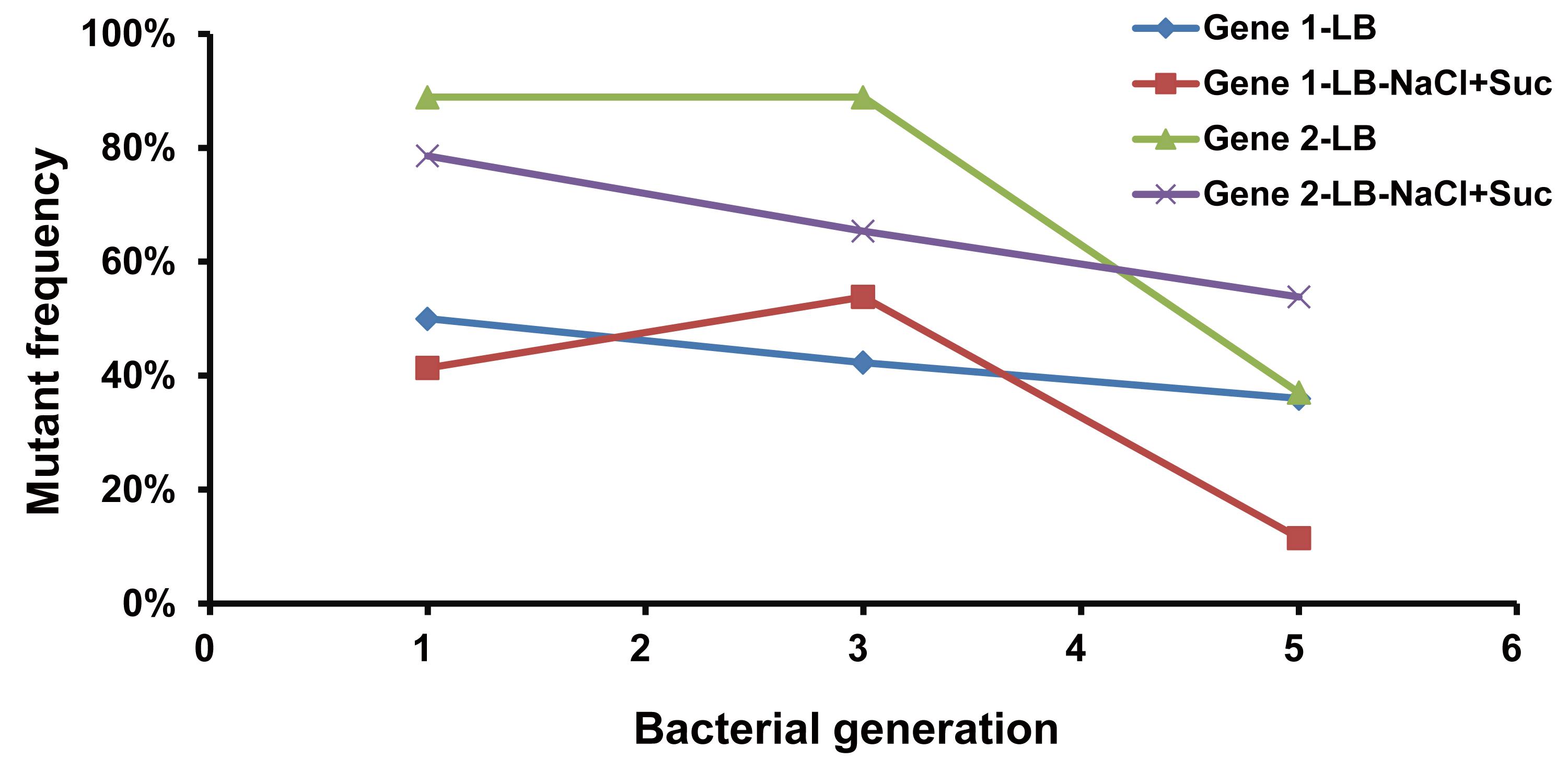
Figure 3. Comparison of different bacterial generations in mutant screening for two genes of P. aeruginosa. Twenty colonies were picked for coPCR and identified by gel electrophoresis. Positive mutants will give a short fragment, and negative mutants will show no bands or a large band (depending on the elongation capacity of the Taq polymerase and the elongation time for fragment screening).
Validation of protocol
The part of the protocol related to the creation of the deletion mutant has been validated in the following research articles:
• Elfarash et al. [17]. The soluble pyocins S2 and S4 from Pseudomonas aeruginosa bind to the same FpvAI receptor. MicrobiologyOpen (Figure 1C). https://doi.org/10.1002/mbo3.27
• Vercammen et al. [16]. Pseudomonas aeruginosa LysR PA4203 regulator NmoR acts as a repressor of the PA4202 nmoA gene, encoding a nitronate monooxygenase. Journal of Bacteriology (Figure 1). https://doi.org/10.1128/jb.01991-14
• Tarighi et al. [13]. The PA4204 gene encodes a periplasmic gluconolactonase (PpgL) which is important for fitness of Pseudomonas aeruginosa. Microbiology (Reading) (Figure 1). https://doi.org/10.1099/mic.0.2008/018465-0
In total, more than 20 unmarked deletion mutants have been successfully constructed in P. aeruginosa PAO1 using this protocol (Table 11).
Table 11. Genes deleted in P. aeruginosa using the method described in this protocol
| PA number | Gene name | Product |
| PA0085 | hcp1 | Type VI secretion system secreted protein Hcp1 |
| PA0291 | oprE | Anaerobically induced outer membrane porin OprE precursor |
| PA0962 | dps | Probable DNA-binding stress protein |
| PA0985 | pys5 | Pyocin S5 |
| PA1150 | pys2 | Pyocin S2 |
| PA1179/PA1180 | phoP/phoQ | Two-component system |
| PA1201 | Probable transcriptional regulator | |
| PA1285* | Probable transcriptional regulator | |
| PA1431 | rsaL | Regulatory protein RsaL |
| PA2020 | mexZ | Probable transcriptional regulator |
| PA2386 | pvdA | L-ornithine N5-oxygenase |
| PA2398 | fpvA | Ferripyoverdine receptor |
| PA2398/PA4168 | fpvA/fpvB | Ferripyoverdine receptors |
| PA2398/PA4221 | fpvA/fptA | Ferripyoverdine receptor/Fe (III)-pyochelin outer membrane receptor precursor |
| PA2398/PA4168/PA4221 | fpvA/fpvB/fptA | Ferripyoverdine receptors/Fe (III)-pyochelin outer membrane receptor precursor |
| PA3126 | ibpA | Heat-shock protein IbpA |
| PA3479 | rhlA | Rhamnosyltransferase chain A |
| PA3866 | Pyocin protein | |
| PA4117 | bphP | Bacterial phytochrome |
| PA4168 | fpvB | Ferripyoverdine receptor |
| PA4168/PA4221 | fpvB/fptA | Ferripyoverdine receptor/Fe (III)-pyochelin outer membrane receptor precursor |
| PA4203 | Probable transcriptional regulator | |
| PA4203-HTH | HTH domain of PA4203 | |
| PA4203-LSBD | LSBD domain of PA4203 | |
| PA4221 | fptA | Fe (III)-pyochelin outer membrane receptor precursor |
| PA5112 | estA | Esterase EstA |
*PA1285 was not successful, probably due to its essentiality in P. aeruginosa; we tried many times, and the control genes were always giving the desired deletion using exactly the same procedures, which was also found in another study [9].
Anticipated results
We have successfully used this protocol to delete one of the LysR-type transcriptional regulators, PA4203, and some other genes in P. aeruginosa under investigation in our lab. In this protocol, we take the PA4203 deletion as an example to show how we performed the experiments (Figure 2). The complete coding sequence of PA4203 was deleted by fusing the upstream 356 bp (product AB) and downstream 539 bp (product CD) to generate a deleted gene copy (895 bp, product AD) by MOE-PCR, which was then digested with SalI and XbaI and ligated into the same cut suicide vector pDM4. The positive recombinant suicide vector was conjugated into the recipient wild-type P. aeruginosa; the resultant merodiploid, selected on an LB agar plate with 300 μg/mL Cm, was resolved by screening in the presence of 10% sucrose without antibiotics or NaCl. Finally, the deletion mutant was confirmed by coPCR and gel electrophoresis analysis, as well as the susceptibility of Cm. Interestingly, there was a very clean background when we reduced the concentration of the internal primers in the first PCR amplification, which greatly facilitated the gel excision procedure. Under this condition, the AB and CD bands are weak or invisible on the gel (Figure 2b), which is expected because the yield of the first PCR is limited by the low concentration of the internal primers (b and c). The small amount of AB and CD is nevertheless sufficient as the template for the MOE-PCR, in which the fused product AD is amplified efficiently by the flanking primers (a and d).
It is noteworthy that the frequency of mutants is not substantially affected by the number of bacterial generations; thus, growth for one generation was often enough, which saves time and simplifies the procedure. As can be seen from Figure 3, the first generation screening showed an advantage over the following generation, probably because of the growth defect compared to wild type when co-culturing (in theory, 50% of wild type will appear after the second crossover). In addition, the screening medium also has no obvious impact on the proportion of positive mutants. However, LB medium gives relatively higher frequency, and the LB-NaCl+Suc medium normally gives well-defined colony morphology and pigment production. Therefore, we recommend the selection of the medium based on the experimental objective. Besides, during our tests with other media such as the PM and CAA, which produce very low amounts of ROSs when compared to LB medium, we found that it is relatively simple to select the mutants with these media, especially for those genes involved in the oxidative stress response.
General notes and troubleshooting
Biosafety
1. P. aeruginosa is classified as a Risk Group 2 organism, and all manipulations of bacterial cultures should be performed under Biosafety Level 2 (BSL-2) conditions. Use a Class II biological safety cabinet rather than a general-purpose laminar flow hood, because the cabinet provides protection for the operator, the sample, and the environment.
2. Wear a laboratory coat, disposable gloves, and safety glasses when handling bacterial cultures, antibiotic solutions, and ethidium bromide. Wash your hands thoroughly after removing the gloves.
3. All bacterial cultures, plates, pipette tips, tubes, and other contaminated materials must be autoclaved at 121 °C for at least 20 min before disposal. Disinfect contaminated surfaces and spills with 1:10 diluted bleach or 70% ethanol.
4. The strains used in this protocol carry antibiotic resistance markers, including chloramphenicol (Cm) and kanamycin (Km) resistance. Cultures of antibiotic-resistant bacteria and all waste in contact with them must be autoclaved before disposal, to prevent the release of resistant organisms and resistance genes into the environment.
5. Ethidium bromide is toxic and mutagenic. Always wear gloves when handling stained gels and solutions. Gels and buffers containing ethidium bromide should be collected in closed, labeled containers and disposed of as chemical waste. Diluted buffer solutions can be decontaminated by filtration through activated charcoal before disposal.
General notes
1. This protocol is optimized for P. aeruginosa PAO1 but can be adapted to other Gram-negative bacteria where the sacB-based counter-selection system is functional, including Acinetobacter baumannii, Klebsiella pneumoniae, and Agrobacterium sp.
2. For genes involved in oxidative stress response, use PM or CAA instead of LB during mutant screening to reduce ROS-mediated toxicity.
Troubleshooting
Problem 1. No or incorrect first-PCR products AB and CD (sections B–C)
| Likely diagnostic observation | Probable cause | Specific corrective action | Expected result after correction |
|---|---|---|---|
| No bands or very weak bands | Expired polymerase or wrong buffer | Use fresh Phusion stored at -20 °C, kept on ice, with its supplied buffer at 1× (sections B–C). | Specific bands of expected size (200–1,000 bp for AB and CD) appear |
| Multiple nonspecific bands or smears | Annealing temperature too low | Run a gradient PCR and keep the annealing temperature above 50 °C (section C, second Critical point). | A single specific band with little background |
| No bands at all | Wrong or degraded template | Check the DNA concentration and quality (A260:A280 ratio of 1.7–1.9, step A3) and include a positive control in the PCR. | Bands appear together with the positive control |
| No bands or multiple bands despite a correct template | Poorly designed primers | Recheck the primer design criteria (≥17 nt match, balanced Tm among primers a–d, GC content >50%, no secondary structures) and reorder HPLC-purified primers. | Specific single bands appear |
| No bands or smears on a GC-rich template | No additive for the high GC content of P. aeruginosa | Add 3% DMSO or use the 5× Phusion GC buffer instead of the HF buffer (section B). | Clear specific bands are obtained |
Problem 2. Incorrect or weak MOE-PCR product AD (sections E–F)
| Likely diagnostic observation | Probable cause | Specific corrective action | Expected result after correction |
|---|---|---|---|
| Smear or high-molecular-weight background in the MOE-PCR | Too much AB and CD template | Reduce the amount of AB and CD added to the MOE-PCR, e.g., start with 5 μL of each and decrease the volume when the bands are strong (section E). | A single AD band of 400–2,000 bp |
| Residual AB and CD bands beside the AD band | Too many flanking primers (a and d) | Reduce the flanking primers, e.g., flanking to internal ratio from 1:1 to 10:1 in the first PCR (section E, second Critical point). | Only the full-length AD product is visible |
| AD band of unexpected size | AB or CD products of the wrong size | Verify the AB and CD sizes (200–1,000 bp each) on a gel before the MOE-PCR (section D). | AD band of the expected 400–2,000 bp |
| Weak AD band | Too many PCR cycles | Reduce the cycle number in the MOE-PCR program (section F). | A stronger and cleaner AD band |
Problem 3. Low transformation efficiency (section J)
| Likely diagnostic observation | Probable cause | Specific corrective action | Expected result after correction |
|---|---|---|---|
| No or very few colonies on the Cm plate | Ineffective competent cells | Prepare fresh RbCl-competent E. coli S17-1 λpir cells and verify the efficiency with a positive control (uncut plasmid, step J6). | Hundreds of colonies on the positive control plate |
| No colonies | Problems in the digestion step (enzyme carryover, expired enzyme, or buffer) | Inactivate the enzymes after digestion or purify the DNA (QIAquick kit, step I1) and use fresh enzymes and buffers (section H). | Colonies appear on the transformation plate |
| Few colonies | Suboptimal insert-to-vector ratio | Measure the DNA concentrations and test several ratios, e.g., 1:1 to 10:1 (step I3). | More colonies at the optimal ratio |
| Few colonies | Incorrect ligation temperature or time | Ligate at 22 °C for 10 min with the Rapid DNA Ligation kit (step I3). | Sufficient colonies appear |
| No colonies or background colonies | Wrong antibiotic or concentration | Use LB agar with Cm 50 μg/mL for E. coli (step J7) and confirm the antibiotic stock. | Only resistant transformants grow |
| Colonies appear, but most lack the insert | Incomplete digestion or religation of the empty vector | Prolong the digestion (e.g., overnight, about 14 h, step H2) and verify the complete linearization of pDM4 on a gel before ligation. | More colonies carry pDM-AD |
Problem 4. Failure of the first crossover or low conjugation efficiency (section L)
| Likely diagnostic observation | Probable cause | Specific corrective action | Expected result after correction |
|---|---|---|---|
| Few or no transconjugants on the Cm 300 μg/mL plate | Recipient cells not heat-treated | Heat the recipient P. aeruginosa culture at 42 °C for at least 30 min before conjugation (step L1). | More transconjugants appear |
| Few transconjugants | Incorrect washing of the cells | Wash the donor, recipient, and helper separately, then wash the combined cells and the conjugated cells twice (step L2). | Higher conjugation efficiency |
| No transconjugants | Cm concentration inappropriate for P. aeruginosa | Select on LB agar with Cm 300 μg/mL (section M), since 50 μg/mL is used for E. coli. | P. aeruginosa transconjugants (mucus, greenish colonies) appear |
| Few transconjugants | Cells too dilute or damaged | Use dense cells and resuspend the lawn in 3 mL of LB (step M1). | Sufficient transconjugant colonies |
Problem 5. Failure of the second crossover or failure to obtain deletion mutants (section O)
| Likely diagnostic observation | Probable cause | Specific corrective action | Expected result after correction |
|---|---|---|---|
| Only merodiploid-like colonies on the sucrose plates | Incubation time too short | Incubate for another 24 h, up to 48 h in total (step O2). | Vector-free colonies appear |
| coPCR gives only the wild-type band | Colony PCR failed, or a wrong template was picked | Set up a new coPCR with fresh cell lysate and a wild-type control (section N). | A mutant band appears in at least one colony |
| All tested colonies are wild type | Too few colonies screened | Screen around 20 colonies with positive and negative controls (section N). | At least one mutant is identified |
| Mutants grow poorly or not at all on LB | Deletion mutants sensitive to oxidative stress | Use PM (King’s A) or CAA medium for counter-selection and screening (section O, General notes 2). | Mutants grow on the PM or CAA plates |
| No mutant after several attempts | Inefficient recombination or an essential gene | Prolong the incubation or use a conditional deletion if the gene is essential. | Mutants obtained or the gene confirmed essential |
Acknowledgments
This work did not receive any specific grant from funding agencies in the public, commercial, or not-for-profit sectors. We are grateful to Dr. Miguel Cámara and Paul Williams for sending us the pDM4 plasmid.
Author contributions
Protocol development, J.Z., P.W., and Q.W.; Protocol optimization or validation, J.Z., X.Z., and P.W.; Writing—original draft, J.Z. and Q.W.; Writing—review & editing, K.C. and Q.W.; Manuscript submission and revision, X.Z.; Supervision, Q.W. and K.C.
Competing interests
The authors declare that they have no competing financial interests.
Ethical considerations
This protocol does not involve human or animal subjects.
References
- Stover, C. K., Pham, X. Q., Erwin, A. L., Mizoguchi, S. D., Warrener, P., Hickey, M. J., Brinkman, F. S., Hufnagle, W. O., Kowalik, D. J., Lagrou, M., et al. (2000). Complete genome sequence of Pseudomonas aeruginosa PAO1, an opportunistic pathogen. Nature. 406(6799): 959–964. https://doi.org/10.1038/35023079
- Jacobs, M. A., Alwood, A., Thaipisuttikul, I., Spencer, D., Haugen, E., Ernst, S., Will, O., Kaul, R., Raymond, C., Levy, R., et al. (2003). Comprehensive transposon mutant library of Pseudomonas aeruginosa. Proc Natl Acad Sci USA. 100(24): 14339–14344. https://doi.org/10.1073/pnas.2036282100
- Lewenza, S., Gardy, J. L., Brinkman, F. S. and Hancock, R. E. (2005). Genome-wide identification of Pseudomonas aeruginosa exported proteins using a consensus computational strategy combined with a laboratory-based PhoA fusion screen. Genome Res. 15(2): 321–329. https://doi.org/10.1101/gr.3257305
- Liberati, N. T., Urbach, J. M., Miyata, S., Lee, D. G., Drenkard, E., Wu, G., Villanueva, J., Wei, T. and Ausubel, F. M. (2006). An ordered, nonredundant library of Pseudomonas aeruginosa strain PA14 transposon insertion mutants. Proc Natl Acad Sci USA. 103(8): 2833–2838. https://doi.org/10.1073/pnas.0511100103
- Barnes, R. J., Leung, K. T., Schraft, H. and Ulanova, M. (2008). Chromosomal gfp labelling of Pseudomonas aeruginosa using a mini-Tn7 transposon: application for studies of bacteria-host interactions. Can J Microbiol. 54(1): 48–57. https://doi.org/10.1139/w07-118
- Lewenza, S., Falsafi, R. K., Winsor, G., Gooderham, W. J., McPhee, J. B., Brinkman, F. S. and Hancock, R. E. (2005). Construction of a mini-Tn5-luxCDABE mutant library in Pseudomonas aeruginosa PAO1: a tool for identifying differentially regulated genes. Genome Res. 15(4): 583–589. https://doi.org/10.1101/gr.3513905
- Wiehlmann, L., Munder, A., Adams, T., Juhas, M., Kolmar, H., Salunkhe, P. and Tümmler, B. (2007). Functional genomics of Pseudomonas aeruginosa to identify habitat-specific determinants of pathogenicity. Int J Med Microbiol. 297(7-8): 615–623. https://doi.org/10.1016/j.ijmm.2007.03.014
- Dennis, J. J. and Zylstra, G. J. (1998). Plasposons: modular self-cloning minitransposon derivatives for rapid genetic analysis of gram-negative bacterial genomes. Appl Environ Microbiol. 64(7): 2710–2715. https://doi.org/10.1128/aem.64.7.2710-2715.1998
- Choi, K. H. and Schweizer, H. P. (2005). An improved method for rapid generation of unmarked Pseudomonas aeruginosa deletion mutants. BMC Microbiol. 5: 30. https://doi.org/10.1186/1471-2180-5-30
- Windgassen, M., Urban, A. and Jaeger, K. E. (2000). Rapid gene inactivation in Pseudomonas aeruginosa. FEMS Microbiol Lett. 193(2): 201–205. https://doi.org/10.1111/j.1574-6968.2000.tb09424.x
- Lesic, B. and Rahme, L. G. (2008). Use of the lambda Red recombinase system to rapidly generate mutants in Pseudomonas aeruginosa. BMC Mol Biol. 9: 20. https://doi.org/10.1186/1471-2199-9-20
- Hmelo, L. R., Borlee, B. R., Almblad, H., Love, M. E., Randall, T. E., Tseng, B. S., Lin, C., Irie, Y., Storek, K. M., Yang, J. J., et al. (2015). Precision-engineering the Pseudomonas aeruginosa genome with two-step allelic exchange. Nat Protoc. 10(11): 1820–1841. https://doi.org/10.1038/nprot.2015.115
- Tarighi, S., Wei, Q., Cámara, M., Williams, P., Fletcher, M. P., Kajander, T. and Cornelis, P. (2008). The PA4204 gene encodes a periplasmic gluconolactonase (PpgL) which is important for fitness of Pseudomonas aeruginosa. Microbiology (Reading). 154(Pt 10): 2979–2990. https://doi.org/10.1099/mic.0.2008/018465-0
- Aendekerk, S., Diggle, S. P., Song, Z., Høiby, N., Cornelis, P., Williams, P. and Cámara, M. (2005). The MexGHI-OpmD multidrug efflux pump controls growth, antibiotic susceptibility and virulence in Pseudomonas aeruginosa via 4-quinolone-dependent cell-to-cell communication. Microbiology (Reading). 151(Pt 4): 1113–1125. https://doi.org/10.1099/mic.0.27631-0
- Aendekerk, S., Ghysels, B., Cornelis, P. and Baysse, C. (2002). Characterization of a new efflux pump, MexGHI-OpmD, from Pseudomonas aeruginosa that confers resistance to vanadium. Microbiology (Reading). 148(Pt 8): 2371–2381. https://doi.org/10.1099/00221287-148-8-2371
- Vercammen, K., Wei, Q., Charlier, D., Dötsch, A., Haüssler, S., Schulz, S., Salvi, F., Gadda, G., Spain, J., Rybtke, M. L., et al. (2015). Pseudomonas aeruginosa LysR PA4203 regulator NmoR acts as a repressor of the PA4202 nmoA gene, encoding a nitronate monooxygenase. J Bacteriol. 197(6): 1026–1039. https://doi.org/10.1128/jb.01991-14
- Elfarash, A., Wei, Q. and Cornelis, P. (2012). The soluble pyocins S2 and S4 from Pseudomonas aeruginosa bind to the same FpvAI receptor. Microbiologyopen. 1(3): 268–275. https://doi.org/10.1002/mbo3.27
- Kim, J. S., Sung, M. H., Kho, D. H. and Lee, J. K. (2005). Induction of manganese-containing superoxide dismutase is required for acid tolerance in Vibrio vulnificus. J Bacteriol. 187(17): 5984–5995. https://doi.org/10.1128/jb.187.17.5984-5995.2005
- Milton, D. L., O'Toole, R., Horstedt, P. and Wolf-Watz, H. (1996). Flagellin A is essential for the virulence of Vibrio anguillarum. J Bacteriol. 178(5): 1310–1319. https://doi.org/10.1128/jb.178.5.1310-1319.1996
- Rampioni, G., Pustelny, C., Fletcher, M. P., Wright, V. J., Bruce, M., Rumbaugh, K. P., Heeb, S., Cámara, M. and Williams, P. (2010). Transcriptomic analysis reveals a global alkyl-quinolone-independent regulatory role for PqsE in facilitating the environmental adaptation of Pseudomonas aeruginosa to plant and animal hosts. Environ Microbiol. 12(6): 1659–1673. https://doi.org/10.1111/j.1462-2920.2010.02214.x
- Choi, K. H., Kumar, A. and Schweizer, H. P. (2006). A 10-min method for preparation of highly electrocompetent Pseudomonas aeruginosa cells: application for DNA fragment transfer between chromosomes and plasmid transformation. J Microbiol Methods. 64(3): 391–397. https://doi.org/10.1016/j.mimet.2005.06.001
- Blomfield, I. C., Vaughn, V., Rest, R. F. and Eisenstein, B. I. (1991). Allelic exchange in Escherichia coli using the Bacillus subtilis sacB gene and a temperature-sensitive pSC101 replicon. Molecular Microbiology. 5(6): 1447–1457. https://doi.org/10.1111/j.1365-2958.1991.tb00791.x
- Steinmetz, M., Le Coq, D., Aymerich, S., Gonzy-Tréboul, G. and Gay, P. (1985). The DNA sequence of the gene for the secreted Bacillus subtilis enzyme levansucrase and its genetic control sites. Mol Gen Genet. 200(2): 220–228. https://doi.org/10.1007/bf00425427
- Gay, P., Le Coq, D., Steinmetz, M., Ferrari, E. and Hoch, J. A. (1983). Cloning structural gene sacB, which codes for exoenzyme levansucrase of Bacillus subtilis: expression of the gene in Escherichia coli. J Bacteriol. 153(3): 1424–1431. https://doi.org/10.1128/jb.153.3.1424-1431.1983
- Reyrat, J. M., Pelicic, V., Gicquel, B. and Rappuoli, R. (1998). Counterselectable markers: untapped tools for bacterial genetics and pathogenesis. Infect Immun. 66(9): 4011–4017. https://doi.org/10.1128/iai.66.9.4011-4017.1998
- Hassett, D. J., Alsabbagh, E., Parvatiyar, K., Howell, M. L., Wilmott, R. W. and Ochsner, U. A. (2000). A protease-resistant catalase, KatA, released upon cell lysis during stationary phase is essential for aerobic survival of a Pseudomonas aeruginosa oxyR mutant at low cell densities. J Bacteriol. 182(16): 4557–4563. https://doi.org/10.1128/jb.182.16.4557-4563.2000
- de Lorenzo, V. and Timmis, K. N. (1994). Analysis and construction of stable phenotypes in gram-negative bacteria with Tn5- and Tn10-derived minitransposons. Methods Enzymol. 235: 386–405. https://doi.org/10.1016/0076-6879(94)35157-0
- Figurski, D. H. and Helinski, D. R. (1979). Replication of an origin-containing derivative of plasmid RK2 dependent on a plasmid function provided in trans. Proc Natl Acad Sci USA. 76(4): 1648–1652. https://doi.org/10.1073/pnas.76.4.1648
- Hanahan, D. (1983). Studies on transformation of Escherichia coli with plasmids. J Mol Biol. 166(4): 557–580. https://doi.org/10.1016/s0022-2836(83)80284-8
- Cohen, S. N., Chang, A. C. and Hsu, L. (1972). Nonchromosomal antibiotic resistance in bacteria: genetic transformation of Escherichia coli by R-factor DNA. Proc Natl Acad Sci USA. 69(8): 2110–2114. https://doi.org/10.1073/pnas.69.8.2110
- Heckman, K. L. and Pease, L. R. (2007). Gene splicing and mutagenesis by PCR-driven overlap extension. Nat Protoc. 2(4): 924–932. https://doi.org/10.1038/nprot.2007.132
- Xiao, Y. H., Yin, M. H., Hou, L., Luo, M. and Pei, Y. (2007). Asymmetric overlap extension PCR method bypassing intermediate purification and the amplification of wild-type template in site-directed mutagenesis. Biotechnol Lett. 29(6): 925–930. https://doi.org/10.1007/s10529-007-9327-4
- An, Y., Ji, J., Wu, W., Lv, A., Huang, R. and Wei, Y. (2005). A rapid and efficient method for multiple-site mutagenesis with a modified overlap extension PCR. Appl Microbiol Biotechnol. 68(6): 774–778. https://doi.org/10.1007/s00253-005-1948-8
- Wurch, T., Lestienne, F. and Pauwels, P. J. (1998). A modified overlap extension PCR method to create chimeric genes in the absence of restriction enzymes. Biotechnology Techniques. 12(9): 653–657. https://doi.org/10.1023/A:1008848517221
- Bryksin, A. V. and Matsumura, I. (2010). Overlap extension PCR cloning: a simple and reliable way to create recombinant plasmids. Biotechniques. 48(6): 463–465. https://doi.org/10.2144/000113418
Article Information
Publication history
Received: Jul 7, 2026
Accepted: Sep 10, 2026
Available online: Sep 23, 2026
Published: Oct 20, 2026
Copyright
© 2026 The Author(s); This is an open access article under the CC BY-NC license (https://creativecommons.org/licenses/by-nc/4.0/).
How to cite
Zhang, J., Wang, P., Zhang, X., Wei, Q. and Cui, K. (2026). An Improved Method for Rapid Unmarked Gene Deletion in Bacteria: Pseudomonas aeruginosa as an Example. Bio-protocol 16(20): e5847. DOI: 10.21769/BioProtoc.5847.

 Download Citation in RIS Format
Category
Microbiology > Microbial genetics > Genome editing
Molecular Biology > DNA > Mutagenesis
Do you have any questions about this protocol?
Post your question to gather feedback from the community. We will also invite the authors of this article to respond.
 Post a Question


