


- Protocols

- Articles and Issues
- For Authors
- About
- Become a Reviewer


PCR-Guided Isolation of Leptospira Strains From Refrigerated Serum Samples for Serogroup and Genomic Characterization

Published: Vol 16, Iss 20, Oct 20, 2026 DOI: 10.21769/BioProtoc.5842 Views: 12
Reviewed by: Vunjia TiongSunanda MallikAnonymous reviewer(s)
 PDF
PDF  Ask a question
Original research article
The authors used this protocol in:

Mar 2025

Protocol Collections
Comprehensive collections of detailed, peer-reviewed protocols focusing on specific topics




Abstract
Leptospirosis is a widespread zoonotic disease caused by pathogenic bacteria of the genus Leptospira. The isolation and comprehensive characterization of circulating strains within a region are essential for understanding the local epidemiology and improving public health surveillance. Historically, whole blood has been the specimen of choice for isolation; however, its efficiency can be limited by the presence of inhibitory substances in the sample, and Leptospira viability may depend on rapid processing and inoculation. Here, we present an in-house culture protocol for the isolation of Leptospira from serum samples previously maintained under refrigeration (i.e., 4–8 °C) for up to 10 days. The protocol employs a real-time PCR-guided strategy by first screening specimens for the lipL32 gene. Positive samples are then inoculated into specialized EMJH media supplemented with AFAS and EMJH+AFAS supplemented with STAFF antibiotic cocktail, followed by incubation at 30 °C for up to six months. Growth is monitored weekly through visual inspection and, once turbid, the presence of Leptospira is determined via dark-field microscopy prior to downstream serogroup and genomic characterization. A significant advantage of this method is the successful recovery of viable Leptospira from non-fresh serum specimens stored under refrigeration, even in samples with low bacterial loads. Additionally, the protocol facilitates broader surveillance by repurposing serum samples already collected for routine serology, increasing the probability of identifying diverse strains without further clinical collection
Key features
• Builds on Chinchilla et al.’s method [1] but targeting lipL32, the preferred diagnostic gene, providing higher sensitivity and specificity than secY for Leptospira detection [2].
• It allows the isolation of Leptospira from human serum samples stored under refrigerated conditions for up to 10 days.
• It enables the isolation of a wide variety of Leptospira serogroups, which is relevant in highly endemic regions.
• Serum is the most commonly referred sample for the diagnosis of leptospirosis; therefore, this method facilitates Leptospira isolation in laboratories worldwide.
Keywords: Leptospira Culture
Isolation
Serum
Costa Rica
Serogroup
MAT
EMJH
STAFF
Culture
Isolation
Serum
Costa Rica
Serogroup
MAT
EMJH
STAFF
Graphical overview
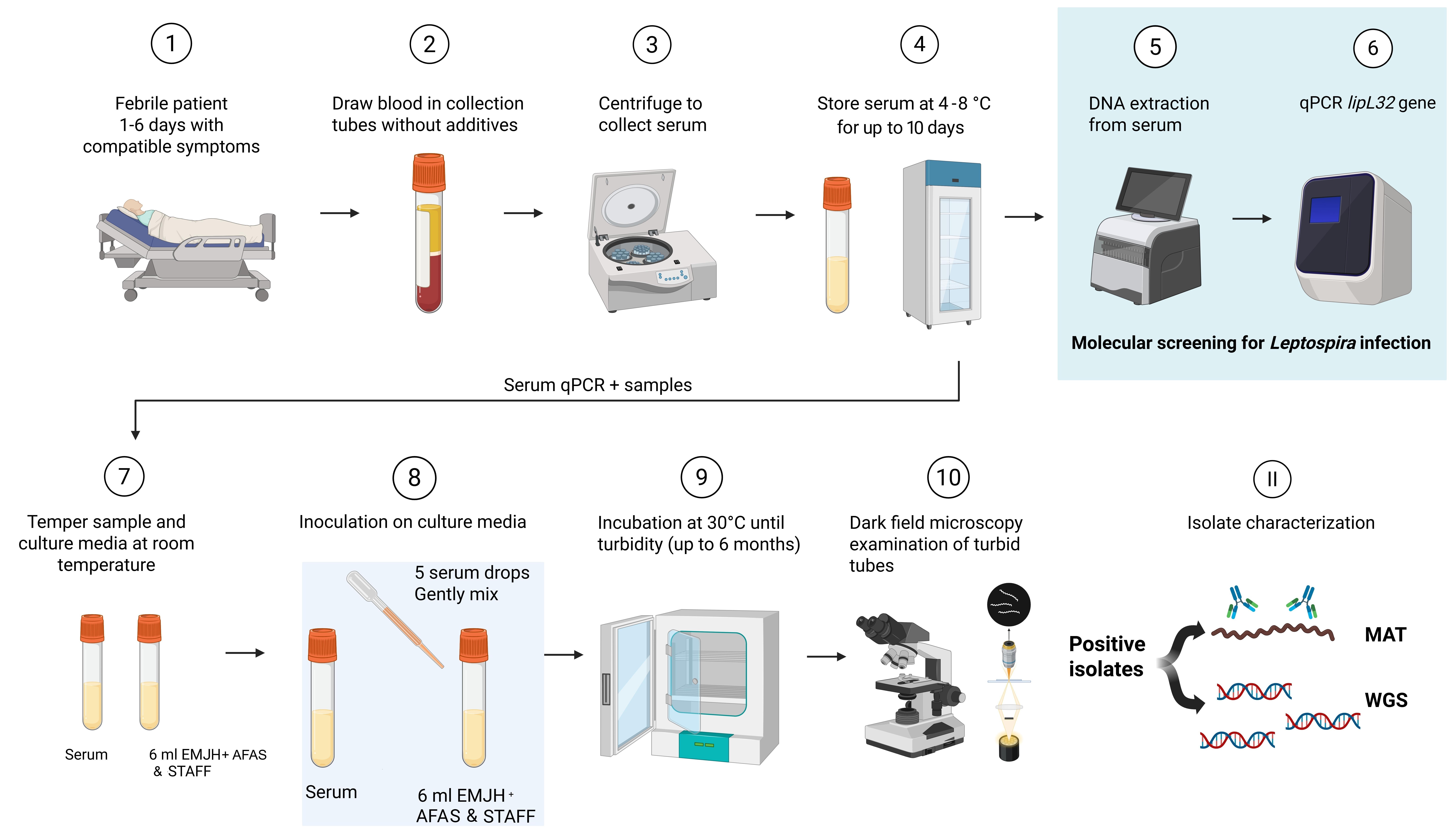
Stepwise protocol for the isolation of Leptospira from human serum samples. Modified from Chinchilla et al. [1]. Media are EMJH (Ellinghausen–McCullough–Johnson–Harris) + AFAS (albumin fatty acid supplement) and STAFF (EMJH+AFAS+ sulfamethoxazole–trimethoprim–amphotericin B–fosfomycin–5-fluorouracil).
Background
Leptospirosis is a widespread zoonotic disease caused by pathogenic bacteria of the genus Leptospira [3,4]. The disease is particularly prevalent in tropical and subtropical regions where abundant rainfall and frequent flooding favor its transmission [5]. The main reservoirs of Leptospira are wild and domestic mammals, with rodents being the most common hosts [6]. In humans, infection occurs through direct contact with the urine or body fluids of infected animals, or indirectly through the exposure of environments, such as soil or water, contaminated with the urine of infected animals [7,8]. In human hosts, the disease presents with a broad spectrum of clinical manifestations, ranging from mild symptoms to severe multisystem disease, known as Weil's syndrome [9].
The isolation and comprehensive characterization of circulating strains are fundamental for understanding the local epidemiology and transmission cycles in a region, identifying animal reservoirs, and improving public health surveillance. Furthermore, these efforts contribute to the optimization of serological panels used for the diagnosis of leptospirosis in humans and animals [10].
The culture of Leptospira spp. is a laborious technique that has historically presented many challenges for laboratories [1]. Whole blood has been the specimen of choice for the isolation of Leptospira during the acute phase of infection, though urine and cerebrospinal fluid are also utilized [11]. Standard isolation protocols typically require freshly obtained clinical samples to ensure bacterial viability, as the success of the culture is often considered dependent on the immediate inoculation of the specimen into specialized media [e.g., Ellinghausen–McCullough–Johnson–Harris (EMJH)] [10]. The primary advantage of the in-house protocol presented in this study is the successful isolation of Leptospira from serum samples, a matrix traditionally reserved for serology rather than culture. Serum offers a significant benefit over whole blood because it contains lower concentrations of inhibitory substances [12], such as hemoglobin [14], IgG immunoglobulins, and lactoferrin, which can reduce the sensitivity of both PCR and isolation [13,15]. Anticoagulants found in whole blood collection tubes, including heparin and EDTA, also have an effect as PCR-inhibitor substances [12]. This protocol demonstrates that Leptospira remains viable in non-fresh samples stored at 4–8 °C for up to 10 days, facilitating isolation in reference laboratories that receive samples after long transit times.
Molecular diagnosis of leptospirosis via real-time PCR is frequently used for rapid detection during the acute phase of the disease; however, leptospiremia is variable and typically declines with increasing time after symptom onset [10]. The use of PCR to detect leptospiral DNA prior to culture, together with the use of selective and specialized culture media such as STAFF [16], can optimize isolation procedures. These approaches are particularly important because bacterial loads during infection are often very low, and Leptospira is a slow-growing organism, making cultures highly susceptible to contamination [13].
This protocol can be used to advance the development of improved serological panels for the microagglutination test (MAT) by incorporating locally circulating isolates. It also provides a framework for enhancing global surveillance by repurposing serum samples already collected for routine serological diagnosis, thereby increasing the opportunity to identify novel serovars and species without additional clinical specimen collection.
Materials and reagents
Biological materials
1. Leptospira interrogans serovar Copenhageni strain M20 (Amsterdam UMC, KIT code: KIT0132)
Reagents
1. STARMag 96 ProPrep extraction kit (Seegene, catalog number: EX00009P)
2. TaqMan Fast Advanced Master Mix (Applied Biosystems, catalog number: 4444963)
3. Forward primer LipL32-f (5′CGCTGAAATGGGAGTTCGTATGATTTCC3′) and reverse primer LipL32-r (5′GGCATTGATTTTTCTTCYGGGGTWGCC3′) at final concentrations of 0.4 μM
4. Probe LipL32-p (5′FAM AGGCGAAATCGGKGARCCAGGCGAYGG3′BHQ1) at a final concentration of 0.2 μM
5. Nuclease-free water for molecular biology
6. Medium base EMJH for Leptospira (BD Difco, catalog number: 274910)
7. Albumin fatty acid supplement for Leptospira growth (AFAS) (Amsterdam UMC, Netherlands); store at -20 °C
8. Sulfamethoxazole (Sigma-Aldrich, catalog number: S7507)
9. Trimethoprim (Sigma-Aldrich, catalog number: 92131)
10. Amphotericin B of Streptomyces (Sigma-Aldrich, catalog number: A2411)
11. Phosphomycin disodium salt (Sigma-Aldrich, catalog number: P5396)
12. 5-Fluorouracil (Sigma-Aldrich, catalog number: F6627)
13. Sterile distilled water
14. Dimethyl sulfoxide (DMSO) (Sigma-Aldrich, catalog number: D2650)
15. Concentrated acetone (JT Baker, catalog number: 9006-3)
16. NaOH (Fluka, catalog number: 71692)
17. HCl concentrated (Fisher Scientific, catalog number: A144C-212)
18. pH indicator paper (Whatman, catalog number: WHA2600100A)
19. QIAmp DNA Mini kit (Qiagen, catalog number: 51304)
20. Absolute ethanol, 200 proof, molecular biology grade (Thermo Fischer, catalog number: T038181000)
21. Tris-HCl, 1 M solution, pH 8.0, molecular biology grade, ultrapure (Thermo Fischer, catalog number: J22638.AE)
22. Phosphate buffered saline (PBS) (Sigma-Aldrich, catalog number: P3813)
23. Polyclonal rabbit antisera (Amsterdam UMC) (Table 1)
Table 1. Panel of polyclonal rabbit anti sera for serogroup typification of Leptospira isolates
| Serogroup | Serovar | Strain |
|---|---|---|
| Australis | Australis | Ballico |
| Australis | Bratislava | Jez Bratislava |
| Autumnalis | Bangkinang | Bangkinang I |
| Autumnalis | Butembo | Butembo |
| Autumnalis | Carlos | 3 C |
| Autumnalis | Rachmati | Rachmat |
| Ballum | Ballum | Mus 127 |
| Ballum | Kenya | Njenga |
| Ballum | Castellonis | Castellon 3 |
| Bataviae | Bataviae | Swart |
| Canicola | Canicola | Hond Utrecht IV |
| Canicola | Schueffneri | VI. 90 C |
| Celledoni | Celledoni | Celledoni |
| Cynopteri | Cynopteri | 3522 C |
| Djasiman | Djasiman | Djasiman |
| Grippotyphosa | Grippotyphosa | Moskva V |
| Grippotyphosa | Huanuco | M 4 |
| Hebdomadis | Hebdomadis | Hebdomadis |
| Hebdomadis | Worsfoldi | Worsfold |
| Icterohaemorrhagiae | Copenhageni | M 20 |
| Icterohaemorrhagiae | Icteroaemorrhagiae | RGA |
| Javanica | Poi | Poi |
| Javanica | Arenal | MAVJ4-01 |
| Louisiana | Louisiana | LSU 1945 |
| Manhao | Manhao | L 60 |
| Mini | Mini | Sari |
| Panama | Panama | CZ 214 K |
| Pomona | Pomona | Pomona |
| Pyrogenes | Pyrogenes | Salinem |
| Pyrogenes | Pyrogenes | Salinem |
| Pyrogenes | Costa Rica | INCIENSA 04 |
| Sarmin | Rio | Rr 5 |
| Sarmin | Weaveri | CZ 390 |
| Sejroe | Hardjo | Hardjoprajitno |
| Sejroe | Saxkoebing | Mus 24 |
| Shermani | Shermani | 1342 K |
| Tarassovi | Bakeri | LT 79 |
| Tarassovi | Chagres | 1913 K |
| Tarassovi | Corredores | JICH |
| Tarassovi | Mogden | Compton |
| Tarassovi | Rama | 316 |
| Tarassovi | Tarassovi | Perepelitsin |
| Semaranga | Patoc | Patoc 1 |
| Fainei | Hurstbridge | BUT 6 |
| Leptonema | Illini | 3055 |
| Ranarum | Ranarum | ICF |
| Genomospecies 1 | Sichuan | 79601 |
24. Monoclonal antibodies for Leptospira serotyping (Amsterdam UMC) (Table 2)
Table 2. Monoclonal antibodies for Leptospira serotyping (Amsterdam UMC)
| Serogroup | Monoclonal antibodies |
|---|---|
| Australis | F81C1 |
| Autumnalis | F69C11, F69C15 |
| Ballum | F74C1 |
| Bataviae | F129C19 |
| Canicola | F152C18 |
| Cynopteri | F69C11, F69C15 |
| Grippotyphosa | F71C9 |
| Hebdomadis | F16H28, F16H140 |
| Icterohaemorrhagiae | F70C14, F89C12, F70C24 |
| Javanica | F98C12 |
| Pomona | F48C6 |
| Pyrogenes | F134C6 |
| Sejroe | F50C3, F16H327, F16H140, F16H28, F13A3-1, F22C1-6 |
| Sarmin | F98C12 |
| Tarassovi | F151C8 |
25. Nextera XT DNA Library Preparation kit (Illumina, catalog number: 15032354)
Solutions
1. EMJH medium base supplemented with AFAS (EMJH+AFAS) (see Recipes)
2. NaOH 10 M (see Recipes)
3. Trimethoprim 20 mg/mL (see Recipes)
4. Phosphomycin 80 mg/mL (see Recipes)
5. Sulfamethoxazole 20 mg/mL (see Recipes)
6. 5-Fluorouracil 20 mg/mL (see Recipes)
7. Amphotericin B 2.5 mg/mL (see Recipes)
8. STAFF cocktail 10× (see Recipes)
9. STAFF media (see Recipes)
10. Tris-HCl elution buffer (see Recipes)
Recipes
1. EMJH+AFAS
| Reagent | Final concentration | Quantity or volume |
|---|---|---|
| Medium base EMJH | 2.3 g/L | 2.3 g |
| Sterile distilled water | n/a | 900 mL |
| AFAS | 10% | 100 mL |
Note: Medium base EMJH can be prepared and stored at 2–8 °C up to six months prior to supplementation with AFAS.
First, prepare the medium base EMJH by adding 2.3 g of EMJH to a 2 L Erlenmeyer flask. Add 900 mL of sterile distilled water and dissolve using a magnetic stirrer. Check that the final pH is 7.5; if necessary, add drops of concentrated HCl or NaOH 10 M to adjust the pH. Transfer to a 2 L Corning storage bottle and autoclave. Store at 2–8 °C until supplementation with AFAS. For supplementation, thaw the frozen AFAS using a water bath at room temperature. Aseptically, add the supplement for a final volume of 1 L of EMJH+AFAS.
Perform a sterility check of the EMJH+AFAS medium by incubating the prepared medium for one week at 37 °C, one week at 30 °C, and two weeks at room temperature to confirm the absence of contamination. After one month of incubation, dispense 6 mL aliquots into Corning culture tubes using a self-refilling laboratory syringe. Store the aliquots at 2–8 °C for up to a year.
Perform a growth control inoculating 100 μL of fully grown Leptospira interrogans serovar Copenhageni strain M20 (grown at 30 °C for a week in EMJH+AFAS) in a Corning culture tube and incubate at 30 °C for a week. The presence of turbidity in the culture tube confirms the usefulness of the medium for the isolation of Leptospira.
2. NaOH 10 M
| Reagent | Final concentration | Quantity or volume |
|---|---|---|
| NaOH | 10 M | 40 g |
| Sterile distilled water | n/a | 100 mL |
3. Trimethoprim 20 mg/mL
| Reagent | Final concentration | Quantity or volume |
|---|---|---|
| Trimethoprim | 20 mg/mL | 1 g |
| DMSO | n/a | 50 mL |
Dissolve and filter using a 0.45 μm filter in a laminar flow cabinet. Store at -20 °C for up to a year protected from light.
Note: Verify the potency of each antibiotic in the Certificate of Analysis of each lot prior to use, to ensure accurate calculation of the amount of antibiotic required to prepare each stock solution.
4. Phosphomycin 80 mg/mL
| Reagent | Final concentration | Quantity or volume |
|---|---|---|
| Phosphomycin disodium salt | 80 mg/mL | 1.06 g |
| Sterile distilled water | n/a | 10 mL |
Dissolve and filter using a 0.45 μm filter in a laminar flow cabinet. Store at -20 °C for up to a year protected from light.
5. Sulfamethoxazole 20 mg/mL
| Reagent | Final concentration | Quantity or volume |
|---|---|---|
| Sulfamethoxazole | 20 mg/mL | 1 g |
| Concentrated acetone | n/a | 50 mL |
Dissolve and filter using a 0.45 μm filter in a laminar flow cabinet. Store at -20 °C for up to a year protected from light.
Note: Store in a sealed container to avoid acetone evaporation.
6. 5-Fluorouracil 20 mg/mL
| Reagent | Final concentration | Quantity or volume |
|---|---|---|
| 5-Fluorouracil | 20 mg/mL | 1 g |
| DMSO | n/a | 50 mL |
Add 1 g of 5-fluorouracil to a 50 mL volumetric flask. Add 40 mL of DMSO. Add a magnetic rod and heat to 56 °C in a stirrer until dissolution. Bring to a final volume of 50 mL using DMSO. Filter using a 0.45 μm filter in a laminar flow cabinet. Store at -20 °C for up to a year protected from light.
7. Amphotericin B 2.5 mg/mL
| Reagent | Final concentration | Quantity or volume |
|---|---|---|
| Amphotericin B | 2.5 mg/mL | 27 mg |
| Sterile distilled water | n/a | 10 mL |
| NaOH 10 M | 20 mM | 1 drop |
Weigh 27 mg of Amphotericin B in an analytical balance and transfer to a 10 mL volumetric flask. Add 5 mL of distilled water and homogenize. Add one drop of NaOH 10 M and mix until the Amphotericin B is completely dissolved, and a dark-yellow, translucent solution is obtained. Bring to a final volume of 10 mL and measure the pH using a pH indicator paper; confirm that the pH exceeds 11. Filter using a 0.45 μm filter in a laminar flow cabinet. Store at -20 °C for up to a year protected from light.
Note: pH must be >11 to dissolve Amphotericin B in water.
8. STAFF cocktail 10×
| Reagent | Final concentration | Quantity or volume |
|---|---|---|
| Trimethoprim 20 mg/mL | 200 μg/mL | 1 mL |
| Phosphomycin 80 mg/mL | 4,000 μg/mL | 5 mL |
| Sulfamethoxazole 20 mg/mL | 400 μg/mL | 2 mL |
| 5-Fluorouracil 20 mg/mL | 1,000 μg/mL | 5 mL |
| Amphotericin B 2.5 mg/mL | 50 μg/mL | 2 mL |
| Sterile distilled water | NA | 80 mL |
Add 50 mL of distilled water to a 100 mL volumetric flask. Add the indicated volumes of each antibiotic solution stock. Add 30 mL of distilled water and bring to a final volume of 100 mL. Sterilize by filtration using a 0.22 μm filter in a laminar flow cabinet. Store at -20 °C for up to a year protected from light.
9. STAFF media
| Reagent | Final concentration | Quantity or volume |
|---|---|---|
| STAFF cocktail 10× | 1× | 100 mL |
| EMJH+AFAS | NA | 900 mL |
Perform the sterility check and the growth control of the prepared medium as previously described for the EMJH+AFAS medium. After one month of incubation of the medium at the indicated temperatures (37 °C, 30 °C, and room temperature), dispense 6 mL aliquots into Corning culture tubes using a self-refilling laboratory syringe. Perform the growth control of Leptospira Copenhageni M20 by inoculating 100 μL of the grown culture in a tube with 6 mL of STAFF media and observing the presence of turbidity after a week of incubation.
10. Tris-HCl elution buffer
| Reagent | Final concentration | Quantity or volume |
|---|---|---|
| Tris-HCl pH 8.0 1 M | 10 mM | 50 μL |
| Nuclease-free water | NA | 4,950 μL |
Use this buffer for the DNA elution step during Leptospira DNA extraction for next-generation sequencing using a column-based extraction kit.
Laboratory supplies
1. MicroAmp fast optical 96-well reaction plate (Applied Biosystems, catalog number: 4346906)
2. MicroAmp optical adhesive film (Applied Biosystems, catalog number: 4311971)
3. MicroAmpTM adhesive film applicator (Thermo Fisher Scientific, catalog number: 4333183)
4. Corning 16 × 125 mm culture tubes, not TC-treated (Corning, catalog number: 430157)
5. 0.5–10 μL, 1–100 μL, and 100–1,000 μL pipettes (Eppendorf, catalog number: EP3123000900)
6. 0.5–10 μL, 1–100 μL, and 100–1,000 μL pipette filtered tips (Eppendorf, catalog numbers: 0030078810, 0030078543, 0030078594)
7. 10–10 μL 12-channel pipette (Eppendorf, catalog number: 3125000044)
8. 2-mL safe-lock micro tube for master mix preparation and DNA extract storage (Eppendorf, catalog number: 0030123344)
9. 2-mL screw-cap micro tube for serum samples storage (Thermo Scientific, catalog number: 3463)
10. 2-L Pyrex Erlenmeyer flasks (Corning, catalog number: 4980-2L)
11. Volumetric flask 10 mL (Fisherbrand, catalog number: FB40010)
12. Volumetric flask 50 mL (Fisherbrand, catalog number: 10-205B)
13. Volumetric flask 100 mL (Fisherbrand, catalog number: 10-205C)
14. Serological pipette 10 mL (Thermo Scientific, catalog number: 170374N)
15. Single-use filter unit 0.22 μm (Sartorius, catalog number: 16534)
16. Disposable sterile syringe filter 0.45 μm (Corning, catalog number: CLS431225)
17. PYREX® round media storage bottles 2 L, with screw cap (Corning, catalog number: 1395-2L)
18. Sterile transfer pipettes (Heathrow Scientific, catalog number: HS206373C)
19. 96-well clear polystyrene microplate round bottom (Corning, catalog number: CLS3367)
20. Disposable pipette basins (Fisherbrand, catalog number: 13-681-509)
21. Microscope slides (Thomas Scientific, catalog number: 6686K20)
Equipment
1. NordicSafe® Class II biological safety cabinet (ESCO, catalog number: NC2-L)
2. Automated nucleic acids extraction system SGprep 32 (Seegene, catalog number: SG71100)
3. 7500 Fast Real-Time PCR system (Applied Biosystems, catalog number: 4351106)
4. Vortex (Fisherbrand, catalog number: 02-215-414)
5. Microcentrifuge (Eppendorf, catalog number: 5425)
6. Laboratory refrigerator (Thermo Scientific, catalog number: TSX5005SA)
7. Incubator (Thermo Scientific, catalog number: 51028066)
8. Analytical balance (Want, model: FA2204G)
9. Precision balance (A&D Weighing, model: FZ-300iWP)
10. pH meter (Thermo Scientific, model: Orion Star A211)
11. Stirrer (Corning, catalog number: PC-410D)
12. Laboratory self-refilling syringe (Socorex, model: Dosys Classic 173, catalog number: 173.0510)
13. Microscope with universal condensator set for dark-field microscopy (Olympus, model: BX53)
14. Laboratory freezer (Thermo Scientific, TSX series, catalog number: TSX2320EA)
15. Orbital shaker (Bioevpeak, model: SHK-0210)
16. Dry bath (Thermo Fischer, catalog number: 88870008)
Software and datasets
1. NextSeq 500 sequencing system (Illumina)
2. Mutualized Platform for Microbiology (P2M) (Institut Pasteur)
3. CLC Genomics Workbench (Qiagen, version 9)
Procedure
A. DNA extraction and real-time PCR amplification
1. DNA extraction from refrigerated serum samples
a. The samples used for the molecular detection of Leptospira DNA correspond to serum specimens collected from patients during the acute phase of the disease (1–6 days after symptom onset). It is not recommended to exclude samples in which the patient had previously received treatment with antibiotics. Serum samples must be stored at 4–8 °C from the time of collection for up to 10 days before further analysis.
b. Prepare the SGprep 32 equipment for use. Select a protocol at least 10 min prior to running the equipment in order for the equipment to reach the target temperature for extraction. Insert the magnetic rod strips into the SGprep32 and correctly position them.
c. Prepare as many UniTubes as necessary according to the number of samples to be extracted and collect all the reagents at the bottom of the UniTube before use.
d. Vortex the serum samples.
e. Insert the UniTube to fit into the position of the individual tube rack and gently remove the metallic seals from the UniTubes.
f. Add 10 μL of proteinase K, 200 μL of sample in Column 1 of the UniTube and mix by pipetting 10 times. After pipetting, incubate at room temperature for 5 min.
Note: Prepare the proteinase K by adding 2.6 mL of proteinase buffer PB into lyophilized proteinase K prior to its use (both reagents are included in the kit). Store the resuspended proteinase K at 4–8 °C. Ensure that the solution appears clear before each use. If any turbidity is observed, discard the reagent.
g. Transfer 650 μL of buffer from column 2 to column 1 and mix by pipetting once.
h. Place the rack with the UniTubes in the heating tray and run the universal extraction protocol.
i. Remove the used magnetic rod strip.
j. Transfer the purified nucleic acids (80–100 μL) from column 6 to clean 2-mL Eppendorf tubes.
k. Turn on the UV lamp for 10 min after the extraction.
2. Real-time PCR amplification
a. In a PCR cabinet, thoroughly mix the TaqMan Fast Advanced Master Mix and thaw the frozen primers and probe on a PCR cooling block.
b. Prepare the master mix for the amplification of lipL32 (Table 3). Each reaction should have a final volume of 25 μL. Calculate three reactions per sample, one positive control, one no-template control (NTC) consisting of nuclease-free water, and an extra reaction to compensate the losses of volume due to variation during pipetting. Prepare the mix in a sterile tube and vortex briefly.
Notes:
1. Each sample must be tested in triplicates; this is important for the interpretation of the results.
2. Protect the master mix and probe from light, as they are light sensitive.
3. The preparation of the master mix and the addition of DNA must be performed in separate, clean rooms to avoid contamination of the PCR.
Table 3. PCR reaction composition
| Reagent | Final concentration | Quantity or volume |
|---|---|---|
| TaqMan® Fast Advanced Master Mix | 1× | 12.5 μL |
| LipL32-f | 0.4 μM | 1 μL |
| LipL32-r | 0.4 μM | 1 μL |
| LipL32-p | 0.2 μM | 0.5 μL |
| Extracted DNA | n/a | 10 μL |
| Total | n/a | 25 μL |
c. Add 15 μL of the master mix to each reaction well on a 96-well plate.
d. Add 10 μL of DNA to the individual PCR wells. Add 10 μL of nuclease-free water to the NTC and 10 μL of extracted DNA of Leptospira interrogans serovar Copenhageni as the positive control. Cover plates and spin to remove liquid from the sides and top of the wells.
e. Program the 7500 Fast real-time thermal cycler on fast mode and perform the PCR (Table 4).
Table 4. PCR cycling conditions in the 7500 Fast Applied Biosystems
| Step | Temperature | Duration (min:s) | Number of cycles |
| UNG incubation | 50.0 °C | 02:00 | 1 |
| Polymerase activation and denaturation | 95.0 °C | 05:00 | 1 |
| Denaturation | 95.0 °C | 00:20 | 45 |
| Annealing/extension | 60.0 °C | 00:30 |
f. After real-time PCR has been run and analyzed, interpret the results as follows:
i. The NTC reaction should not exhibit amplification curves that cross the threshold line within 43 cycles.
ii. Leptospira positive control should produce a positive result each time the assay is performed. The amplicon size for lipL32 is 114 pb.
iii. When all controls meet the stated requirements and the sample threshold result is within 43 cycles, a sample is considered positive if 3 out of 3, or 2 out of 3 replicates show amplification. If only 1 out of 3 replicates is positive, repeat the run the next day using the same extraction. Consider the sample positive if in the next run, 1 or more replicates show amplification. If the repeat run does not show amplification, report the sample as negative, but perform isolation by culture on the sample.
C. Isolation of Leptospira spp.
1. Select for culture all the PCR-positive specimens that had been stored under refrigeration temperatures from the time of collection for up to 10 days. See General note 1.
Notes:
1. Culture inoculation can be performed regardless of whether the patient had recently received antibiotic treatment, as viable leptospires may still be present if the minimum inhibitory concentration (MIC) has not yet been reached.
2. Storage periods longer than 10 days have not been evaluated in our setting.
2. Remove the samples and culture media from the refrigerator and allow them to equilibrate to room temperature.
Critical: Let the samples and the media reach room temperature before inoculation and incubation to minimize thermal shock, as Leptospira are sensitive to rapid temperature fluctuations.
3. Gently vortex the serum sample and, using a sterile disposable pipette, transfer five drops (200 μL) of serum into a culture tube containing 6 mL of EMJH+AFAS. Repeat using a tube containing 6 mL of STAFF media.
4. Gently mix the culture tubes and incubate at 30 °C up to six months.
5. Monitor the culture tubes weekly for the appearance of visible turbidity, which may indicate Leptospira growth (Figure 1B). Once turbid, examine a 5 μL aliquot by dark-field microscopy to determine the presence of spirochaetes compatible with Leptospira (Figure 2).
Notes:
1. The appearance of growth in STAFF media is the same as that observed in Figure 1 for EMJH+AFAS.
2. The absence of turbidity in the medium corresponds to a negative culture. All tubes must be incubated for six months.
Pause point: If slight turbidity is observed in the culture tube and the presence of Leptospira is confirmed by dark-field microscopy, continue incubation at 30 °C until the culture medium exhibits more pronounced turbidity, indicative of a fully developed Leptospira culture (Figure 1C).
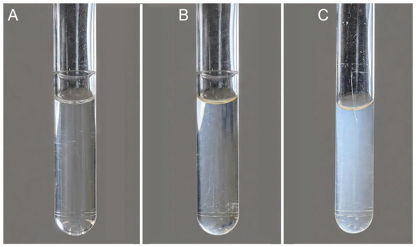
Figure 1. EMJH+AFAS culture tubes showing Leptospira growth. (A) No turbidity, consistent with no leptospiral growth. (B) Slight turbidity, consistent with a low bacterial density in the culture medium. (C) Dense turbidity, consistent with a fully grown Leptospira culture. The appearance of growth in STAFF media is the same as that observed in EMJH+AFAS.
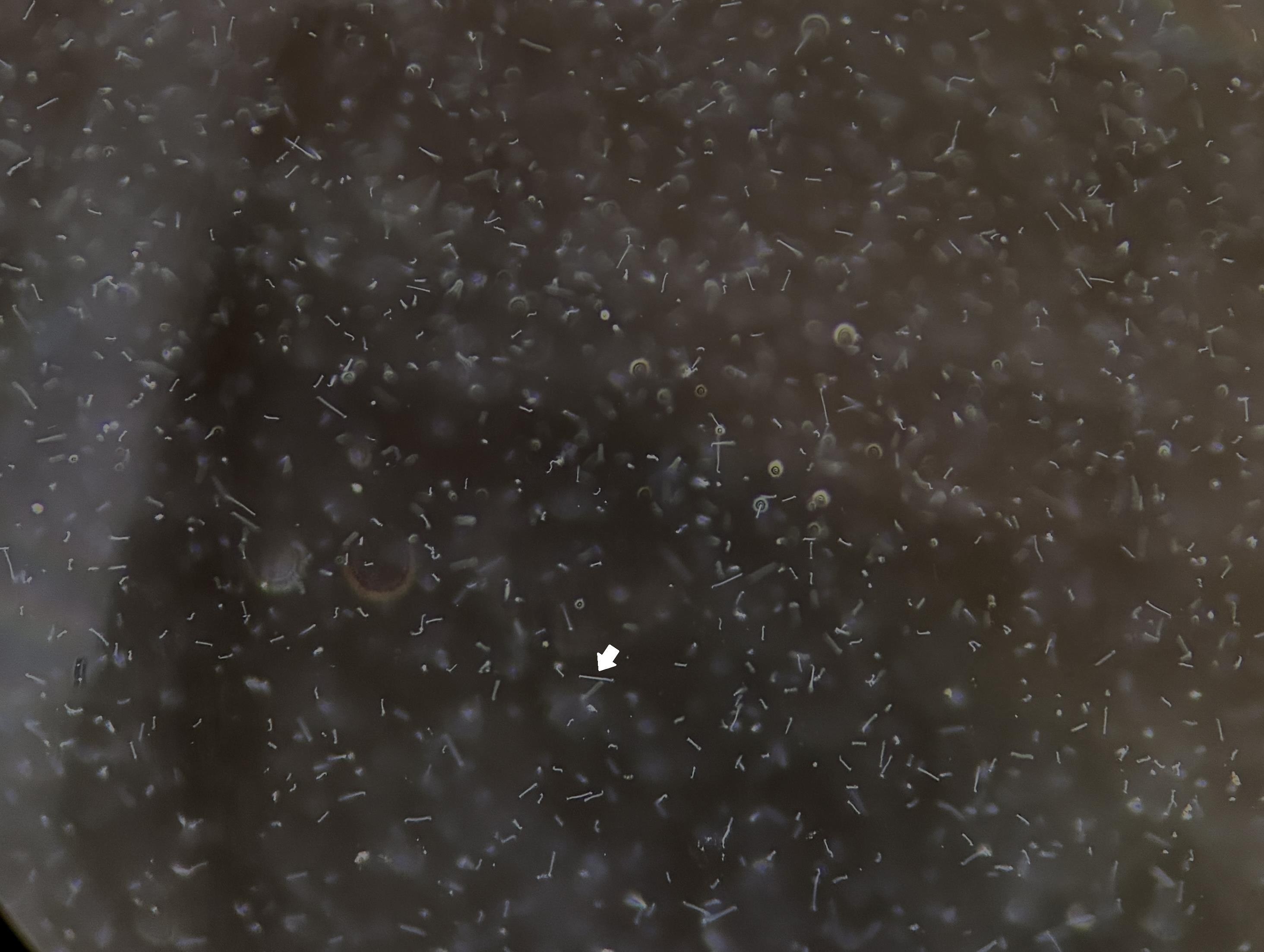
Figure 2. Dark-field microscopy showing the spirochetal morphology (arrow) of Leptospira. Magnification: 10×.
6. Once the culture tube shows dense turbidity, inoculate three tubes of EMJH+AFAS for propagation and for serotyping. See General note 2.
7. After one week, check that the subcultured tubes show dense turbidity and perform the serotyping with rabbit antiserum and monoclonal antibodies.
D. Serotyping of Leptospira using polyclonal rabbit antiserum and monoclonal antibodies by inverted microagglutination test (MAT)
1. Dilute the isolated strain 1:3 with PBS, mixing 12 mL of grown culture with 24 mL of PBS in a disposable pipette basin.
2. Prepare the panel of rabbit antisera and monoclonal antibodies for serotyping. Reconstitute the lyophilized vial with the volume of nuclease-free water indicated on the label. Once reconstituted, divide the solution into small aliquots and store at -20 °C.
3. First, evaluate the isolate against the 44 rabbit antisera panel to identify the reacting serogroup or serogroups. Prepare and label the necessary round-bottom microplates (see Figure 3) to include 8 rabbit antisera per plate and testing dilutions from 1:20 to 1:20,480 for each antiserum.
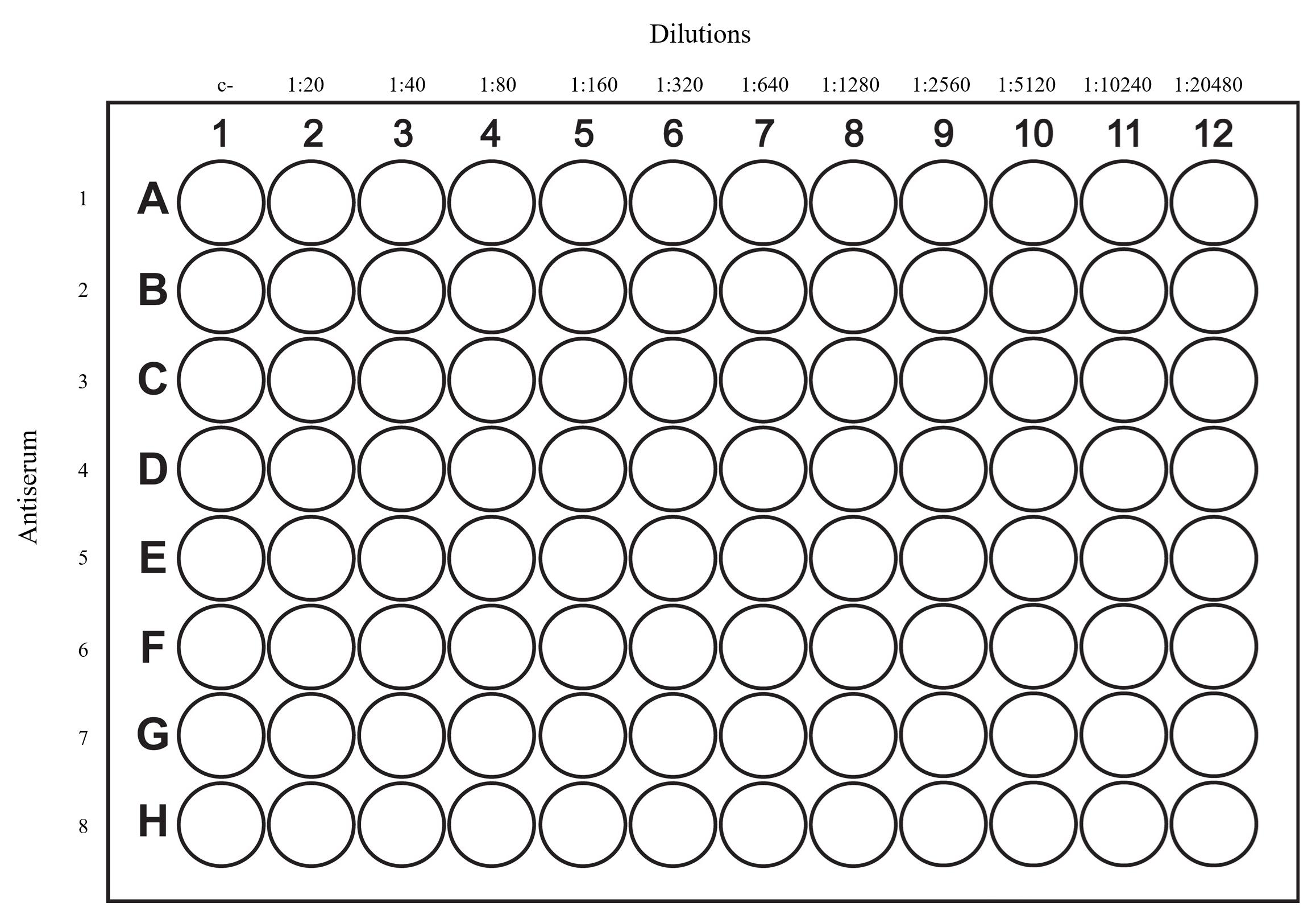
Figure 3. Microplate layout for Leptospira serotyping
4. Using a multichannel pipette, add 50 μL of PBS to each well on the plate.
5. Add 40 μL of PBS to column 2.
6. Add 10 μL of rabbit antiserum to the second well of each row of the plate (1 serum per row) and mix thoroughly.
7. Perform two-fold serial dilutions from column 2 to column 12, transferring 50 μL of each dilution to the next well. Discard the remaining 50 μL after the last column.
8. Add 50 μL of the diluted Leptospira isolate to each well on the plate.
9. Mix in an orbital shaker for 30 s at 150 rpm.
10. Incubate at 37 °C for 1 h.
11. Transfer 5 μL of each well to a microscope slide and observe in dark-field microscopy for the presence of agglutination of the live leptospires. The highest serum dilution that agglutinates 50% or more of the leptospires is considered the antibody titer (Figure 4).
Notes:
1. The observation of leptospires by dark-field microscopy and the determination of the endpoint corresponding to 50% agglutination are inherently subjective. Extensive training of personnel in dark-field microscopy is recommended to improve proficiency and consistency in the interpretation of the observed agglutination patterns.
2. Anti-Leptospira antibodies can react with antigens of multiple serovars across serogroups. During serotyping, reactivity may be observed in antisera raised against either different strains of the same serogroup (homologous reactions) or strains from different serogroups (heterologous reactions).
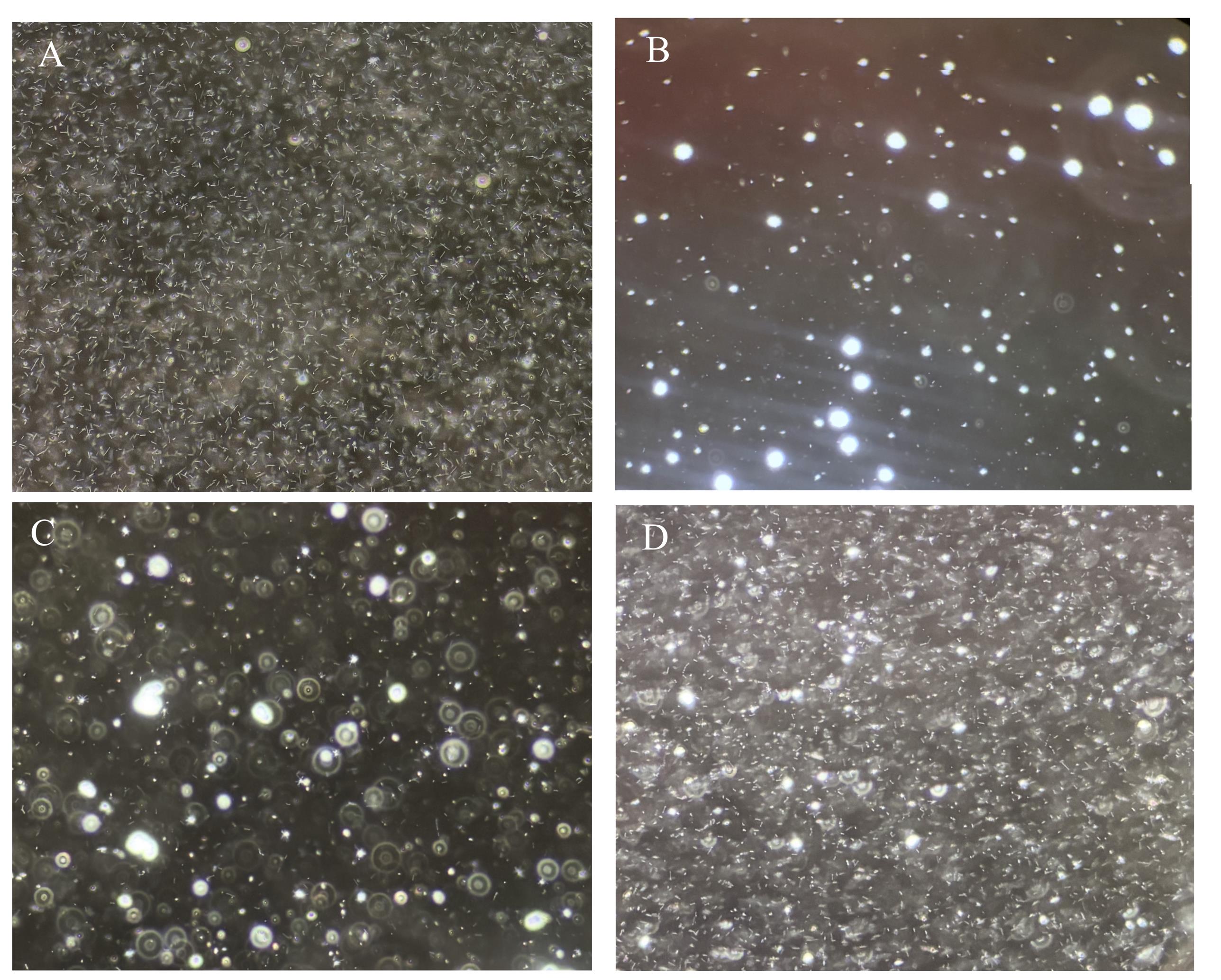
Figure 4. Dark-field microscopy for the microscopic agglutination test (MAT) of an isolate of Leptospira serogroup Icterohaemorrhagiae and its homologous antiserum. (A) Negative control, showing 100% free leptospires. (B) 1:320 titer, showing 100% agglutinated leptospires. (C) 1:1,280 titer, showing 90% agglutinated leptospires. (D) Endpoint titer of 1:5,120, showing 50% free leptospires and 50% agglutinated. Magnification 10×.
12. Identify the serogroup that shows the highest titer of antibodies. In most cases, this corresponds to the serogroup of the isolate. If reactivity is observed against different serogroups, analyze the strain using the available monoclonal antibodies for each serogroup under study. Perform the titration as previously described.
Note: The titers obtained with the monoclonal antibodies provide guidance on the infecting serogroup. Analyze the results based on the expected values specified in the quality control certificates for each monoclonal antibody. The highest titers are associated with the serogroup of the isolate under study.
E. Whole-genome characterization
1. DNA extraction of Leptospira isolates using the QIAmp DNA Mini Kit
a. Transfer 10 μL of a grown Leptospira culture into a 2 mL microtube containing 990 μL of PBS.
b. Centrifuge the tube at 37,732× g for 15 min to pellet the leptospires. Discard the supernatant.
c. Resuspend the pellet in 180 μL of ATL buffer and vortex for 15 s.
d. Add 20 μL of proteinase K and pulse-vortex for 15 s.
e. Incubate at 56 °C in a heating block for 1 h.
f. After incubating, perform a quick spin in order to recover the drops on the lid of the tube.
g. Add 200 μL of buffer AL and pulse-vortex for 15 s. Briefly spin down the tube contents.
h. Incubate at 70 °C for 10 min. Briefly spin down the tube contents.
i. Add 200 μL of absolute ethanol (molecular grade) and pulse vortex for 15 s. Briefly spin down the tube contents.
j. Transfer the content to a column and centrifuge at 10,733× g for 1 min.
k. Discard the filtrate and transfer the column to a clean collection tube.
l. Add 500 μL of buffer AW1 and centrifuge at 10,733× g for 1 min.
m. Discard the filtrate and transfer the column to a clean collection tube.
n. Add 500 μL of buffer AW2 and centrifuge at 32,869× g for 3 min.
Optional: Discard the used collection tube and transfer the column to a new collection tube. Perform an additional centrifugation at 32,869× g for 1 min to improve the elimination of buffer from the column. For this, it is necessary to purchase additional collection tubes.
o. Discard the filtrate and transfer the column to a clean 2 mL microtube.
p. Add 100 μL of Tris-HCl elution buffer (see Recipes), preheated to 56 °C, to the column and incubate for 3 min.
q. Centrifuge at 28,341× g for 3 min, collect the eluate, and discard the column.
2. Next-generation sequencing and genomic analysis
a. Perform the NGS using the Nextera XT DNA Library Preparation kit and the NextSeq 500 sequencing systems (Illumina).
Note: Genomes meeting quality requirements, such as i) sequencing coverage >30×, ii) number of contigs <600, iii) cumulative contigs length within the typical range of Leptospira genomes (3.6–6 Mb), iv) GC content within the typical range of Leptospira genomes (35%–48%), and v) <100 uncalled cgMLST alleles out of the 545 pre-defined core genes, are to be selected for further analyses.
Note: A comprehensive description of the methods used for Leptospira genome analysis is provided in the Materials and Methods section of [4].
Validation of protocol
This culture method enables the isolation of genomically diverse Leptospira strains belonging to a wide range of serogroups (Table 5). In addition, it supports the recovery of Leptospira from serum samples—a specimen type rarely used for Leptospira isolation—even after refrigeration for up to 10 days.
Table 5. Serological and molecular characterization of Leptospira isolates and their isolation conditions.
| Isolate | BIGSdb ID | Species | Serogroup | cgST | Serum Ct value | Days stored at 4 °C before culturing |
|---|---|---|---|---|---|---|
| CR2020 | 1251 | L. santarosai | Hebdomadis | 1025 | 39.23 | 3 |
| CR2120 | 1252 | L. santarosai | Shermani | 1026;1098 | 39.53 | 4 |
| CR0521 | 1254 | L. santarosai | Tarassovi | 1028;1099 | 37.71 | 9 |
| CR1421 | 1256 | L. santarosai | Pyrogenes | 1030;2206 | 34.95 | 5 |
| CR1821 | 1257 | L. santarosai | Grippotyphosa | 2289;1034 | 33.49 | 9 |
| CR2021 | 1258 | L. borgpetersenii | Ballum | 578;1043;1096;1855 | 38.77 | 5 |
| CR2621 | 1259 | L. santarosai | Grippotyphosa | 1031 | 40.25 | 3 |
This protocol (or parts of it) has been used and validated in the following research articles:
• Chinchilla et al. [4]. Phylogenomics of Leptospira santarosai, a prevalent pathogenic species in the Americas. PLoS Neglected Tropical Diseases (Figure 4).
• Chinchilla et al. [1]. In-house isolation protocol from human serum samples demonstrates the circulating of a broad diversity of Leptospira serogroups in Costa Rica. Scientific Reports (Table 2, Supplemental Table 4, Supplemental Figures 1–2).
General notes and troubleshooting
General notes
1. Isolation is possible even in samples with very high Ct values (Ct 40–41). A high Ct value correlates with a low number of Leptospira in the sample; nonetheless, viability may be maintained even at low bacterial loads, allowing successful recovery by culture.
2. For Leptospira spp., an optical density (OD) of 0.25 corresponds approximately to a density of 2 × 10⁸ leptospires/mL, which is consistent with a fully grown Leptospira culture. However, we choose to use visual observation rather than restrict culture propagation to a specific optical density (OD) value for two main reasons:
a. Leptospira is a slow-growing microorganism that is highly susceptible to contamination, as contaminating microorganisms generally grow at a faster rate than leptospires in culture media. Repeatedly opening a culture tube to monitor its optical density substantially increases the likelihood of contamination during the growth process.
b. Successful propagation of Leptospira cultures can be achieved from cultures with either low or high bacterial densities. The main difference is the time required for the subculture to reach confluent turbidity. When a fully grown culture is used as the inoculum, the resulting subculture generally reaches sufficient growth within approximately one week. In contrast, when a culture with limited growth or a lower initial bacterial density is used, the subculture may require approximately 2–3 weeks to reach comparable growth. Since the initial bacterial density is not critical for the successful propagation of the culture, our group has chosen to rely on visual assessment. This approach minimizes the risk of contamination associated with repeated opening of culture tubes while ensuring that a subculture can be obtained within a timely period, generally 1–2 weeks.
3. Historically, the isolation of Leptospira from clinical samples has been challenging, which is one of the reasons why culture is infrequently attempted in clinical laboratories worldwide. In our laboratory, the isolation rate from PCR-positive clinical samples has historically ranged from 10% to 30% since we implemented the culture of PCR-positive serum samples.
Troubleshooting
Problem 1: Turbidity in the EMJH+AFAS but not in STAFF media. Upon examination by dark-field microscopy, microorganisms with a morphology distinct from that of spirochetes are observed.
Possible cause: Contamination of EMJH+AFAS medium by a microorganism that is inhibited by the antibiotics present in STAFF media.
Solution: Discard the EMJH+AFAS and continue observing the STAFF media.
Problem 2: Turbidity in EMJH+AFAS and STAFF media. Dark-field microscopy reveals microorganisms with a morphology inconsistent with that of spirochetes.
Possible cause: Contamination of media with a microorganism not inhibited by the antibiotics present in STAFF media.
Solution: Discard both media.
Problem 3: Turbidity in EMJH+AFAS and STAFF media. Dark-field microscopy reveals microorganisms with different morphologies, including spirochetes consistent with Leptospira.
Possible cause: Co-growth of Leptospira and a contaminating microorganism not inhibited by the antibiotics present in STAFF media.
Solution: Filter 4 mL of the contaminated culture medium through a 0.2 μm membrane filter. Inoculate 1 mL of the filtrate into a fresh tube of EMJH+AFAS medium and a fresh tube of STAFF media to try to recover the leptospires, which can pass through a 0.2 μm filter. Only a small number of Leptospira cells pass through the filter; however, this may be sufficient to support growth in fresh culture medium.
Problem 4: Presence of cloudiness or particulate matter that interferes with the observation of leptospires and the assessment of agglutination during the reading of Leptospira serotyping by the microscopic agglutination test (MAT).
Possible cause: Contamination of the resuspended rabbit antisera or the monoclonal antibodies.
Solution: Filter the resuspended rabbit antisera and monoclonal antibodies using a 0.22 μm filter. This will significantly reduce the volume of reagent but will allow observation in dark-field microscopy.
Acknowledgments
The authors thank the Government of Costa Rica and the Ministry of Health, the Caja Costarricense del Seguro Social de Costa Rica for the referral of the clinical samples, and the Institute Pasteur for the whole genome sequencing of the strains. Graphical overview created with BioRender.
Author contributions
Conceptualization, D.C., I.S.; Methodology, D.C., I.S., D.M., B.Q.; Investigation, D.C.; Writing—Original Draft, D.C; Writing—Review & Editing, D.C.
Competing interests
The authors declare no conflicts of interest.
Ethical considerations
According to decree number 40556-s of the General Health Law of Costa Rica, epidemiological studies that incorporate the review of clinical records do not require the approval of an Ethics Scientific Committee. Human samples were anonymized, and collection of the samples was conducted according to the Declaration of Helsinki.
The need to obtain informed consent was waived by the Costa Rican Institute for Research and Education in Nutrition and Health (INCIENSA) Scientific Committee.
References
- Chinchilla, D., Sánchez, I., Montero, D., Picardeau, M. and Gutiérrez, R. (2025). In-house isolation protocol from human serum samples demonstrates the circulating of a broad diversity of Leptospira serogroups in Costa Rica. Sci Rep. 15(1): e1038/s41598–025–93301–0. https://doi.org/10.1038/s41598-025-93301-0
- Bourhy, P., Bremont, S., Zinini, F., Giry, C. and Picardeau, M. (2011). Comparison of Real-Time PCR Assays for Detection of Pathogenic Leptospira spp. in Blood and Identification of Variations in Target Sequences. J Clin Microbiol. 49(6): 2154–2160. https://doi.org/10.1128/jcm.02452-10
- Levett, P. N. (2001). Leptospirosis. Clin Microbiol Rev. 14(2): 296–326. https://doi.org/10.1128/CMR.14.2.296-326.2001/ASSET/9D6DEA4D-2B9C-4F1B-9FB2-44FB040B64E3/ASSETS/GRAPHIC/CM0210021003.JPEG
- Chinchilla, D., Nieves, C., Gutiérrez, R., Sordoillet, V., Veyrier, F. J. and Picardeau, M. (2023). Phylogenomics of Leptospira santarosai, a prevalent pathogenic species in the Americas. PLoS NeglTrop Dis. 17(11): e0011733. https://doi.org/10.1371/journal.pntd.0011733
- Valente, M., Bramugy, J., Keddie, S. H., Hopkins, H., Bassat, Q., Baerenbold, O., Bradley, J., Falconer, J., Keogh, R. H., Newton, P. N., et al. (2024). Diagnosis of human leptospirosis: systematic review and meta-analysis of the diagnostic accuracy of the Leptospira microscopic agglutination test, PCR targeting Lfb1, and IgM ELISA to Leptospira fainei serovar Hurstbridge. BMC Infect Dis. 24(1): 1–22. https://doi.org/10.1186/s12879-023-08935-0
- Gomard, Y., Dellagi, K., Goodman, S. M., Mavingui, P. and Tortosa, P. (2021). Tracking Animal Reservoirs of Pathogenic Leptospira: The Right Test for the Right Claim. Trop Med Infect Dis. 6(4): 205.https://doi.org/10.3390/tropicalmed6040205
- Brito Monteiro, M., Egídio de Sousa, I., Piteira, M., Coelho, S. and Freitas, P. (2021). Leptospirosis, a Re-emerging Threat. Cureus. 13(4): e14295. https://doi.org/10.7759/cureus.14295
- Faine, S., Adler, B., Bolin, C. and Perolat, P. (1999). “Leptospira” and leptospirosis. In Leptospira and Leptospirosis (2nd ed.). Melbourne, Australia: MediSci. Retrieved from https://research.monash.edu/en/publications/leptospira-and-leptospirosis
- McBride, A. J. A., Athanazio, D. A., Reis, M. G. and Ko, A. I. (2005). Leptospirosis. Curr Opin Infect Dis. 18(5): 376–386. https://doi.org/10.1097/01.QCO.0000178824.05715.2C
- Sykes, J. E., Reagan, K. L., Nally, J. E., Galloway, R. L. and Haake, D. A. (2022). Role of Diagnostics in Epidemiology, Management, Surveillance, and Control of Leptospirosis. Pathogens. 11(4): 395. https://doi.org/10.3390/pathogens11040395
- Levett, P. (2016). Leptospira Culture. In Clinical Microbiology Procedures Handbook (4th ed.). Washington, DC: ASM Press.
- Schrader, C., Schielke, A., Ellerbroek, L. and Johne, R. (2012). PCR inhibitors - occurrence, properties and removal. J Appl Microbiol. 113(5): 1014–1026. https://doi.org/10.1111/j.1365-2672.2012.05384.x
- Wuthiekanun, V., Chierakul, W., Limmathurotsakul, D., Smythe, L. D., Symonds, M. L., Dohnt, M. F., Slack, A. T., Limpaiboon, R., Suputtamongkol, Y., White, N. J., et al. (2007). Optimization of Culture of Leptospira from Humans with Leptospirosis. J Clin Microbiol. 45(4): 1363–1365. https://doi.org/10.1128/jcm.02430-06
- Hoffman, R., Benz, E. J., Silberstein, L. E., Heslop, H. E., Weitz, J. I., Anastasi, J., Salama, M. E. and Abutalib, S. (2017). Hematology: Basic Principles and Practice. Hematology: Basic Principles and Practice. 1–2374. https://doi.org/10.1016/C2013-0-23355-9
- Mgode, G. F., Machang’u, R. S., Mhamphi, G. G., Katakweba, A., Mulungu, L. S., Durnez, L., Leirs, H., Hartskeerl, R. A. and Belmain, S. R. (2015). Leptospira Serovars for Diagnosis of Leptospirosis in Humans and Animals in Africa: Common Leptospira Isolates and Reservoir Hosts. PLoS NeglTrop Dis. 9(12): e0004251. https://doi.org/10.1371/journal.pntd.0004251
- Chakraborty, A., Miyahara, S., Villanueva, S. Y., Saito, M., Gloriani, N. G. and Yoshida, S. I. (2011). A novel combination of selective agents for isolation of Leptospira species. Microbiol Immunol. 55(7): 494–501. https://doi.org/10.1111/j.1348-0421.2011.00347.x
Article Information
Publication history
Received: Jul 13, 2026
Accepted: Sep 6, 2026
Available online: Sep 22, 2026
Published: Oct 20, 2026
Copyright
© 2026 The Author(s); This is an open access article under the CC BY-NC license (https://creativecommons.org/licenses/by-nc/4.0/).
How to cite
Chinchilla, D., Sánchez, I., Montero, D., Cháves, D., Quirós, B. and Gutiérrez, R. (2026). PCR-Guided Isolation of Leptospira Strains From Refrigerated Serum Samples for Serogroup and Genomic Characterization. Bio-protocol 16(20): e5842. DOI: 10.21769/BioProtoc.5842.

 Download Citation in RIS Format
Category
Microbiology > Pathogen detection > PCR
Molecular Biology > DNA > PCR
Do you have any questions about this protocol?
Post your question to gather feedback from the community. We will also invite the authors of this article to respond.
 Post a Question


