


- Protocols

- Articles and Issues
- For Authors
- About
- Become a Reviewer


Optimized Phenol–Chloroform–Isoamyl DNA Extraction Protocol for Single Fish Eggs

(*contributed equally to this work) Published: Vol 16, Iss 20, Oct 20, 2026 DOI: 10.21769/BioProtoc.5839 Views: 29
Reviewed by: Amr Galal Abdelraheem IbrahimAnonymous reviewer(s)
 PDF
PDF  Ask a question
Original research article
The authors used this protocol in:

Aug 2026

Protocol Collections
Comprehensive collections of detailed, peer-reviewed protocols focusing on specific topics




Abstract
Reliable DNA extraction is essential for genetic research on marine species; however, obtaining sufficient DNA from single fish eggs remains challenging. Existing protocols often require optimization to achieve high PCR efficiency. The optimized phenol–chloroform–isoamyl extraction protocol presented in this paper improves DNA yield and quality from individual eggs of Atlantic bluefin tuna (Thunnus thynnus), bogue (Boops bops), saddled seabream (Oblada melanura), and painted comber (Serranus scriba) by modifying buffer volumes, incubation times, and washing steps, following prior micropuncturing of eggs on a glass slide. DNA quality is confirmed by spectrophotometry, PCR amplification of the mitochondrial COI gene, electrophoresis, and Sanger sequencing. This method provides a low-cost and effective approach for species identification from individual fish eggs.
Key features
• Effective and low-cost DNA extraction method from fish eggs.
• Uses only standard reagents and equipment, allowing wide application in all laboratories.
• High PCR efficiency.
Keywords: Atlantic bluefin tuna (Thunnus thynnus) Fish eggs
Phenol–chloroform DNA extraction
Mitochondrial COI gene
Species identification
Fish eggs
Phenol–chloroform DNA extraction
Mitochondrial COI gene
Species identification
Graphical overview
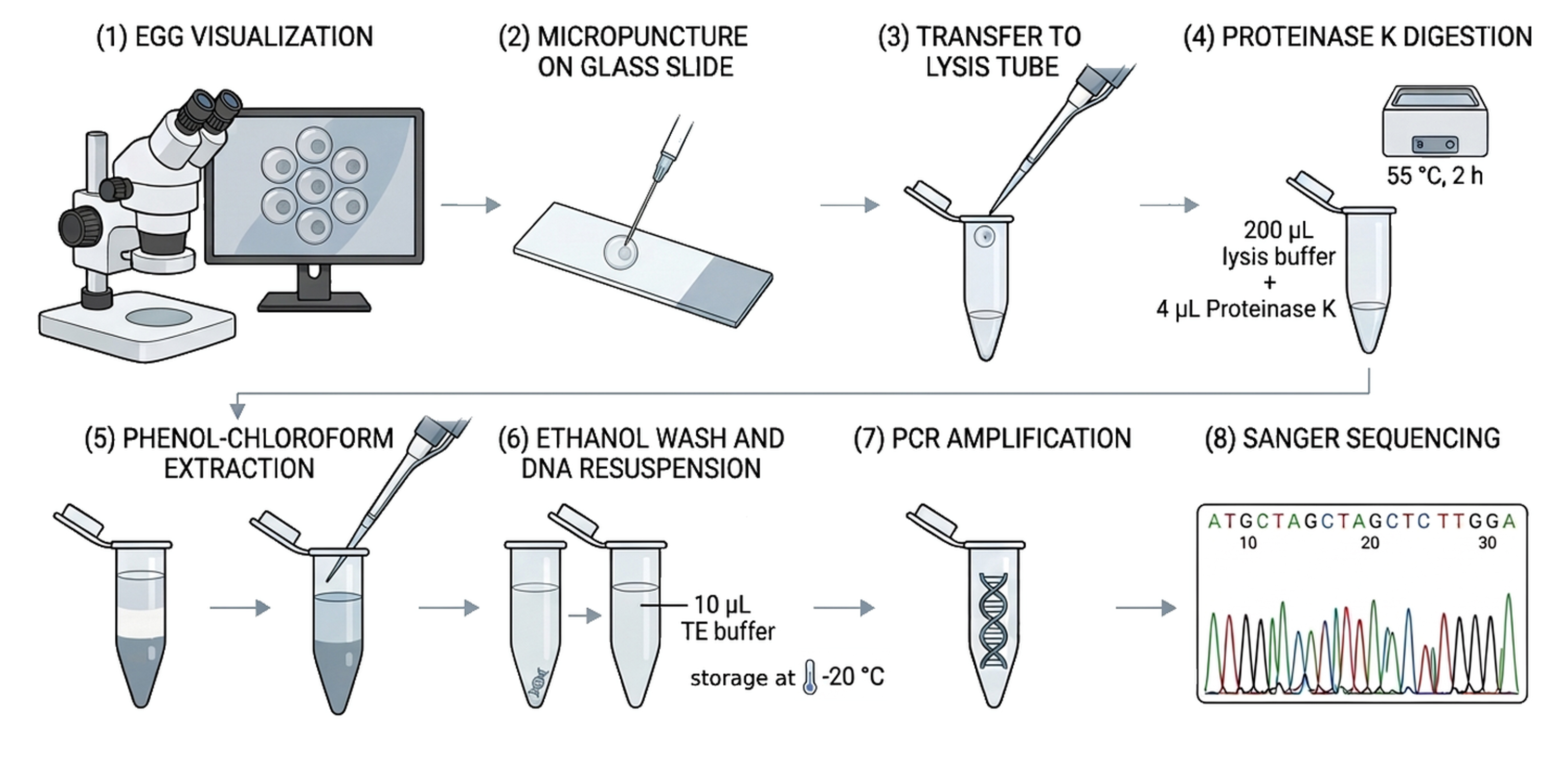
Optimized phenol–chloroform–isoamyl protocol for DNA extraction from single fish eggs. Includes egg visualization under a stereomicroscope, micropuncture on a glass slide, transfer to lysis buffer, Proteinase K digestion, phenol–chloroform extraction, ethanol washing, DNA resuspension and storage at -20 °C, PCR amplification, and Sanger sequencing.
Background
Today, almost all methods for stock research on marine species rely on genetic studies. To obtain high-quality DNA, effective DNA extraction is required. There are many reliable protocols and commercial kits for isolating DNA from tissue, which provide high yields and excellent quality DNA. However, DNA extraction from a single fish egg may result in lower yields due to uniform cell stages. Aranishi (2006) [1] used a one-tube method for DNA extraction, while Golotin et al. (2023) [2] described a low-cost, non-enzymatic protocol for isolating embryonic cells from fish eggs. In our samples, the existing protocols did not consistently provide sufficient DNA yield for reliable PCR amplification from individual fish eggs. Therefore, further optimization was required to achieve reliable PCR amplification and to develop an optimized protocol for DNA extraction from individual fish eggs collected in the vicinity of Atlantic bluefin tuna (Thunnus thynnus) aquaculture farms. The primary aim of the sampling was to confirm the occurrence of T. thynnus spawning within the aquaculture cages. However, the area surrounding aquaculture facilities is also inhabited by a diverse assemblage of wild fish species, as aquaculture cages can attract wild fish by providing increased food availability and serving as functional marine protected areas [3]. Consequently, eggs of other teleost fish species were also collected during sampling conducted around the cages. These eggs were initially selected under a stereomicroscope based on their morphological characteristics and size, while their species identity was subsequently confirmed by molecular analysis. Because eggs of different fish species may occur simultaneously in the vicinity of aquaculture cages, and morphological characteristics alone may not be sufficient for reliable species identification, an efficient DNA extraction protocol for individual fish eggs is required. The protocol described here incorporates micropuncturing of eggs on a glass slide (see General note 1), reducing the volume of lysis buffer to 200 μL, adjusting the volume of phenol–chloroform–isoamyl alcohol, optimizing the incubation period (2 h), and extending the washing period to overnight.
Materials and reagents
Biological materials
1. Fish eggs
Reagents
1. 96% alcohol (Sigma-Aldrich, catalog number: 32294), storage: 4 °C
2. Phenol–chloroform–isoamyl alcohol mixture (Sigma-Aldrich, catalog number: 77617), storage: 4 °C
3. Proteinase K, 20 mg/mL (Thermo Fisher, catalog number: AM2542), storage: -20 °C
4. TE buffer (Thermo Fisher, catalog number: 12090015), storage: 4 °C
5. HotStartTaq DNA Polymerase kit (Qiagen, catalog number: 203203), storage: -20 °C
6. SYBR Safe (Thermo Fisher, catalog number: S33102), storage: room temperature
7. Agarose (Sigma-Aldrich, catalog number: 9012-36-6), storage: room temperature
8. DNA ladder (Cleaver Scientific Ltd., catalog number: CSL-MDNA-100BP), storage: 4 °C
9. GelPilot DNA loading dye, 5× (Qiagen, catalog number: 239901), storage: 4 °C
10. Milli-Q water (MQH2O), storage: room temperature
11. 1 M Tris-HCl, pH 8.0 (Thermo Fisher, catalog number: J22638.AP), storage: room temperature
12. 0.5 M EDTA, pH 8.0 (Thermo Fisher, catalog number: R1021), storage: room temperature
13. 5 M NaCl, RNase-free (Invitrogen, catalog number: AM9760G), storage: room temperature
14. 10% SDS (Invitrogen, catalog number: 15553027), storage: room temperature
Solutions
1. Lysis buffer (see Recipes)
2. 70% alcohol (see Recipes)
3. Proteinase K, 20 mg/mL (see Recipes)
Recipes
1. Lysis buffer
| Reagent | Final concentration | Quantity or volume |
|---|---|---|
| 1 M Tris-HCl | 10 mM | 2 mL |
| 0.5 M EDTA | 1.25 mM | 0.5 mL |
| 5 M NaCl | 150 mM | 6 mL |
| 10% SDS | 0.2% | 4 mL |
| H2O | n/a | 184 mL |
| Total | n/a | 200 mL |
Store the prepared lysis buffer at room temperature and use within 12 months.
2. 70% alcohol
| Reagent | Final concentration | Quantity or volume |
|---|---|---|
| Ethanol (96%) | 70% | 36.5 mL |
| Distilled H2O | to a final volume of 50 mL | |
| Total | n/a | 50 mL |
Store the prepared 70% alcohol at 4 °C.
3. Proteinase K, 20 mg/mL
| Reagent | Final concentration | Quantity or volume |
|---|---|---|
| Proteinase K powder | 20 mg/mL | 100 mg |
| MQH2O | 5 mL | |
| Total | n/a | 5 mL |
Add MQH2O to the powder and mix gently by inversion or pipetting. Avoid vigorous vortexing to prevent shearing the enzyme. Divide the 5-mL solution into small working aliquots (e.g., from 100 μL to 1 mL) to avoid repeated freeze/thaw cycles. Store liquid aliquots at -20 °C.
Laboratory supplies
1. Safe-lock 1.5 mL tubes (Eppendorf, catalog number: 0030120086)
2. PCR tubes (Eppendorf, catalog number: 0030124332)
3. Falcon 50 mL (Eppendorf, catalog number: 0030122178)
4. Pipette tips, 1–20 µL, 20–200 µL, 200–1,000 µL (Gilson, catalog numbers: F171203, F171503, F171703)
5. Laboratory gloves
6. Laboratory scissors
7. Histology glass slides (Thermo Scientific, catalog number: 960004)
8. Medical sterile needles: 22G × 1+14″ (0.7 × 30 mm) (black) and 21G × 1 + 12″ (0.8 × 40 mm) (green) (Sterican®, B. Braun, Germany; catalog numbers: 4657624 and 4657527)
Note: Both 21G and 22G needles are suitable for egg micropuncture; therefore, no specific gauge recommendation based on egg size is required.
Equipment
1. Pipettes (Gilson, model: Pipetman L, FA10002M; FA10005M)
2. UV air recirculator (BioSan, model: UVR-Mi)
3. Stereomicroscope (Olympus, model: SZX12) with DeltaPix camera (model: HDMI16AMDPX)
4. Laboratory rack
5. Isolation and PCR cabinet (BioSan, model: UVT-S-AR)
6. Thermoshaker (BioSan, model: TS-100c)
7. Vortex (BioSan, model: V-1 plus)
8. Centrifuge (Eppendorf, model: 5430 R)
9. Mini centrifuge (Boeco, model: M-6)
10. Spectrophotometer (IMPLEN, model: N50)
11. PCR (Eppendorf, model: nexus GX2)
12. Electrophoresis system (Cleaver Scientific, model: microDOC-CSL-MDOCUV254)
Software and datasets
1. BioEdit-Biological sequence alignment editor, version 5.0.9 (Hall, 1999) [4]
2. BLAST (Basic Local Alignment Search Tool) (National Center for Biotechnology Information; free to use); https://blast.ncbi.nlm.nih.gov/Blast.cgi
Procedure
A. Laboratory preparation
1. To prevent potential contamination, decontaminate the laboratory and equipment with UV light for 60 min.
2. Use negative and positive controls at the PCR stage (see General note 2).
Note: The procedure for obtaining genetic information from fish eggs consists of five main stages: laboratory and equipment sterilization, DNA extraction, DNA quality control, PCR amplification, and Sanger sequencing. Detailed instructions for each stage are provided in the following sections of the protocol.
B. Sample preparation
1. Clean all eggs with MQH2O and place each egg separately into a 1.5 mL Eppendorf tube (see General note 3).
2. To record egg size prior to subsequent PCR analysis, take a photograph under a stereomicroscope (Figure 1; see General note 4).
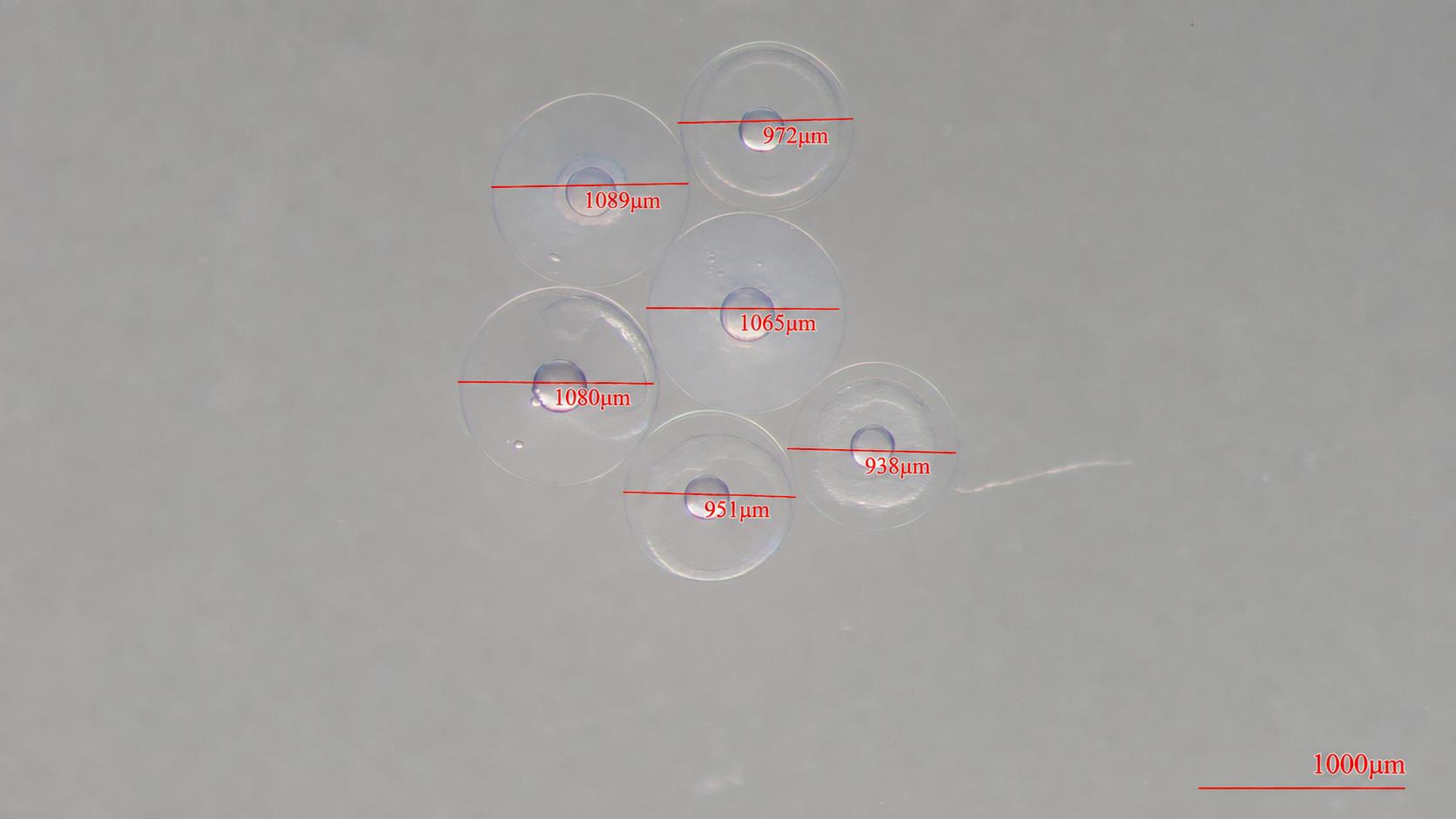
Figure 1. Fish eggs photographed under a stereomicroscope equipped with a DeltaPix digital camera
C. DNA extraction: modified phenol–chloroform–isoamyl genomic DNA extraction protocol
1. For each extraction, use a new, clean histological glass slide and a sterile medical needle on each egg.
2. Use clean laboratory scissors to cut the ends of pipette tips for collecting eggs stored in 96% alcohol after field sampling.
3. Pierce each individual egg on a histological glass slide using a sterile medical needle. Immediately after piercing, use a pipette containing a small volume of lysis buffer to gently collect the entire egg content from the glass slide and transfer it into a 1.5 mL Eppendorf safe-lock tube, minimizing the loss of biological material (see General note 1).
4. Isolation steps:
a. In a 1.5 mL safe-lock tube, add 200 μL of lysis buffer.
b. Transfer the egg content placed on the histology glass (see General note 1).
Critical: Ensure that the entire egg content is collected from the glass slide to minimize loss of biological material.
c. Add 4 μL of Proteinase K (20 mg/mL).
d. Vortex vigorously for 1 min.
e. Incubate for 2 h in a thermomixer at 55 °C with rotation at 650 rpm.
f. Add 200 μL of phenol–chloroform–isoamyl alcohol (25:24:1).
Caution: Phenol–chloroform–isoamyl alcohol is hazardous and should be handled in a chemical fume hood using appropriate personal protective equipment
g. Vortex vigorously for a few seconds to mix well and form an emulsion.
h. Centrifuge at high speed (17,949 rcf, 13,000 rpm) for 5 min at RT.
i. Remove approximately 120–150 μL of the top aqueous phase and transfer to a new tube.
Critical: Carefully transfer only the upper aqueous phase without disturbing the interface (see General note 5).
j. Add an equal volume of 96% ethanol and mix slowly by inverting 4–5 times. No additional salt or sodium acetate is added at this step; precipitation is achieved using only 96% ethanol.
k. Centrifuge at high speed (17,949 rcf, 13,000 rpm) for 15 min at RT.
l. Carefully remove the supernatant by inverting the tube.
m. Wash with 70% ethanol and leave overnight at 4 °C. All ethanol must be completely removed before proceeding to the next step. If necessary, re-centrifuge and remove the residual ethanol thoroughly by pipetting.
n. Air dry, but do not overdry.
o. Resuspend in 10 μL of TE buffer (see General note 6).
p. Store the DNA at -20 °C for long-term preservation.
Pause point: Extracted DNA can be stored at -20 °C until further PCR analysis.
Data analysis
The concentration and purity of the extracted DNA were measured using a spectrophotometer, as described in General notes 7. PCR amplification was confirmed by agarose gel electrophoresis, and the resulting gel images were examined to verify the presence and expected size of the amplified DNA fragments (Figure 2). The PCR products were subsequently sequenced, and the resulting sequences were compared with reference sequences in the NCBI BLAST database for molecular species identification (Figure 2).
A. Polymerase chain reaction and sequencing
A fragment of the mitochondrial cytochrome c oxidase subunit I (COI) gene was amplified for a subset of samples using the universal primers FishF1 and FishR1 [5]. PCR reactions were performed in a total volume of 25 μL containing 1 μL of template DNA, 0.125 μL of HotStarTaq DNA Polymerase (Qiagen; 5 U/μL), 2.5 μL of 10× PCR buffer, 1 μL of MgCl2 (25 mM), 0.5 μL of dNTPs (0.25 mM each), 0.5 μL of each primer (10 μM), and nuclease-free water to volume. Amplification was carried out under the following conditions: initial denaturation at 95 °C for 15 min; 35 cycles of 94 °C for 45 s, 54 °C for 45 s, and 72 °C for 1 min; and a final extension at 72 °C for 10 min. PCR products were verified on a 1% agarose gel under UV illumination. Sequencing was performed by Macrogen Inc. (Amsterdam, Netherlands) using an ABI 3730 automated sequencer.
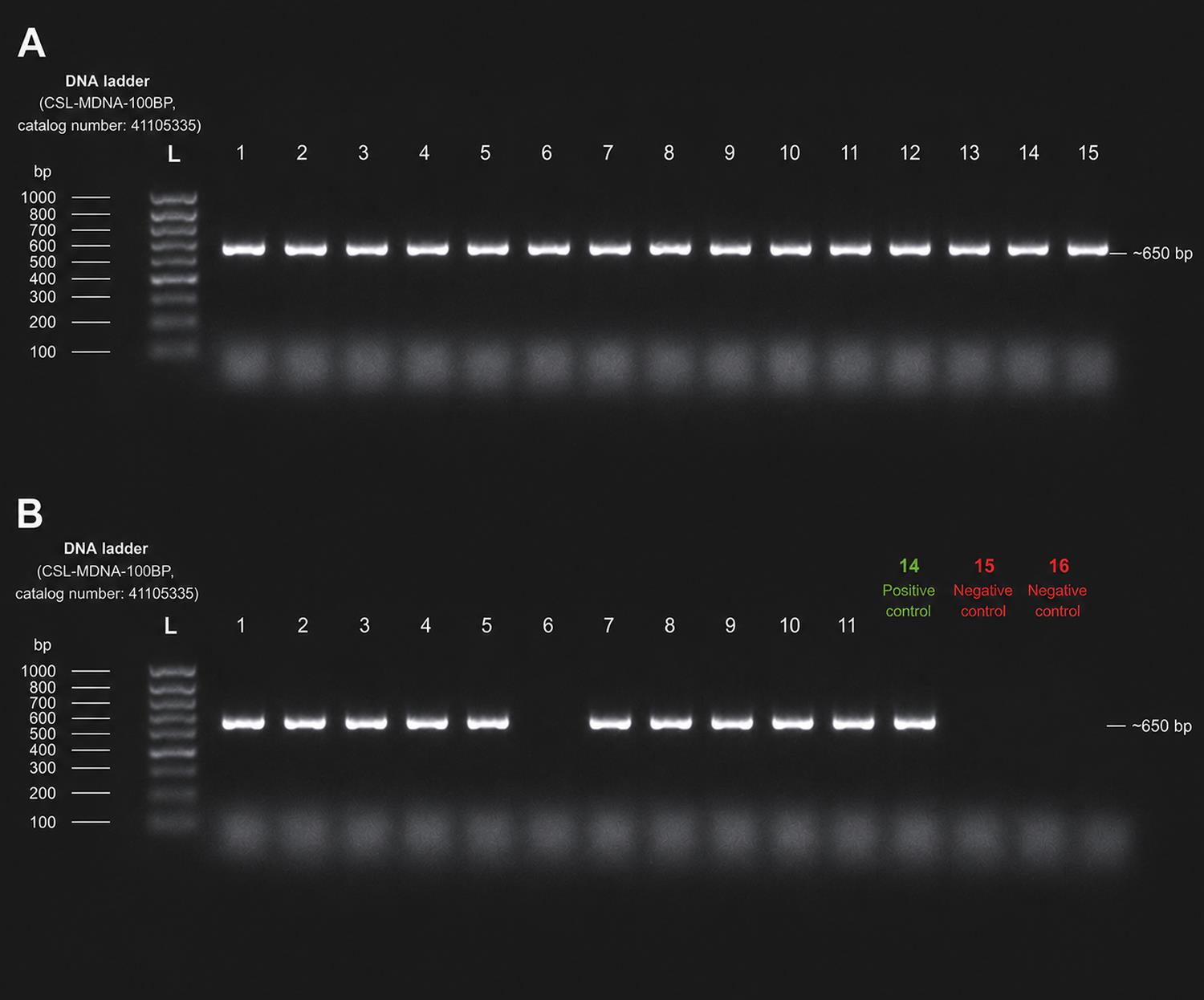
Figure 2. Agarose gel electrophoresis analysis of PCR-amplified DNA products. (A) Detection of 15 successfully amplified PCR constructs. (B) Eleven successfully amplified constructs. The last three lanes represent controls, including one positive control and the last two negative controls. The amplified fragment was approximately 650 bp in size.
B. Genetic analyses
Sequences were checked and edited in BioEdit [4], while taxonomic assignment was performed using BLASTn against the NCBI nucleotide database (nr/nt). Species identification was based on ≥99% sequence identity (Figure 3).
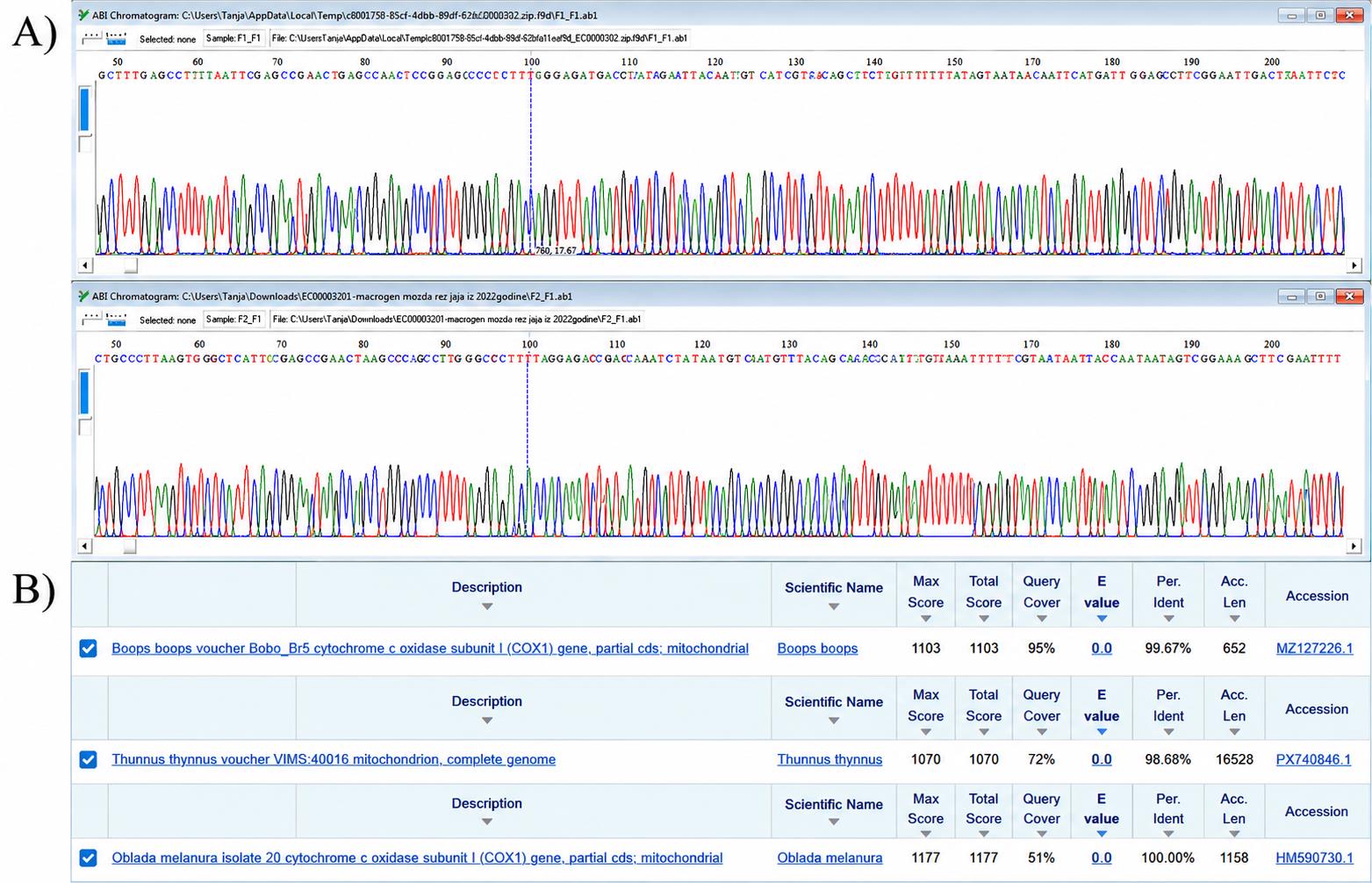
Figure 3. Representative chromatogram snippets from two individually collected fish eggs submitted for sequencing, following sequence quality assessment using BioEdit (A), and NCBI BLAST results used for molecular species identification based on sequence similarity to reference sequences (B)
Validation of protocol
This protocol is currently used for DNA isolation from fish eggs collected near Atlantic bluefin tuna (Thunnus thynnus) farms under the project Enhancing Environmental Performance of Net-Pen Marine Aquaculture (EpoMariNet), funded by the Croatian Science Foundation Research Project IP-2022–10-7232.
More than 200 individual fish eggs were processed using the optimized extraction protocol. Due to the low DNA yield expected from individual eggs and the limited final DNA volume (10 μL), DNA concentration was measured only in a subset of samples to preserve sufficient template for downstream PCR amplification and sequencing. The PCR amplification success rate for DNA isolated using this protocol was >98%, and the resulting sequences were successfully used for species identification by comparison with reference sequences in the NCBI database using BLAST.
This protocol has been used and validated in the following research article:
Žuvić et al. [6] Scalable juvenile small-tuna identification and new insights into spawning and population structure in the Adriatic Sea. Estuarine Coastal Shelf Sci.
General notes and troubleshooting
General notes
1. Particular care should be taken during the micropuncture and transfer of individual fish eggs, as the amount of biological material available from a single egg is limited. Both 21G and 22G needles are suitable for egg micropuncture; therefore, the needle gauge does not need to be selected according to egg size and can instead be chosen based on the researcher’s preference and ease of handling. The needle should be carefully and gently brought into contact with the egg to avoid excessive disruption, as the egg contents can easily disperse over the glass slide. Following micropuncture, the entire egg contents should be collected from the glass slide using a pipette containing a small volume of lysis buffer. Particular care should be taken to recover the entire egg contents and minimize the loss of biological material during transfer.
2. PCR controls were included specifically at the PCR stage and were not used during DNA extraction. A known DNA sample from Atlantic bluefin tuna (Thunnus thynnus) served as the positive control, while Milli-Q water was used as the negative control (no-template control, NTC). The positive control confirmed successful PCR amplification, whereas the negative control monitored potential contamination or nonspecific amplification in the absence of template DNA.
3. Upon returning from the field, carefully rinse the collected fish eggs with Milli-Q water to remove unwanted organic material, including seaweed and other debris, before proceeding with recording egg and DNA extraction.
4. Recording egg size is optional; for us, it was important to record the egg size of the target species to observe differences between species.
5. During phase separation following phenol–chloroform–isoamyl alcohol extraction, the upper aqueous phase should be carefully transferred without disturbing the interface to minimize carryover of organic solvents, which may interfere with subsequent PCR amplification
6. Due to the small amount of starting material, the final DNA is resuspended in 10 μL of TE buffer.
7. The DNA concentration obtained from individual fish eggs ranged from 10 to 45 ng/μL. DNA purity, assessed by spectrophotometric absorbance ratios, ranged from 1.2 to 1.9 for A260/A280 and from 1.8 to 2.1 for A260/A230.
Troubleshooting
Problem 1: Low DNA yield.
Possible cause: Loss of biological material during micropuncture and transfer.
Solution: After micropuncture, gently collect the entire egg content from the glass slide using a pipette containing a small volume of lysis buffer, minimizing the loss of biological material.
Problem 2: Incomplete lysis.
Possible causes: Incomplete disruption of the egg or inadequate digestion.
Solution: Ensure complete micropuncture of the egg and thorough mixing with lysis buffer and Proteinase K. Incubate for 2 h at 55 °C and 650 rpm; prolonged incubation should be avoided due to the low amount of DNA available from a single egg.
Problem 3: PCR inhibition.
Possible causes: Carryover of phenol–chloroform–isoamyl alcohol during transfer of the aqueous phase or residual ethanol after the washing step.
Solution: Carefully transfer only the upper aqueous phase without disturbing the interface to minimize carryover of organic solvents. Completely remove residual ethanol before DNA resuspension. If necessary, re-centrifuge and carefully remove the remaining ethanol by pipetting.
Acknowledgments
This research was funded by the Croatian Science Foundation under the project HRZZ-IP-2022- 10-7232 (EpoMariNet).
This protocol is used in [6]. The authors would like to thank the reviewers for their valuable comments and suggestions, which helped improve the quality and final presentation of this protocol.
Author contributions
Conceptualization, I.Z.V.; Investigation, I.Z.V. and I.L.P.; Writing—Original Draft, I.Z.V. and K.I.; Writing—Review & Editing, I.Z.V., K.I., and I.L.P.; Funding acquisition, T.S.B.
Competing interests
The authors declare no conflicts of interest.
References
- Aranishi, F. (2006). Single Fish Egg DNA Extraction for PCR Amplification. Conserv Genet. 7(1): 153–156. https://doi.org/10.1007/s10592-005-5387-y
- Golotin, V., Lyutikov, A., Filatova, T., Sharoyko, V. and Apalikova, O. (2023). A Rapid and Simple Procedure for the Isolation of Embryonic Cells from Fish Eggs. Bio Protoc. 13(19): e4836. https://doi.org/10.21769/bioprotoc.4836
- Stagličić, N., Šegvić-Bubić, T., Ugarković, P., Talijančić, I., Žužul, I., Tičina, V. and Grubišić, L. (2017). Ecological role of bluefin tuna (Thunnus thynnus) fish farms for associated wild fish assemblages in the Mediterranean Sea. Mar Environ Res. 132: 79–93. https://doi.org/10.1016/j.marenvres.2017.10.015
- Hall, T. A. (1999). BioEdit: a user-friendly biological sequence alignment editor and analysis program. Nucleic Acids Symposium Series. 41: 95–98.
- Ward, R. D., Zemlak, T. S., Innes, B. H., Last, P. R. and Hebert, P. D. (2005). DNA barcoding Australia's fish species. Philos Trans R Soc Lond B Biol Sci. 360(1462): 1847–1857. https://doi.org/10.1098/rstb.2005.1716
- Žuvić, L., Talijančić, I., Zorica, B., Bušelić, I., Pleić, I. L., Vrgoč, I. Å., Grubišić, L. and Šegvić-Bubić, T. (2026). Scalable juvenile small-tuna identification and new insights into spawning and population structure in the Adriatic Sea. Estuarine Coastal Shelf Sci. 336: 109895. https://doi.org/10.1016/j.ecss.2026.109895
Article Information
Publication history
Received: May 30, 2026
Accepted: Sep 7, 2026
Available online: Sep 15, 2026
Published: Oct 20, 2026
Copyright
© 2026 The Author(s); This is an open access article under the CC BY-NC license (https://creativecommons.org/licenses/by-nc/4.0/).
How to cite
Žužul Vrgoč, I., Lepen Pleić, I., Ivanišević, K. and Šegvić-Bubić, T. (2026). Optimized Phenol–Chloroform–Isoamyl DNA Extraction Protocol for Single Fish Eggs. Bio-protocol 16(20): e5839. DOI: 10.21769/BioProtoc.5839.

 Download Citation in RIS Format
Category
Environmental science > Marine vertebrates
Molecular Biology
Do you have any questions about this protocol?
Post your question to gather feedback from the community. We will also invite the authors of this article to respond.
 Post a Question


