


- Protocols

- Articles and Issues
- For Authors
- About
- Become a Reviewer


A SPAAC-Based Bioorthogonal Method for Verifying Protein Palmitoylation

(*contributed equally to this work) Published: Vol 16, Iss 20, Oct 20, 2026 DOI: 10.21769/BioProtoc.5835 Views: 16
Reviewed by: Catherine HurdBhanu JagilinkiJoyce Chiu
 PDF
PDF  Ask a question
Original research article
The authors used this protocol in:
May 2025

Protocol Collections
Comprehensive collections of detailed, peer-reviewed protocols focusing on specific topics




Related protocols
Abstract
Palmitoylation is a crucial post-translational modification, and bioorthogonal chemistry based on azide-alkyne cycloaddition is typically used to verify protein palmitoylation. Traditional copper-catalyzed click chemistry (CuAAC) proceeds with fast kinetics and is widely used, but it requires a copper catalyst and suffers from copper-induced toxicity and nonspecific labeling. By contrast, strain-promoted click chemistry (SPAAC) has slower kinetics but is catalyst-free, offering high specificity, low cytotoxicity, and simple operation. However, SPAAC is mostly applied to live-cell labeling and imaging of known palmitoylated proteins in the field of palmitoylation, and its use in identifying novel palmitoylated proteins is still limited. Here, we present a SPAAC-based method for detecting endogenous protein palmitoylation. Compared with CuAAC, this method eliminates the need for copper catalysts and reducing agents, thereby simplifying the procedure and reducing reagent usage.
Key features
• A bioorthogonal method for identifying endogenous protein palmitoylation in cells.
• Metabolical labeling of palmitoylated proteins in cells using azido palmitic acid.
• The method requires simple steps and minimal reagents, reducing hands-on time and cost.
Keywords: Click chemistry CuAAC
SPAAC
Palmitoylation
Protein tagging
Post-translational modifications
CuAAC
SPAAC
Palmitoylation
Protein tagging
Post-translational modifications
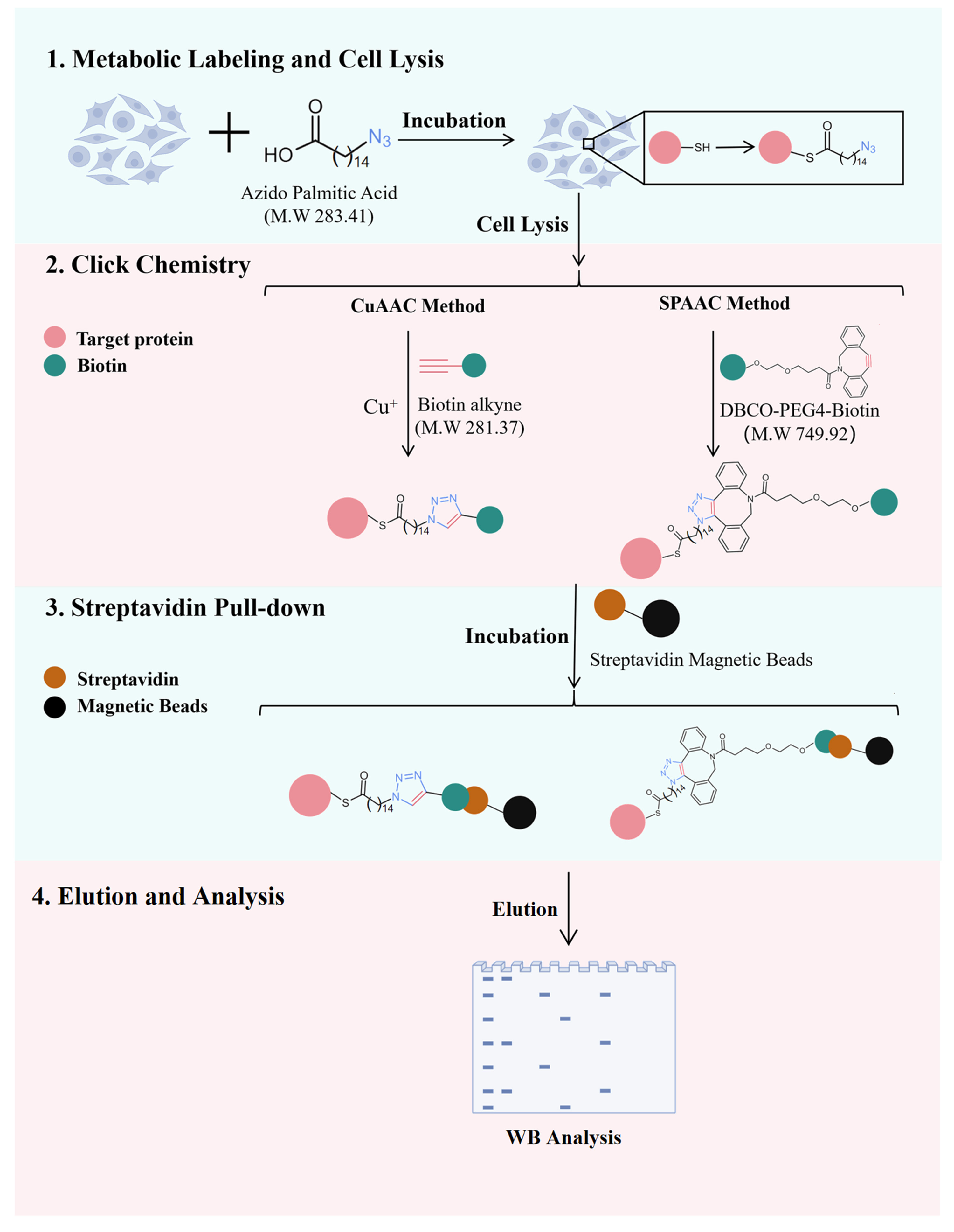
Graphical overview
Background
Palmitoylation is a reversible post-translational modification that involves the covalent attachment of palmitic acid (C16:0) to cysteine residues via a thioester bond. It dynamically regulates protein membrane localization, stability, and signal transduction [1–3]. Aberrant palmitoylation is closely associated with cancer, neurodegenerative diseases, and infectious diseases [3–6]. Therefore, identifying and validating palmitoylation modifications is crucial for elucidating their biological functions and pathogenic mechanisms.
Methods for verifying palmitoylation are mainly divided into two categories: non-metabolic labeling and metabolic labeling [7]. The acyl-biotin exchange (ABE) method is a non-metabolic labeling strategy that offers direct applicability to tissue samples, high sensitivity, and good compatibility with downstream mass spectrometry analysis [8]. Nonetheless, it cannot readily distinguish between different lipid types, requires a complex procedure, and is susceptible to false-positive results [9,10]. Currently, metabolic labeling based on bioorthogonal chemistry is the most widely used strategy. In this approach, azide-bearing palmitic acid analogs (e.g., 15-azido pentadecanoic acid) are incorporated into palmitoylated proteins through cellular metabolism, followed by azide-alkyne cycloaddition to conjugate reporter groups (e.g., fluorophores or biotin) for detection, enrichment, and identification [10,11]. This strategy primarily includes copper-catalyzed click chemistry (CuAAC) and strain-promoted click chemistry (SPAAC). Traditional copper-catalyzed azide-alkyne cycloaddition (CuAAC) proceeds through the Cu(I)-mediated activation of a terminal alkyne to form a copper–acetylide intermediate, which then reacts with azides to yield a triazole. Although CuAAC offers fast reaction kinetics and is the most commonly used click reaction, it requires a copper ion catalyst, which can lead to cytotoxicity and nonspecific labeling [12]. This is partly because Cu(I) ions can coordinate with thiol groups of cysteine residues and imidazole groups of histidine residues in proteins and also generate reactive oxygen species (ROS), resulting in off-target modifications and cellular stress. In contrast, strain-promoted azide–alkyne cycloaddition (SPAAC) relies on the ring strain of cyclooctyne derivatives to drive a catalyst-free [3+2] cycloaddition with azides. Although SPAAC exhibits relatively slower reaction kinetics than CuAAC, it requires no catalyst and has become widely adopted owing to its high specificity, low cytotoxicity, and operational simplicity [13,14]. However, in the context of palmitoylation research, SPAAC is mainly applied to live-cell labeling and imaging for real-time tracking and localization of palmitoylated proteins, whereas few studies have applied it to the identification of previously unreported palmitoylated proteins.
Here, we describe a SPAAC-based protocol that eliminates copper catalyst usage, simplifies the workflow, and provides comparable sensitivity to CuAAC for endogenous palmitoylation detection. Moreover, it can also be extended to other applications, including the enrichment and mass spectrometry identification of palmitoylated proteins and the determination of palmitoylation sites.
Materials and reagents
Biological materials
1. HEK293T cell line
Reagents
1. 30% Acrylamide:Bisacrylamide (Acr/Bis) (29:1) (Solarbio, catalog number: A1010)
2. AEG-1 polyclonal antibody (Proteintech, catalog number: 13860-1-AP)
3. Ammonium persulfate (APS) (Aladdin, catalog number: A112450)
4. Azido palmitic acid (MCE, catalog number: HY-151656)
5. β-actin polyclonal antibody (Proteintech, catalog number: 20536-1-AP)
6. Biotin alkyne (MCE, catalog number: HY-138749)
7. Bovine serum albumin (BSA) (fatty acid free) (MCE, catalog number: HY-D0842A)
8. BCA Protein Assay kit (Biosharp, catalog number: 143179)
9. Chloroform (Aladdin, catalog number: C1506334)
10. Glycerol (Aladdin, catalog number: G116206)
11. CuSO4 (Aladdin, catalog number: C573445)
12. DBCO-PEG4-biotin (MCE, catalog number: HY-130809)
13. Dulbecco's modified Eagle medium (DMEM) (Vivacell, catalog number: C3130)
14. Dimethyl sulfoxide (DMSO) (Aladdin, catalog number: D103277)
15. Fetal bovine serum (FBS) (Vivacell, catalog number: C04001)
16. Fetal bovine serum (lipid depleted) (Vivacell, catalog number: C3840)
17. GPX4 polyclonal antibody (Proteintech, catalog number: 30388-1-AP)
18. Methanol (Aladdin, catalog number: M116115)
19. Non-fat milk (Solarbio, catalog number: D3840)
20. NaCl (Aladdin, catalog number: S433743)
21. NP-40 (Solarbio, catalog number: N8030)
22. Penicillin-streptomycin solution, 100× (Vivacell, catalog number: C3421)
23. Phosphate-buffered saline (PBS) (Biosharp, catalog number: BL601A)
24. Protease inhibitor cocktail (EDTA-free, 100×) (MCE, catalog number: HY-K0010)
25. PVDF membrane (Millipore, catalog number: IPVH00010)
26. Sodium dodecyl sulfate (SDS) (Aladdin, catalog number: S432158)
27. SDS-PAGE loading buffer (4×) (Proteintech, catalog number: PR20003)
28. Semi-dry membrane transfer solution (1×) (Biosharp, catalog number: BL1310A)
29. L-Ascorbic acid sodium salt (Aladdin, catalog number: S105026)
30. Streptavidin magnetic beads (Beyotime, catalog number: P2151)
31. N,N,N',N'-Tetramethylethylenediamine (TEMED) (Aladdin, catalog number: T105496)
32. Tris(3-hydroxypropyltriazolylmethyl)amine (THPTA) (Aladdin, catalog number: T405015)
33. Tris-buffered saline (1×) (Biosharp, catalog number: BL602A)
34. Tris-glycine-SDS running buffer (Biosharp, catalog number: BL603A)
35. Tris-HCl (1 M, pH 6.8) (Solarbio, catalog number: T1020)
36. Tris-HCl (1.5 M, pH 8.8) (Solarbio, catalog number: T1010)
37. Tween 20 (Solarbio, catalog number: T8820)
38. Trypsin EDTA solution (0.25%) (Vivacell, catalog number: C3530)
39. Ultrasensitive ECL Detection kit (Proteintech, catalog number: PK10003)
Solutions
1. DMEM medium (10% FBS) (see Recipes)
2. DMEM medium (5% lipid-depleted FBS) (see Recipes)
3. Fatty acid-free BSA solution (10%) (see Recipes)
4. Azido palmitic acid (50 mM) (see Recipes)
5. Azido-containing labeling medium (see Recipes)
6. NaCl solution (1.5 M) (see Recipes)
7. Cell lysis buffer (see Recipes)
8. Biotin alkyne (1 mM) (see Recipes)
9. DBCO-PEG4-biotin (10 mM) (see Recipes)
10. CuSO4 solution (20 mM) (see Recipes)
11. THPTA (100 mM) (see Recipes)
12. Sodium L-ascorbate solution (300 mM) (see Recipes)
13. TBST (see Recipes)
14. Washing buffer (see Recipes)
15. Spacer gel (see Recipes)
16. Stacking gel (see Recipes)
17. Blocking buffer (5% non-fat milk) (see Recipes)
Recipes
Note: In the following recipes, H2O is deionized water, and DMSO is cell culture grade.
1. DMEM medium (10% FBS)
| Reagent | Final concentration | Quantity or volume |
|---|---|---|
| DMEM | 89% | 44.5 mL |
| FBS | 10% | 5 mL |
| Penicillin-Streptomycin Solution, 100× | 1× | 500 μL |
| Total | n/a | 50 mL |
Store at 4 °C for a week.
2. DMEM medium (5% lipid-depleted FBS)
| Reagent | Final concentration | Quantity or volume |
|---|---|---|
| DMEM | 95% | 47.5 mL |
| Lipid-depleted FBS | 5% | 2.5 mL |
| Total | n/a | 50 mL |
Store at 4 °C for 4 weeks.
3. Fatty acid-free BSA solution (10%)
| Reagent | Final concentration | Quantity or volume |
|---|---|---|
| Fatty acid-free BSA | 10% | 1 g |
| PBS | n/a | 10 mL |
| Total | n/a | 10 mL |
Slowly stir with a magnetic stirrer at 4 °C until dissolved. Divide into 1 mL aliquots and store at -20 °C after filtering through a 0.22 μm filter in a biosafety cabinet.
4. Azido palmitic acid (50 mM)
| Reagent | Final concentration | Quantity or volume |
|---|---|---|
| Azido palmitic acid | 50 mM | 14.2 mg |
| DMSO | n/a | 1 mL |
| Total | n/a | 1 mL |
Divide into 200 μL aliquots in amber centrifuge tubes and store at -80 °C for 6 months.
5. Azido-containing labeling medium
| Reagent | Final concentration | Quantity or volume |
|---|---|---|
| Azido palmitic acid | 50 μM | 10 μL |
| 10% fatty acid-free BSA solution | 1% | 1 mL |
| DMEM medium (5% lipid-depleted FBS) | n/a | up to 10 mL |
| Total | n/a | 10 mL |
Prepare the reagents in the following order: first, combine azido palmitic acid with 10% fatty acid-free BSA solution in a sterile 15 mL centrifuge tube. Mix by inverting the tube or by gently pipetting up and down. Then, incubate the tube in a 37 °C water bath for 0.5–1 h. Finally, add DMEM medium (5% lipid-depleted FBS) and mix by pipetting. Use immediately after preparation. The volumes can be scaled up or down as needed.
Note: The purpose of this step is to allow azido palmitic acid to mix with fatty acid-free BSA, which facilitates cellular uptake of azido palmitic acid.
6. NaCl solution (1.5 M)
| Reagent | Final concentration | Quantity or volume |
|---|---|---|
| NaCl | 1.5 M | 8.8 g |
| H2O | n/a | 100 mL |
| Total | n/a | 100 mL |
7. Cell lysis buffer
| Reagent | Final concentration | Quantity or volume |
|---|---|---|
| NP-40 | 0.5% | 200 μL |
| Glycerol | 10% | 4 mL |
| Protease inhibitor cocktail (100×) | 1× | 400 μL |
| PBS, pH 7.4 | n/a | up to 40 mL |
| Total | n/a | 40 mL |
Aliquot into 10 mL aliquots and store at -20 °C.
Critical: The buffer must not contain Tris or EDTA, as they inhibit the click chemistry reaction [15].
8. Biotin alkyne (5 mM)
| Reagent | Final concentration | Quantity or volume |
|---|---|---|
| Biotin alkyne | 5 mM | 1.4 mg |
| DMSO | n/a | 1 mL |
| Total | n/a | 1 mL |
Aliquot into 500 μL aliquots in amber centrifuge tubes and store at -80 °C for 6 months.
9. DBCO-PEG4-biotin (10 mM)
| Reagent | Final concentration | Quantity or volume |
|---|---|---|
| DBCO-PEG4-biotin | 10 mM | 7.5 mg |
| DMSO | n/a | 1 mL |
| Total | n/a | 1 mL |
Divide into 200 μL aliquots in amber centrifuge tubes and store at -80 °C for 6 months.
10. CuSO4 solution (20 mM)
| Reagent | Final concentration | Quantity or volume |
|---|---|---|
| CuSO4 | 20 mM | 31.9 mg |
| H2O | n/a | 10 mL |
| Total | n/a | 10 mL |
Aliquot into 1 mL aliquots and store at -20 °C.
11. THPTA (100 mM)
| Reagent | Final concentration | Quantity or volume |
|---|---|---|
| THPTA | 100 mM | 43.5 mg |
| H2O | n/a | up to 1 mL |
| Total | n/a | 1 mL |
Divide into 200 μL aliquots in amber centrifuge tubes and store at -20 °C for 3 months.
12. Sodium L-ascorbate solution (300 mM)
| Reagent | Final concentration | Quantity or volume |
|---|---|---|
| L-ascorbate acid sodium salt | 300 mM | 594.3 mg |
| H2O | n/a | 10 mL |
| Total | n/a | 10 mL |
Aliquot into 1 mL aliquots and store at -20 °C protected from light.
13. TBST
| Reagent | Final concentration | Quantity or volume |
|---|---|---|
| Tween 20 | 0.05% | 500 μL |
| Tris-buffered saline (1×) | n/a | up to 1 L |
| Total | n/a | 1 L |
14. Washing buffer
| Reagent | Final concentration | Quantity or volume |
|---|---|---|
| Tween-20 | 0.05% | 50 μL |
| Fatty acid-free BSA solution (10%) | 0.1% | 1 mL |
| PBS | 98.95% | up to 100 mL |
| Total | n/a | 100 mL |
Use immediately after preparation.
15. Spacer gel
| Reagent | Final concentration | Quantity or volume |
|---|---|---|
| 30% Acr/Bis (29:1) | 10% | 3.3 mL |
| 1.5 M Tris-HCl, pH 8.8 | 375 mM | 2.5 mL |
| 10% SDS | 0.1% | 100 μL |
| 10% APS | 0.1% | 100 μL |
| TEMED | 0.2% | 20 μL |
| H2O | n/a | up to 10 mL |
| Total | n/a | 10 mL |
16. Stacking gel
| Reagent | Final concentration | Quantity or volume |
|---|---|---|
| 30% Acr/Bis (29:1) | 5% | 830 μL |
| 1 M Tris-HCl, pH 6.8 | 125 mM | 625 μL |
| 10% SDS | 0.1% | 50 μL |
| 10% APS | 0.15% | 75 μL |
| TEMED | 0.2% | 10 μL |
| H2O | n/a | up to 5 mL |
| Total | n/a | 5 mL |
17. Blocking buffer (5% non-fat milk)
| Reagent | Final concentration | Quantity or volume |
|---|---|---|
| Non-fat milk | 5% | 2 g |
| TBST | n/a | 40 mL |
| Total | n/a | 40 mL |
Laboratory supplies
1. 100 mm TC-treated culture dish (NEST, catalog number: 704202)
2. 1.5 mL microfuge tube (Biosharp, catalog number: BS-15-M)
3. 1.5 mL amber microfuge tube (Biosharp, catalog number: BS-15-A)
4. 2.0 mL microfuge tube (Biosharp, catalog number: BS-20-M)
5. 0.22 μm filters (Biosharp, catalog number: BS-NY13-22-S)
6. Cell scraper (Labselect, catalog number: 1601)
7. Pipette tips (Biosharp, catalog numbers: BS-10-T, BS-200-T, BS-1000-T)
Equipment
1. 4 °C and -20 °C freezer (Haier, model: BCD-390WGHC2B6W9U1)
2. -80 °C freezer (Haier, model: DW-86L486)
3. Automated chemiluminescence/fluorescence image analysis system (Tanon, model: 4600)
4. Biosafety cabinet (Thermo, model: MSC1)
5. CO2 cell culture incubator (Thermo, model: Heracell-150i)
6. Cell sonicator (Branson, model: S-450D)
7. Disc rotator (Scilogex, model: SCI-RD-E)
8. Inverted microscope (Leica, model: DM IL)
9. Magnetic stirrer (IKA, model: RET basic)
10. Metal bath heater (Yuejin, model: HDB1)
11. Orbital shaker (Scilogex, model: SLK-O3000-S)
12. Pipette (Thermo, catalog numbers: 4640110, 4640000, 4640030, 4640050, 4640060, 4640100)
13. Protein electrophoresis system (Bio-Rad, model: Mini-PROTEAN Tetra)
14. Refrigerated centrifuge (Thermo, model: ST16R)
15. Semi-dry transfer apparatus (Bio-Rad, model: Trans-Blot Turbo)
16. Vortex mixer (Scilogex, model: MX-S)
17. Water purification system (Neo Lab, model: Lab UP/RO)
Software and datasets
1. ImageJ (NIH, V14.3)
2. GraphPad Prism (GraphPad Software, V9.1)
Procedure
A. Cell culture
1. When HEK 293T cells reach 80%–90% confluence in the 100 mm culture dish, aspirate the spent medium and rinse the cells twice with 2 mL of prewarmed PBS (37 °C).
2. Add 1 mL of Trypsin EDTA solution to the dish and gently swirl to ensure the reagent covers the entire monolayer.
3. Incubate the dish at 37 °C until the cells begin to round up and intercellular spaces become apparent. Then, add 3 mL of DMEM medium (10% FBS) to neutralize the trypsin.
4. Gently pipette the cells to detach them and obtain a single-cell suspension. Transfer the suspension to a centrifuge tube and centrifuge at 90× g for 3 min.
5. Discard the supernatant and gently resuspend the cell pellet in 2 mL of DMEM medium (containing 10% FBS). Count the cells using a hemocytometer, then dilute the suspension with additional DMEM medium to achieve a final cell density of 2 × 10⁶ cells/mL before seeding.
6. Seed 1 mL of the cell suspension into each 100 mm culture dish, then add 6 mL of DMEM medium (10% FBS) per dish. Swirl gently to ensure an even distribution (the final volume is 7 mL).
7. Incubate the dishes at 37 °C in a 5% CO2 incubator until the cells reach 70%–80% confluence.
Notes:
1. Using 60 mm dishes or six-well plates for culturing is also acceptable. Ensure that the total protein content in the lysate exceeds 300 μg after cell lysis.
2. To determine the palmitoylation sites of a protein by overexpressing the wild-type and cysteine point mutant proteins in cells, perform metabolic labeling with azido palmitic acid at 18 or 36 h post-transfection of the overexpression plasmid.
B. Metabolic labeling and cell lysis
1. Remove the original medium, wash cells twice with 2 mL of prewarmed PBS (37 °C), then add 5 mL of DMEM containing 5% lipid-depleted FBS and continue culturing at 37 °C for 2 h.
Critical: The purpose of using DMEM with lipid-depleted FBS is to deplete endogenous fatty acids in cells under normal culture conditions, thereby promoting increased cellular uptake of azido palmitic acid.
Note: Serum-free DMEM or Opti-MEM can be used instead of DMEM with 5% lipid-depleted FBS for this step.
2. Remove the medium, wash cells twice with 2 mL of prewarmed PBS, then add 5 mL of azido-containing labeling medium per dish and incubate for 6 h.
Notes:
1. This step aims to label palmitoylatable proteins with azido palmitic acid. The incubation time is not fixed and depends on the palmitoylation half-life of the target protein.
2. Perform a time-course experiment over 3–12 h (e.g., at 3, 6, 9, and 12 h) to determine the optimal incubation time.
3. Incubation exceeding 16 h may lead to β-oxidation of azido palmitic acid, reducing its labeling efficiency.
3. Remove the medium and wash cells twice with 4 °C pre-chilled PBS. Add 1 mL of pre-chilled PBS per dish and scrape off the cells using a cell scraper. Gently pipette to mix the cell suspension and transfer it to a 2 mL centrifuge tube. Rinse the dish twice with 500 μL of PBS each time, collect the remaining cells, and combine them into the same tube.
4. Centrifuge at 90× g for 5 min and discard the supernatant. Add 1 mL of pre-chilled PBS to resuspend the cells, centrifuge again, and discard the supernatant.
Note: This step removes residual extracellular azido palmitic acid to minimize interference with subsequent experiments.
5. Add 1,000 μL of 4 °C pre-chilled cell lysis buffer to each tube, mix by pipetting, and place the tubes on ice.
6. Disrupt the cells on ice using a cell sonicator with the following settings: power 180 W, 3 s on, 3 s off, total sonication time 1 min, alarm temperature 4 °C.
Note: A completely disrupted cell lysate appears clear and transparent without transparent clumps. If the lysate does not meet this standard, repeat the sonication.
7. Centrifuge the cell lysate at 16,000× g for 10 min at 4 °C and then collect the supernatant.
8. Measure the protein concentration of the cell lysate using the BCA Protein Assay kit, then add cell lysis buffer to adjust the protein concentration to 1 mg/mL.
Pause point: Samples can be stored at -80 °C or in liquid nitrogen for up to 1 week.
Note: No dilution is required if the protein concentration is below 1 mg/mL.
C. Click chemistry (CuAAC method)
1. Before starting the click chemistry reaction, take 90 μg of protein as the input sample.
a. Add 90 μL of cell lysate (adjusted to 1 mg/mL protein concentration) to a 1.5 mL centrifuge tube.
b. Add 30 μL of 4× SDS-PAGE loading buffer and mix by pipetting.
c. Place the tube on a metal bath heater and heat at 95 °C for 5 min.
Note: For membrane protein detection, heat first at 50 °C for 5 min, then at 70 °C for 5 min.
2. Perform the click chemistry reaction (total reaction volume: 500 μL):
a. Transfer 415 μL of cell lysate into a 1.5 mL amber centrifuge tube (or a 1.5 mL centrifuge tube wrapped with aluminum foil).
b. Add 10 μL of 5 mM biotin alkyne solution (final concentration 100 μM) and vortex for 3 s.
c. Add 25 μL of 100 mM THPTA solution (final concentration 5 mM) and vortex for 3 s.
d. Add 25 μL of 20 mM CuSO4 solution (final concentration 1 mM) and vortex for 3 s.
e. Add 25 μL of 300 mM sodium L-ascorbate solution (final concentration 15 mM) and vortex for 3 s.
f. Mix the solution on a rotator for 1 h at room temperature or for 3 h at 4 °C.
Notes:
1. Perform the entire reaction under light-protected conditions to avoid direct exposure of reagents and the mixture to light.
2. The total reaction volume is flexible and can be scaled up or down as needed.
3. The total protein amount in the reaction should ideally be no less than 300 μg.
3. Precipitate the protein after the click chemistry reaction:
a. Transfer 500 μL of the post-reaction mixture into a 5 mL centrifuge tube and place the tube on ice.
b. Add 1.6 mL of -20 °C pre-chilled methanol and vortex thoroughly to mix.
c. Add 400 μL of chloroform and vortex to mix.
d. Add 1.2 mL of deionized water and vortex to mix (total volume = 3.7 mL).
e. Transfer the 3.7 mL mixture to the same 2 mL centrifuge tube in three aliquots (≤1.5 mL each). After each transfer, centrifuge at 16,000× g for 10 min at 4 °C. Carefully aspirate and discard the upper aqueous phase while keeping the middle protein layer and the lower organic phase intact.
f. Repeat step C3e until the entire mixture has been processed.
g. Add 800 μL of pre-chilled methanol to the tube, vortex to mix, then centrifuge at 16,000× g for 10 min at 4 °C.
h. Discard the supernatant and repeat step C3g once.
i. Air-dry the tube in a fume hood for 5–10 min until the pellet loses its obvious liquid sheen.
Critical:
1. This step aims to efficiently remove THPTA, unreacted biotin alkyne, and copper ions from the reaction system, thereby preventing them from interfering with the subsequent streptavidin magnetic beads pull-down results.
2. Do not over-dry the pellet, as it will become difficult to redissolve.
4. Add 500 μL of cell lysis buffer to the centrifuge tube and rotate to mix at 4 °C for 30 min to completely dissolve the protein.
Note: Extend the rotation mixing time or place the tube in a water bath sonicator to assist dissolution if the protein does not dissolve completely after 30 min.
Pause point: Samples can be stored at -80 °C or in liquid nitrogen for up to 1 week.
D. Click chemistry (SPAAC method)
1. Before starting the click chemistry reaction, take 90 μg of protein as the input sample (as in step C1).
2. Perform the click chemistry reaction (total reaction volume: 500 μL):
a. Transfer 415 μL of cell lysate and 80 μL of cell lysis buffer into a 1.5 mL amber centrifuge tube.
Note: This step only serves to match the protein amount used in the click chemistry reaction in section C. In practice, you can directly add 495 μL of cell lysate.
b. Add 5 μL of 10 mM DBCO-PEG4-biotin (final concentration 100 μM) and vortex for 3 s.
c. Mix the solution on a rotator for 1 h at room temperature or for 3 h at 4 °C.
Notes:
1. Perform the entire reaction under light-protected conditions to avoid direct exposure of reagents and the mixture to light.
2. The total reaction volume is flexible and can be scaled up or down as needed.
3. The total protein amount in the reaction should ideally be no less than 300 μg.
3. Precipitate the protein after the click chemistry reaction (as in step C3).
Notes:
1. The SPAAC reaction contains no THPTA or copper ions, which could interfere with streptavidin magnetic beads. Therefore, the precipitation step can be omitted to reduce product loss and shorten the workflow.
2. Using 1.5-fold excess of streptavidin beads removes any interference from residual DBCO-PEG4-biotin.
4. Dissolve the protein (as in step C4).
Note: Ignore this step if you skipped step D3.
E. Streptavidin pull-down and elution
1. Transfer 80 μL of streptavidin magnetic beads to a 1.5 mL centrifuge tube. Place the tube on a magnetic stand for 30 s, then remove the supernatant.
Notes:
1. Cut off 1 mm from the tip of the pipette tip to avoid damaging the beads when pipetting them.
2. Transfer 120 μL of streptavidin magnetic beads to a 1.5 mL centrifuge tube if you skipped step D3.
2. Add 500 μL of 1× TBS. Gently pipette up and down to resuspend the streptavidin magnetic beads. Place the tube on the magnetic stand for 30 s to separate the beads and then remove the supernatant.
3. Repeat step E2 twice.
4. Add 500 μL of the protein solution to the tube containing the beads. Mix by pipetting, then place the tube on a rotator and incubate for 1 h at room temperature or for 4 h at 4 °C.
5. Place the tube on the magnetic stand for 30 s to separate the beads and then remove the supernatant.
6. Add 1 mL of washing buffer to the tube and rotate on a rotator at 4 °C for 8 min. Place the tube on the magnetic stand for 30 s to separate the beads and then remove the supernatant.
7. Repeat step E6 three more times.
Note: A micro-volume spectrophotometer can be used to measure the A280 of the wash fractions to determine if washing is complete. Washing is considered complete when the difference in A280 between two consecutive wash fractions and the washing buffer is less than 0.01.
8. Add 120 μL of washing buffer and 40 μL of 4× SDS-PAGE loading buffer, then mix by pipetting.
9. Place the tube in a metal bath heater and heat at 95 °C for 5 min.
Pause point: Samples can be stored at -20 or -80 °C.
F. Western blot analysis
1. Prepare a 10% SDS-PAGE gel according to Recipes 15 and 16 and assemble the electrophoresis tank. Ensure the inner chamber is filled to the top with Tris-glycine-SDS running buffer before loading the sample.
2. Load 10 μL of sample into each well of the gel and fill the outer chamber with Tris-glycine-SDS running buffer to the appropriate level according to the instrument manual.
3. Run the gel at 80 V for 40 min, then increase the voltage to 130 V for another 40 min.
4. Twenty minutes prior to the completion of electrophoresis, place the transfer filter papers into a clean container and saturate them thoroughly with semi-dry membrane transfer solution. Ensure the papers are fully immersed and remain wet until needed for the transfer step.
5. Activate a PVDF membrane matching the gel size by immersing it in anhydrous methanol for 1 min after electrophoresis.
Notes:
1. The activation of PVDF membranes with methanol serves to render their inherently hydrophobic surface hydrophilic, thereby facilitating efficient protein binding.
2. The activation duration should be strictly controlled and generally limited to 2 min. Prolonged exposure may otherwise lead to membrane brittleness or excessive shrinkage.
6. Immediately after activation, wash the membrane by incubating it in deionized water (or double-distilled water) for 1–2 min. Then, transfer the membrane to semi-dry membrane transfer solution and equilibrate for at least 5 min.
Note: This step removes residual methanol from the membrane. Residual methanol may affect the composition of the subsequent transfer buffer and the transfer efficiency.
7. Place the transfer filter papers, activated PVDF membrane, gel, and another set of transfer filter papers onto the semi-dry transfer apparatus in this order. Set the voltage to 18 V and transfer for 1 h.
8. Immediately after transfer, add 15 mL of pre-prepared blocking buffer to the incubation box, ensuring it covers the PVDF membrane surface. Place the membrane into the buffer and incubate on an orbital shaker at 75 rpm for 1 h at room temperature.
Note: According to the requirements of the subsequent antibody, you may substitute BSA or a commercial blocking buffer for 5% non-fat milk at this step.
9. Wash the PVDF membrane three times with 1× TBST at 200 rpm on an orbital shaker, 8 min per wash.
10. Determine the target protein region according to the protein marker. Cut the corresponding PVDF membrane strip using a ceramic blade.
Note: Use ceramic blades or scissors instead of metal tools. Compared with metal blades, ceramic blades are less likely to tear the PVDF membrane during cutting. Moreover, ceramic is an excellent insulator and generates almost no static electricity during cutting, whereas metal blades may attract impurities via static electricity and contaminate the target protein region on the membrane.
11. Place the trimmed PVDF membrane into the primary antibody solution prepared in blocking buffer. Incubate on an orbital shaker at 75 rpm for 2 h at room temperature or overnight (12 h) at 4 °C.
Note: The primary antibody solutions used in this experiment (see Reagents 2, 5, and 17) were prepared in 5% non-fat milk at a 1:4,000 dilution. The choice of antibody is left to the user and should follow the manufacturer's instructions.
12. Wash the PVDF membrane five times with 1× TBST at 200 rpm on an orbital shaker, 8 min per wash.
13. Incubate the PVDF membrane in the corresponding secondary antibody solution on an orbital shaker at 75 rpm for 2 h at room temperature.
14. Wash the PVDF membrane five times with 1× TBST at 200 rpm on an orbital shaker, 8 min per wash.
15. Visualize the western blot using a chemiluminescence imaging system after treatment with an Ultrasensitive ECL Detection kit.
16. Process the images using ImageJ:
a. Open the pull-down image and convert it to 8-bit grayscale.
b. Subtract the background to minimize nonspecific signal interference on the membrane.
c. Use rectangular selections of identical size to frame each target band.
d. Measure the gray value to obtain the integrated density, then normalize it to the input protein band to calculate the relative expression level.
Note: The size of the selection boxes and the background subtraction parameters for all bands must be kept consistent throughout the entire analysis process to ensure that the results are comparable.
17. Generate graphs using GraphPad Prism:
a. Use GraphPad Prism to normalize the relative expression levels of the three target protein bands in the CuAAC group to 1.
b. Calculate the relative fold change of the corresponding proteins in the SPAAC group.
c. Generate a bar chart based on the values from three independent biological replicates and perform a t-test to evaluate the statistical significance of the difference between the two groups.
Validation of protocol
Azido palmitic acid was pre-incubated with BSA and then added to HEK293T cells. Metabolic labeling was performed for 6 h. After cell lysis, the lysates were divided and subjected to click chemistry reactions using either CuAAC or SPAAC to conjugate biotin. The biotin-tagged proteins were then pulled down with streptavidin magnetic beads and eluted. It has been reported that AEG1, GPX4, and β-actin are palmitoylated proteins [16–18]. Accordingly, their relative levels were measured and compared in the pull-down eluates by western blot to assess the two click chemistry approaches.
As shown in Figure 1A, under the BSA negative control condition, none of the three proteins displayed detectable palmitoylation signals. Both CuAAC and SPAAC readily detected palmitoylation of AEG1, GPX4, and β-actin, with no significant difference between the two methods (Figure 1B). In conclusion, both CuAAC and SPAAC efficiently detected the palmitoylation of these proteins, with no significant difference between them, confirming that the SPAAC method can serve as a reliable copper-free click chemistry alternative for the verification of endogenous protein palmitoylation.
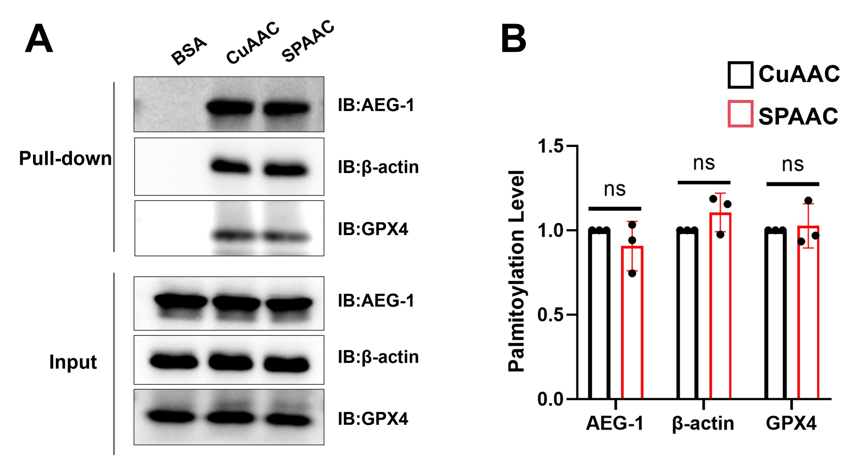
Figure 1. Comparison of palmitoylation detection efficiency between CuAAC and SPAAC methods. (A) Western blot detection of palmitoylation of AEG-1, GPX4, and β-actin. (B) Quantification by ImageJ (n = 3 biological replicates). Error bars represent the standard deviation (SD) of three biological replicates. Statistical analysis was performed using a t-test to compare the two methods. Palmitoylation levels were normalized to those of the CuAAC group. ns, p > 0.05.
This protocol, or parts of it, has been used and validated in the following research article:
Zhou et al. [19]. Palmitoylation of GPX4 via the targetable ZDHHC8 determines ferroptosis sensitivity and antitumor immunity. Nat Cancer. https://doi.org/10.1038/s43018-025-00937-y
General notes and troubleshooting
General notes
1. The SPAAC method is not applicable when using alkynyl fatty acids (e.g., alkynyl palmitic acid) for labeling.
2. Azido palmitic acid is cytotoxic, and its maximum tolerable concentration varies among different cell lines. Determine the maximum working concentration of azido palmitic acid using methods such as MTT or CCK-8 assays before conducting experiments.
3. Researchers can extend the application of this protocol beyond conventional immunoblotting by analyzing SPAAC-enriched proteins via mass spectrometry to identify and quantify palmitoylated proteins under different conditions.
Troubleshooting
Problem 1: Excessive cell death occurs after adding azido palmitic acid to cells.
Possible cause: The concentration of azido palmitic acid is too high.
Solution: Reduce the working concentration of azido palmitic acid and/or shorten the labeling time.
Problem 2: Prolonged incubation or sonication fails to completely dissolve the protein pellet.
Possible cause: Residual chloroform remains in the protein pellet.
Solution:
a. Increase the number of methanol washes.
b. Vacuum-dry the pellet at room temperature for 10–15 min to remove chloroform.
c. Dissolve the pellet completely in 2% SDS buffer (2% SDS in 50 mM Tris-HCl, pH 8.0), then dilute the SDS to 0.1% with cell lysis buffer.
Problem 3: No protein pellet forms after chloroform-methanol precipitation.
Possible cause: The amount of protein participating in the click reaction is too low.
Solution: After measuring the total protein concentration of the cell lysate, use at least 300 μg of protein for the click reaction.
Problem 4: In the western blot result, the BSA control lane in the pull-down shows a distinct target protein band.
Possible cause: Nonspecific binding of the protein to streptavidin magnetic beads during the pull-down process.
Solution: Extend both the washing time and the number of washes for the beads.
Problem 5: Target protein bands are present in all input lanes but are not detected in any pull-down lane.
Possible causes:
a. Exposure of azido palmitic acid to light, which destroys its structure and prevents the click reaction.
b. Insufficient incubation time with azido palmitic acid, resulting in poor labeling of the target protein.
c. Incomplete click reaction due to short incubation time.
d. Insufficient amount of streptavidin magnetic beads, leading to low binding efficiency.
e. Presence of SDS in the sample (>0.5%), which disrupts the streptavidin structure.
f. The target protein might not undergo palmitoylation under the present assay conditions.
Solutions:
a. Store all reagents under light-protected conditions.
b. Perform a time-course experiment for azido palmitic acid incubation to determine the optimal labeling time.
c. Extend the click reaction time (e.g., incubate for 5 h at 4 °C).
d. Increase the bead-to-protein ratio by using more beads or less protein.
e. Reduce the SDS concentration in the sample before performing the pull-down.
f. Use other detection methods, for example, acyl-biotin exchange (ABE) or mass spectrometric analysis of the target protein labeled with isotopically labeled fatty acids.
Acknowledgments
This study was supported by the 2025 Anhui Institute for Advanced Study Industry-University Collaborative Research and Talent Cultivation Project. This protocol was adapted from previous work published in Nature Cancer [19].
Author contributions
Investigation, Yaoyu Zhang and Jiahui Li. Experiments, Yaoyu Zhang and Jiahui Li; Writing—Original Draft, Yaoyu Zhang and Jiahui Li; Writing—Review & Editing, Xinxin Pei, Tengfei Ma, Yi Wang, Buchang Zhang; Supervision, Changzhi Xu.
Competing interests
The authors declare no conflicts of interest.
Ethical considerations
The study did not involve human subjects or animal work.
References
- Qian, Y., Zhao, Y. and Zhang, F. (2025). Protein palmitoylation: biological functions, disease, and therapeutic targets. MedComm. 6(3): e70096. https://doi.org/10.1002/mco2.70096
- Yang, Y., Zhang, E., Mao, X., Liu, G. and Pan, Y. (2026). Targeting palmitoylation: A novel frontier in cancer biology and immunotherapy. Biochimica et Biophysica Acta (BBA) - Reviews on Cancer. 1881(1): 189509. https://doi.org/10.1016/j.bbcan.2025.189509
- Song, P., Jiang, Q., Wu, X., Bu, L., Xie, W., Wei, W., Xing, X. and Guo, J. (2025). Palmitic acid and palmitoylation in cancer: Understanding, insights, and challenges. Innovation. 6(8): 100918. https://doi.org/10.1016/j.xinn.2025.100918
- Li, W., Shen, J., Zhuang, A., Wang, R., Li, Q., Rabata, A., Zhang, Y. and Cao, D. (2025). Palmitoylation: an emerging therapeutic target bridging physiology and disease. Cell Mol Biol Lett. 30(1): 98. https://doi.org/10.1186/s11658-025-00776-w
- B. S., P. and Talwar, P. (2025). Influence of palmitoylation in axonal transport mechanisms in neurodegenerative diseases. Front Cell Neurosci. 19: e1613379. https://doi.org/10.3389/fncel.2025.1613379
- Cao, S., Xiao, W., Pan, S., Hua, T. and Yang, M. (2025). Exploring the role of palmitoylation in sepsis: mechanistic insights and future perspectives. Mol Med. 31(1): 217. https://doi.org/10.1186/s10020-025-01284-5
- Zhou, B., Wang, Y., Yan, Y., Mariscal, J., Di Vizio, D., Freeman, M. R. and Yang, W. (2019). Low-Background Acyl-Biotinyl Exchange Largely Eliminates the Coisolation of Non-S-Acylated Proteins and Enables Deep S-Acylproteomic Analysis. Anal Chem. 91(15): 9858–9866. https://doi.org/10.1021/acs.analchem.9b01520
- Ji, X., Xu, C., Luo, J., Hu, X. and Wu, X. (2025). S-palmitoylation in bone biology: emerging insights and therapeutic potential. Biochem Pharmacol. 242: 117232. https://doi.org/10.1016/j.bcp.2025.117232
- Nederstigt, A. E., Sardana, S. and Baggelaar, M. P. (2025). Deciphering protein long-chain S-acylation using mass spectrometry proteomics strategies. RSC Chem Biol. 6(10): 1532–1545. https://doi.org/10.1039/d5cb00146c
- Gao, X. and Hannoush, R. N. (2018). A Decade of Click Chemistry in Protein Palmitoylation: Impact on Discovery and New Biology. Cell Chem Biol. 25(3): 236–246. https://doi.org/10.1016/j.chembiol.2017.12.002
- Martin, B. R. and Cravatt, B. F. (2009). Large-scale profiling of protein palmitoylation in mammalian cells. Nat Methods. 6(2): 135–138. https://doi.org/10.1038/nmeth.1293
- Suazo, K. F., Park, K. Y. and Distefano, M. D. (2021). A Not-So-Ancient Grease History: Click Chemistry and Protein Lipid Modifications. Chem Rev. 121(12): 7178–7248. https://doi.org/10.1021/acs.chemrev.0c01108
- Macias-Contreras, M., He, H., Little, K. N., Lee, J. P., Campbell, R. P., Royzen, M. and Zhu, L. (2020). SNAP/CLIP-Tags and Strain-Promoted Azide–Alkyne Cycloaddition (SPAAC)/Inverse Electron Demand Diels–Alder (IEDDA) for Intracellular Orthogonal/Bioorthogonal Labeling. Bioconjugate Chem. 31(5): 1370–1381. https://doi.org/10.1021/acs.bioconjchem.0c00107
- Fu, Y., Qian, H., Yang, Y., Li, J. and Xie, G. (2024). Enhanced imaging of protein-specific palmitoylation with HCR-based cis-membrane multi-FRET. Talanta. 266: 124972. https://doi.org/10.1016/j.talanta.2023.124972
- Parker, C. G. and Pratt, M. R. (2020). Click Chemistry in Proteomic Investigations. Cell. 180(4): 605–632. https://doi.org/10.1016/j.cell.2020.01.025
- Zhou, B., Wang, Y., Zhang, L., Shi, X., Kong, H., Zhang, M., Liu, Y., Shao, X., Liu, Z., Song, H., et al. (2022). The palmitoylation of AEG-1 dynamically modulates the progression of hepatocellular carcinoma. Theranostics. 12(16): 6898–6914. https://doi.org/10.7150/thno.78377
- Ren, J. (2019). The Verification and Functional Study of β-actin Palmitoylation. Master's thesis. Xinxiang Medical University.
- Huang, B., Wang, H., Liu, S., Hao, M., Luo, D., Zhou, Y., Huang, Y., Nian, Y., Zhang, L., Chu, B., et al. (2025). Palmitoylation-dependent regulation of GPX4 suppresses ferroptosis. Nat Commun. 16(1): 867. https://doi.org/10.1038/s41467-025-56344-5
- Zhou, L., Lian, G., Zhou, T., Cai, Z., Yang, S., Li, W., Cheng, L., Ye, Y., He, M., Lu, J., et al. (2025). Palmitoylation of GPX4 via the targetable ZDHHC8 determines ferroptosis sensitivity and antitumor immunity. Nat Cancer. 6(5): 768–785. https://doi.org/10.1038/s43018-025-00937-y
Article Information
Publication history
Received: Jun 5, 2026
Accepted: Sep 1, 2026
Available online: Sep 15, 2026
Published: Oct 20, 2026
Copyright
© 2026 The Author(s); This is an open access article under the CC BY-NC license (https://creativecommons.org/licenses/by-nc/4.0/).
How to cite
Zhang, Y., Li, J., Pei, X., Ma, T., Wang, Y., Zhang, B. and Xu, C. (2026). A SPAAC-Based Bioorthogonal Method for Verifying Protein Palmitoylation. Bio-protocol 16(20): e5835. DOI: 10.21769/BioProtoc.5835.

 Download Citation in RIS Format
Category
Cancer Biology > Cancer biochemistry > Protein
Biochemistry > Protein > Posttranslational modification
Do you have any questions about this protocol?
Post your question to gather feedback from the community. We will also invite the authors of this article to respond.
 Post a Question


