


- Protocols

- Articles and Issues
- For Authors
- About
- Become a Reviewer


Efficient Generation of Fetal Hepatic Stellate Cells from hiPSC

Published: Vol 16, Iss 19, Oct 5, 2026 DOI: 10.21769/BioProtoc.5827 Views: 19
Reviewed by: Samantha HallerAniruddha Vijay SavargaonkarSravanthi S P Nadiminti
 PDF
PDF  Ask a question
Original research article
The authors used this protocol in:

Jan 2026

Protocol Collections
Comprehensive collections of detailed, peer-reviewed protocols focusing on specific topics




Abstract
Human induced pluripotent stem cell (hiPSC)-derived liver organoids have emerged as valuable models for studying human liver development. However, existing organoid systems often lack developmentally matched cell populations, particularly fetal hepatic stellate cells (HSCs), limiting their ability to recapitulate key developmental processes. Current approaches for generating HSCs rely on primary cells, immortalized cell lines, or hiPSC differentiation methods that frequently produce activated HSC-like cells and often require cell sorting. Here, we describe an efficient protocol for generating expandable fetal-like HSCs from hiPSCs through a stepwise differentiation strategy that mimics embryonic HSC development. The resulting cells can be robustly expanded while maintaining characteristic molecular and functional features of fetal HSCs. This protocol provides a reproducible and scalable source of fetal-like HSCs without cell sorting and supports the generation of multicellular liver organoids containing developmentally relevant stromal components. Beyond the validation of the protocol in studies of liver maturation and vascularization, it can be applied to investigations of HSC biology and congenital liver diseases.
Key features
• Efficient generation of fetal-like HSCs from hiPSCs without cell sorting.
• Robust expansion of hiPSC-derived HSCs in vitro while maintaining fetal HSC characteristics.
• Generation of developmentally relevant HSCs for multicellular liver organoid construction.
• Applicable to studies of HSC biology, liver development, vascularization, and cell–cell interactions; successful implementation requires experience in hiPSC maintenance and differentiation.
Keywords: Human induced pluripotent stem cell Hepatic stellate cell
Differentiation
Expansion culture
Liver development
Hepatic stellate cell
Differentiation
Expansion culture
Liver development
Graphical overview
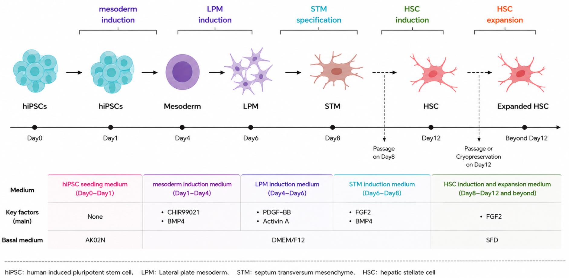
Representation of the experimental design to generate human induced pluripotent stem cell (hiPSC)-derived hepatic stellate cells (HSCs) using the indicated combination of small molecules and cytokines
Background
Hepatic stellate cells (HSCs) are important components of the developing liver microenvironment. Studies in animal models and single-cell RNA sequencing (scRNA-seq) datasets suggest that HSCs promote hepatoblast differentiation and support the maintenance of hematopoietic stem cells during liver development [1,2]. However, the functions of HSCs during human liver organogenesis remain incompletely understood.
Human induced pluripotent stem cell (hiPSC)-derived liver organoids (LOs) provide a useful platform for modeling human liver development in vitro [3]. Nevertheless, the existing LO systems lack key cell types, particularly developmentally stage-matched HSCs. The absence of fetal-like HSCs may limit the ability of these models to recapitulate the multicellular microenvironment required for liver maturation and vascularization.
Previously reported methods for differentiating HSC-like cells from hiPSCs often generate activated HSC-like cells or require cell sorting to enrich the target population [4–6]. These limitations increase experimental complexity and may reduce the scalability and reproducibility of HSC production. Here, we describe a robust protocol for generating expandable fetal-like HSCs from hiPSCs by sequentially mimicking key stages of embryonic HSC development through stage-specific cytokine modulation. The resulting cells retain features of fetal HSCs and can be expanded to support large-scale downstream applications.
Beyond investigating HSC-mediated regulation of liver maturation and vascularization in a multicellular organoid system [7], this protocol may facilitate studies of HSC biology, human liver organogenesis, congenital liver disorders, drug safety testing, and regenerative medicine. The scalable generation of fetal-like HSCs also provides a valuable cell source for engineering more physiologically relevant human liver models.
Materials and reagents
Biological materials
1. Human iPSC line (M48) (Kyoto University, catalog number: N/A)
Reagents
1. StemFit AKO2N (Ajinomoto Co., Inc, catalog number: AJ100)
2. Dulbecco’s modified Eagle medium/nutrient mixture F-12 (DMEM/F12) (Thermo Fisher Scientific, catalog number: 11320033)
3. D-MEM (high glucose) with L-glutamine and phenol red (Fujifilm Wako, catalog number: 043-30085)
4. Iscove’s modified Dulbecco’s medium (IMDM) (Invitrogen, catalog number: 12440-053)
5. Ham’s F-12 K medium (Invitrogen, catalog number: 21127-022)
6. D-PBS (Nacalai Tesque. Inc, catalog number: 14249-24)
7. Glutamax (Thermo Fisher Scientific, catalog number: 35050061)
8. B27 supplement (S50X) (Thermo Fisher Scientific, catalog number: 17504001)
9. N2 supplement (S100X) (Invitrogen, catalog number: 175020-01)
10. Bovine serum albumin (Sigma-Aldrich, catalog number: A9647-100G)
11. L-glutamine (Wako, catalog number: 073-05391)
12. Penicillin-streptomycin (Thermo Fisher Scientific, catalog number: 15140122)
13. Monothioglycerol solution (Fujifilm Wako, catalog number: 195-15791)
14. L-ascorbic acid (Sigma-Aldrich, catalog number: A4544-25G)
15. Fetal bovine serum (FBS) (Funakoshi, catalog number: S1820)
16. Sodium butyrate (Sigma-Aldrich, catalog number: B5887)
17. Y-27632 dihydrochloride (Wako, catalog number: 034-24024)
18. iMatrix-511TM (Nippi, Inc, catalog number: 381-07363)
19. TrypLE Express Enzyme (1×), no phenol red (Thermo Fisher Scientific, catalog number: 12604013)
20. CHIR-99021 (Cayman, catalog number: 13122)
21. Bone Morphogenetic Protein 4 (BMP4) (R&D Systems, catalog number: RSD-314-BP-050-50)
22. Platelet-derived growth factor-BB (PDGF-BB) (Peprotech, catalog number: 100-14B)
23. ACTIVIN A (Ajinomoto Inc, catalog number: P08476)
24. Fibroblast growth factor 2 (FGF2) (Wako Pure Chemical Industries, catalog number: 060-04543)
25. WNT3a (R&D Systems, catalog number: 5036-WN-500)
26. Forskolin (Cayman, catalog number: 11018)
27. Vascular endothelial growth factor (VEGF) (R&D, catalog number: 293-VE-01M)
28. Dexamethasone (DEX) (Sigma, catalog number: D2915-100MG)
29. Oncostatin M (OSM) (R&D System, Minneapolis, Minnesota, USA, catalog number: 295-OM)
30. N-acetyl cysteine (NAC) (Sigma-Aldrich, catalog number: A7256)
31. DAPI (Nacalai Tesque, catalog number: 19178-91)
32. Antifade mounting medium (Abcam, catalog number: ab104135)
33. ECL blocking reagent, human (Roche, catalog number: RPN418)
34. 4% paraformaldehyde (PFA) (Fujifilm, catalog number: WK16320145)
35. Triton X-100 (Sigma, catalog number: T8787)
36. STEM-CELLBANKER® (Amsbio, catalog number: 11924)
37. Propidium iodide (PI) (Sigma, catalog number: P4170-10MG)
38. Retinol (Sigma, catalog number: R2625-50mg)
39. Antibodies (flow cytometry and immunostaining):
a. PE mouse anti-human CD166 (ALCAM) (BD Pharmingen, catalog number: 559263; working dilution 1:20)
b. APC mouse anti-human CD73 (BD Pharmingen, catalog number: 560847; working dilution 1:20)
c. PE labeled anti-human PDGFRα (BD Pharmingen, catalog number: 556002; working dilution 1:20)
d. PE mouse anti-human PDGFRβ (BD Pharmingen, catalog number: 558821; working dilution 1:20)
e. APC mouse anti-human CD71 (BD Pharmingen, catalog number: 551374; working dilution 1:20)
f. Mouse anti-human α-SMA (Sigma, catalog number: 2547; working dilution 1:100)
g. Rabbit anti-human collagen I α1 (Novus Biological, catalog number: NB600-408; working dilution 1:200)
h. Rabbit anti-human fibronectin (Abcam, catalog number: ab2413; working dilution 1:200)
i. Mouse anti-human vimentin (Abcam, catalog number: ab8069; working dilution 1:200)
j. Donkey anti-mouse IgG (H+L) Alexa Fluor 488 (Invitrogen, catalog number: A-21202; working dilution 1:500)
k. Donkey anti-goat IgG (H+L) Alexa Fluor 555 (Invitrogen, catalog number: A-21432; working dilution 1:500)
l. Donkey anti-rabbit IgG (H+L) Alexa Fluor 647 (Invitrogen, catalog number: A-31573; working dilution 1:500)
Solutions
1. Plate coating solution (see Recipes)
2. AK02N complete medium (see Recipes)
3. hiPSC seeding medium (see Recipes)
4. Mesoderm induction medium (see Recipes)
5. Lateral plate mesoderm induction medium (see Recipes)
6. Septum transversum mesenchyme (STM) induction medium (see Recipes)
7. Serum-free defined medium (SFDM) (see Recipes)
8. HSC induction and expansion medium (see Recipes)
9. Quench medium (see Recipes)
10. FACS buffer (see Recipes)
11. Blocking buffer (see Recipes)
12. DAPI working solution (see Recipes)
Recipes
1. Plate coating solution
| Reagent | Final concentration | Quantity or volume |
|---|---|---|
| D-PBS | N/A | 10 mL |
| iMatrix-511 | 2.5 μg/mL | 50 μL |
Prepare fresh.
2. AK02N complete medium
| Reagent | Final concentration | Quantity or volume |
|---|---|---|
| StemFit AK02N basal medium A | 79.60% (v/v) | 398 mL |
| Liquid B | 20% (v/v) | 100 mL |
| Liquid C | 0.40% (v/v) | 2 mL |
Mix well by inverting the bottle 5 times. The mixed medium can be stored at 4 °C for up to two weeks. Alternatively, aliquot the medium and store at -20 °C for up to 6 months.
3. hiPSC seeding medium
| Reagent | Final concentration | Quantity or volume |
|---|---|---|
| AK02N complete medium (Recipe 2) | N/A | 10 mL |
| Y-27632 dihydrochloride (10 mM) | 10 μM | 10 μL |
| iMatrix-511 | 2.5 μg/mL | 50 μL |
Prepare fresh.
4. Mesoderm induction medium
| Reagent | Final concentration | Quantity or volume |
|---|---|---|
| DMEM/F12 | N/A | 25 mL |
| B27 | 1% | 250 μL |
| Glutamax | 1% | 250 μL |
| CHIR-99021 (2 mM) | 8 μM | 100 μL |
| BMP4 (25 μg/mL) | 25 ng/mL | 25 μL |
Store at 4 °C for up to 1 week.
5. Lateral plate mesoderm induction medium
| Reagent | Final concentration | Quantity or volume |
|---|---|---|
| DMEM/F12 | N/A | 25 mL |
| B27 | 1% | 250 μL |
| Glutamax | 1% | 250 μL |
| PDGFBB (10 μg/mL) | 10 ng/mL | 25 μL |
| ACTIVIN A (33.3 μg/mL) | 2 ng/mL | 1.5 μL |
Store at 4 °C for up to 1 week.
6. STM induction medium
| Reagent | Final concentration | Quantity or volume |
|---|---|---|
| DMEM/F12 | N/A | 25 mL |
| B27 | 1% | 250 μL |
| Glutamax | 1% | 250 μL |
| FGF2 (5 μg/mL) | 10 ng/mL | 50 μL |
| BMP4 (25 μg/mL) | 12 ng/mL | 12 μL |
Store at 4 °C for up to 1 week.
7. SFDM
| Reagent | Final concentration | Quantity or volume |
|---|---|---|
| IMDM | 75% (v/v) | 375 mL |
| Ham’s F-12 K medium | 25% (v/v) | 125 mL |
| B27 | 1% | 5 mL |
| N2 | 0.5% | 2.5 mL |
| Bovine serum albumin (50 mg/mL) | 0.5 mg/mL | 5 mL |
| L-glutamine (200 mM) | 2 mM | 5 mL |
| Penicillin–streptomycin (100×) | 1% | 5 mL |
| Monothioglycerol solution | 0.45 mM | 4.5 mL |
| L-ascorbic acid (125 mM) | 0.5 mM | 2 mL |
| EGF (20 μg/mL) | 10 ng/mL | 250 μL |
| VEGF (50 μg/mL) | 10 ng/mL | 100 μL |
| FGF2 (5 μg/mL) | 10 ng/mL | 1,000 μL |
| HGF (5 μg/mL) | 20 ng/mL | 2,000 μL |
| Dexamethasone (100 μM) | 100 nM | 500 μL |
Store at 4 °C for up to 1 week.
8. HSC induction and expansion medium
| Reagent | Final concentration | Quantity or volume |
|---|---|---|
| SFDM | N/A | 100 mL |
| FBS | 5% | 5 mL |
| FGF2 (5 μg/mL) | 10 ng/mL | 200 μL |
| NAC (5 mM) | 10 μM | 200 μL |
Store at 4 °C for up to 1 week.
9. Quench medium
| Reagent | Final concentration | Quantity or volume |
|---|---|---|
| D-MEM (high glucose) | N/A | 500 mL |
| Penicillin-streptomycin | 1% | 5 mL |
| FBS | 5% | 25 mL |
Store at 4 °C for up to 1 month.
10. FACS buffer
| Reagent | Final concentration | Quantity or volume |
|---|---|---|
| D-PBS | N/A | 100 mL |
| FBS | 2% | 2 mL |
| PI (1 mg/mL) | 1 μg/mL | 100 μL |
Store at 4 °C in the dark for up to 1 month.
11. Blocking buffer
| Reagent | Final concentration | Quantity or volume |
|---|---|---|
| D-PBS | N/A | 100 mL |
| ECL Prime blocking agent | 10% | 10 g |
| Triton X-100 | 0.3% | 300 μL |
Store at 4 °C for up to 1 month.
12. DAPI working solution
| Reagent | Final concentration | Quantity or volume |
|---|---|---|
| DAPI | 1 μg/mL | 10 μL |
| Antifade mounting medium | N/A | 10 mL |
Store at 4 °C in the dark for up to 1 month.
Laboratory supplies
1. Falcon 12-well clear-bottom TC-treated multiwell cell culture plate (Corning, catalog number: 353043)
2. Falcon 100 mm TC-treated cell culture dish (Corning, catalog number: 353003)
3. Falcon® 15 mL high clarity PP centrifuge tube (Corning, catalog number: 352096)
4. Falcon® 50 mL high clarity PP centrifuge tube (Corning, catalog number: 352070)
5. Falcon® 5 mL round-bottom polystyrene test tube (Corning, catalog number: 352008)
6. Millex-GP syringe filter unit, 0.22, polyethersulfone, 33 mm, gamma sterilized (Millipore, catalog number: SLGPR33RB)
7. Bio freezing vessel, Bicell (NFZ, catalog number: 4100)
Equipment
1. Laboratory centrifuge with rotors for 15 and 50 mL conical tubes (Kubota, catalog number: 4000)
2. Inverted phase contrast microscope (Nikon, model: ECLIPSE TS100)
3. Leica Microsystems fluorescence microscope (Leica, model: THUNDER Imager Tissue)
4. BD FACSCelesta Cell Analyzer (BD Biosciences, Mississauga, Canada)
5. Tomy Capsulfuge Centrifuge (TOMY KOGYO, model: PMC-060)
Software and datasets
1. FlowJo software (BD Biosciences, version: 10.8.1)
Procedure
A. Differentiation of hiPSC-derived fetal HSCs
Notes:
1. The following procedure uses a 12-well plate as an example. When using other culture vessels, refer to the General notes for the recommended volumes of plate coating solution and culture medium.
2. The hiPSC culture and harvest procedure could be found in our detailed protocol published previously [9].
(Day 0) Step 1: hiPSC seeding
1. Add 1 mL of hiPSC seeding medium (see Recipe 3) to each well of a 12-well plate and allow it to warm to room temperature.
2. When hiPSCs reach 50%–60% confluence (Figure 1), harvest hiPSCs and resuspend them in hiPSC seeding medium.
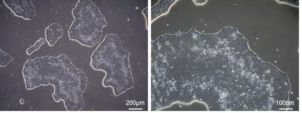
Figure 1. Representative morphology of human induced pluripotent stem cells (hiPSCs) prior to fetal hepatic stellate cell (HSC) differentiation. hiPSCs exhibit compact colonies with well-defined borders and a high nucleus-to-cytoplasm ratio before initiating HSC differentiation.
3. Seed 1.32 × 10⁴ hiPSCs/cm2 into each well and gently move the culture plate back and forth in perpendicular directions to ensure even cell distribution.
4. Incubate the cells at 37 °C with 5% CO2 for 24 h prior to differentiation induction.
Notes:
1. For the initial induction of hiPSC-HSCs on day 0, no precoating of the culture plate is required.
2. Cell density is a critical determinant of successful differentiation. Low seeding densities may result in extensive cell loss during induction, whereas excessively high densities may promote cell aggregation and spheroid formation. For initial optimization, four seeding densities (0.79 × 10⁴, 1.32 × 10⁴, 1.84 × 10⁴, and 2.63 × 10⁵ cells/cm2) are recommended.
(Days 1–3) Step 2: Mesoderm induction
5. After 24 h, allow the mesoderm induction medium (see Recipe 4) to equilibrate to room temperature. Then, replace the medium with 1 mL of the mesoderm induction medium.
6. Culture the cells for 3 days and replace the medium again on day 3 to induce mesoderm formation (Figure 2).
Note: After 24 h of seeding, initiate mesoderm induction. At this time point, the hiPSCs have not yet formed typical colonies. The expected cell morphology can be compared with the day-1 hiPSCs shown in Figure 2.
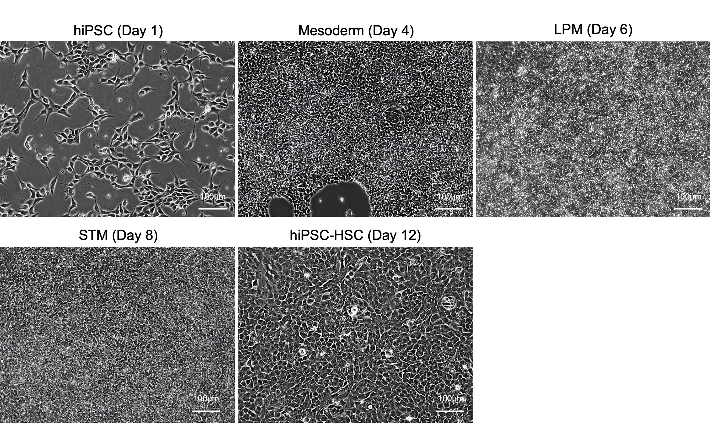
Figure 2. Cell morphology of human induced pluripotent stem cells (hiPSCs), mesoderm, lateral plate mesoderm (LPM), septum transversum mesenchyme (STM), and hiPSC-derived fetal hepatic stellate cells (HSC). Reprinted and adapted with permission from Yang et al. [7].
(Days 4–5) Step 3: Lateral plate mesoderm induction
7. On day 4, replace the medium with 1 mL of lateral plate mesoderm induction medium (see Recipe 5), pre-equilibrated to room temperature.
8. On day 5, replace the medium with 1 mL of lateral plate mesoderm induction medium (see Recipe 5), pre-equilibrated to room temperature.
(Days 6–7) Step 4: STM induction
9. On day 6, replace the medium with 1 mL of STM induction medium (see Recipe 6), pre-equilibrated to room temperature.
10. On day 7, replace the medium with 1 mL of STM induction medium (see Recipe 6), pre-equilibrated to room temperature.
(Day 8-12) Step 5: Cell passaging and fetal HSC induction
Note: The following procedure uses a 10-cm dish as an example. When using other culture vessels, refer to the General notes for the recommended volumes of plate coating solution, TrypLETM Express Enzyme (1×), quench medium, and culture medium, as well as the recommended cell seeding numbers.
11. Prepare an iMatrix-coated 10-cm dish (see General note 1).
12. Prewarm the HSC induction and expansion medium (see Recipe 8), quench medium (see Recipe 9), and IMDM containing 1% penicillin-streptomycin to room temperature.
13. Aspirate the medium from the induction plate.
14. Add 0.3 mL of TrypLE to each well and incubate at 37 °C with 5% CO2 for 30 s.
15. Add 0.3 mL of quench medium to each well and collect the cells into a 15-mL conical tube containing 5 mL of quench medium.
16. Rinse each well with an additional 1 mL of quench medium and combine with the collected cells.
17. Centrifuge at 200× g for 4 min at 4 °C.
18. Aspirate the supernatant and resuspend the pellet in 5 mL of IMDM containing 1% penicillin–streptomycin.
19. Centrifuge again at 200× g for 4 min at 4 °C.
20. Aspirate the supernatant and resuspend the cells in HSC induction and expansion medium.
21. Determine cell number and viability using Trypan Blue staining.
22. Aspirate the D-PBS in the iMatrix-coated 10-cm dish and add 10 mL of HSC induction and expansion medium supplemented with 10 μM Y-27632 dihydrochloride.
23. Seed 4.5 × 10³ cells/cm2 in the dish and gently move the dish back and forth in perpendicular directions to ensure even cell distribution.
24. Incubate the cells at 37 °C with 5% CO2 for 24 h.
25. On the following day, replace the medium with fresh HSC induction and expansion medium.
26. Continue culturing the cells for an additional three days to induce fetal HSCs (Figure 2), replacing the medium every other day.
B. Expansion of hiPSC-derived fetal HSCs
Note: The following procedure uses a 10-cm dish as an example. When using other culture vessels, refer to the General notes for the recommended volumes of plate coating solution, TrypLETM Express Enzyme (1×), quench medium, and culture medium, as well as the recommended cell seeding numbers.
1. Prepare an iMatrix-coated 10-cm dish (see General note 1).
2. Prewarm the HSC induction and expansion medium, quench medium, and IMDM containing 1% penicillin–streptomycin to room temperature.
3. Aspirate the culture medium and add 2 mL of TrypLETM Express Enzyme (1×) to the dish.
4. Incubate at 37 °C with 5% CO2 for 2–5 min.
5. Beginning within 2 min, monitor cell detachment under a microscope. Once approximately 90% of cells have detached, add 2 mL of quench medium (see Recipe 9) to stop the enzymatic reaction.
6. Collect the cells into a 15-mL conical tube.
7. Rinse the dish with an additional 2 mL of quench medium and combine with the collected cells.
8. Centrifuge at 200× g for 4 min at 4 °C.
9. Aspirate the supernatant and resuspend the pellet in 5 mL of IMDM containing 1% penicillin–streptomycin.
10. Centrifuge again at 200× g for 4 min at 4 °C.
11. Aspirate the supernatant and resuspend the cells in HSC induction and expansion medium (see Recipe 8).
12. Count viable cells using Trypan Blue staining.
Note: At this stage, the hiPSC-HSCs are ready for cryopreservation. Detailed cryopreservation and thawing procedures are provided in the following section.
13. Seed 4.5 × 10³ cells/cm2 into an iMatrix-coated 10-cm dish containing 10 mL of HSC induction and expansion medium supplemented with 10 μM Y-27632 dihydrochloride.
14. Incubate the cells at 37 °C with 5% CO2 for 24 h.
15. On the following day (Figure 3A), replace the medium with fresh HSC induction and expansion medium and continue culturing the cells with medium changes every other day until cells reach 90%–100% confluence (Figure 3B).
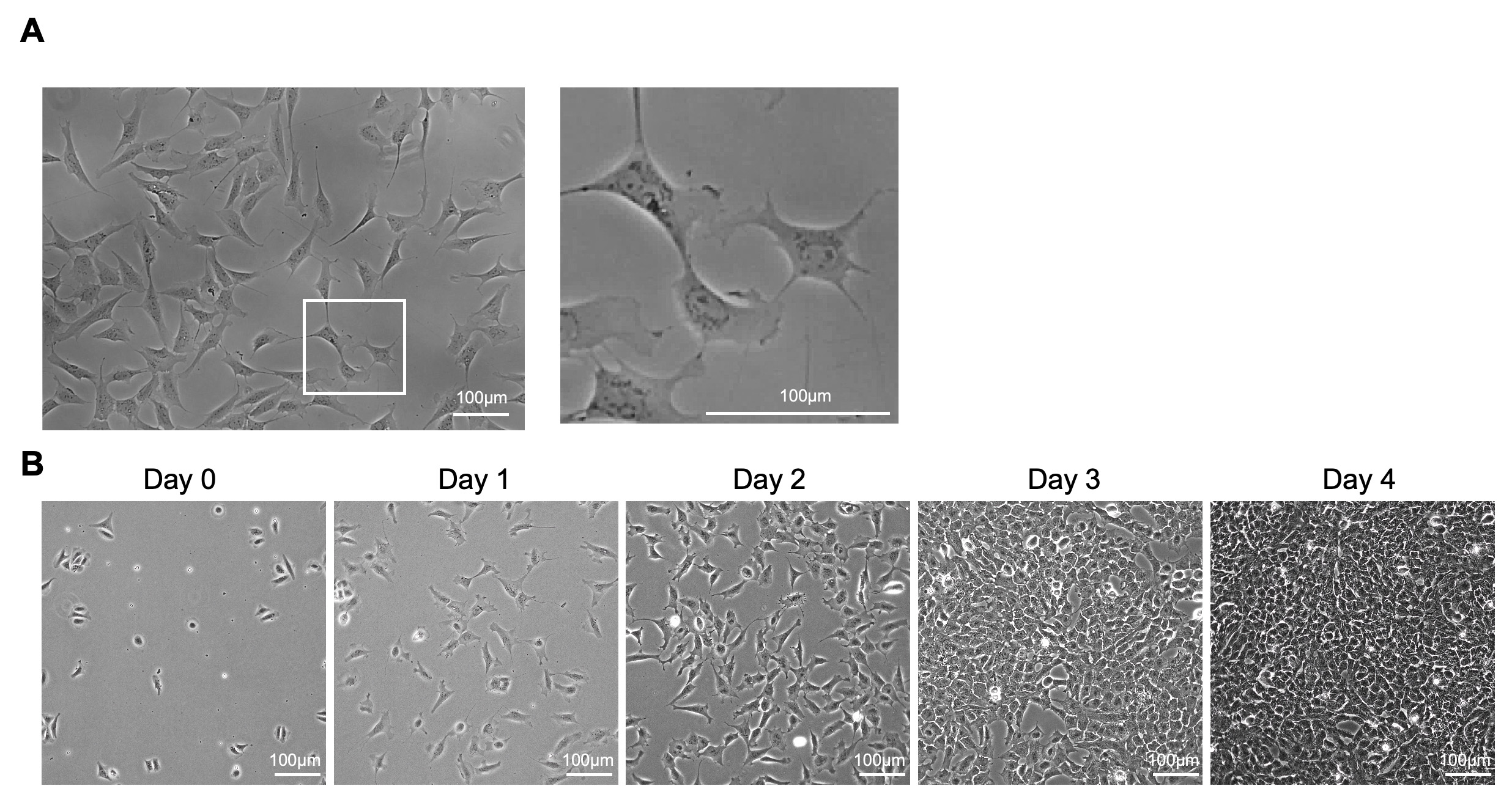
Figure 3. Cell morphology of human induced pluripotent stem cell (hiPSC)–hepatic stellate cells (HSCs). (A) Cell morphology of hiPSC-HSCs with low cell confluence. (B) Cell morphology of hiPSC-HSCs during expansion. Reprinted and adapted with permission from Yang et al. [7].
C. Cryopreservation of hiPSC-derived fetal HSCs
1. Harvest the cells, determine the cell number (see section B), and centrifuge at 200× g for 4 min at 4 °C.
2. Carefully aspirate the supernatant as completely as possible.
3. Gently resuspend the cell pellet in STEM-CELLBANKER cryopreservation medium at a density of 5 × 10⁵–5 × 10⁶ cells/mL. The cryopreservation medium is stored at 4 °C and can be used directly without prewarming.
4. Dispense 1 mL of the cell suspension into each labeled cryovial. Label each cryovial with the cell name, cell concentration, passage number, date, and other relevant information.
5. Place the cryovials in Bicell and directly transfer to a -80 °C freezer. For long-term storage, transfer the cryovials to a liquid nitrogen storage tank after they have been stored at -80 °C for at least 24 h.
D. Thawing of hiPSC-derived fetal HSCs
1. Prepare an iMatrix-coated 10-cm dish (see General note 1).
2. Prewarm the HSC induction and expansion medium and IMDM containing 1% penicillin–streptomycin to room temperature.
3. Thaw a cryovial of hiPSC-HSCs rapidly in a 37 °C water bath until the contents are completely thawed.
4. Transfer the cell suspension to a 15-mL conical tube containing 7 mL of IMDM supplemented with 1% penicillin–streptomycin.
5. Centrifuge the cells at 200× g for 4 min at 4 °C.
6. Carefully aspirate the supernatant and gently resuspend the cell pellet in 1 mL of HSC induction and expansion medium supplemented with 10 μM Y-27632 dihydrochloride.
7. Determine the viable cell number using Trypan Blue exclusion.
8. Seed 4.5 × 10³ viable cells/cm2 into the iMatrix-coated 10-cm dish containing 10 mL of HSC induction and expansion medium supplemented with 10 μM Y-27632 dihydrochloride.
9. Incubate the cells at 37 °C in a humidified incubator with 5% CO2 for 24 h.
10. On the following day, replace the medium with fresh HSC induction and expansion medium. Thereafter, change the medium every other day.
E. Characterization of hiPSC-derived fetal HSCs
E1. Flow cytometry analysis of cell surface markers
1. Harvest hiPSC-HSCs as single-cell suspensions according to section B.
2. Transfer 5 × 10⁵ cells into a 1.5 mL Eppendorf tube.
3. Wash cells with 0.5 mL of D-PBS by gentle pipetting, followed immediately by centrifugation with a Tomy Capsulfuge Centrifuge for 1–3 min to pellet cells. Carefully aspirate the supernatant.
4. Repeat step E1.3.
5. Resuspend the cell pellet in 50 μL of D-PBS.
6. Incubate cells with primary antibodies (PDGFRβ, ALCAM, CD71, CD73, and PDGFRα; see Reagents for working dilutions) for 30 min on ice in the dark.
7. Wash cells with 0.5 mL of D-PBS by gentle pipetting, immediately centrifuge for 1–3 min using a Tomy Capsulfuge Centrifuge, and carefully remove the supernatant.
8. Repeat step E1.7.
9. Resuspend cells in 0.5 mL of FACS buffer (see Recipe 10) and transfer to FACS tubes. Keep samples on ice and protected from light until analysis.
10. Perform instrument startup and fluidics cleaning procedures according to the manufacturer's instructions before sample acquisition.
11. For the initial assay setup, establish the acquisition and analysis settings, including fluorescence compensation, and save the settings as an experiment template in BD FACSDiva. For subsequent experiments, load the established experiment template before sample acquisition.
12. Acquire 10,000 events for each sample.
13. Gate cells sequentially based on forward scatter (FSC-A) and side scatter (SSC-A) to identify the main cell population. Perform singlet discrimination using FSC-H vs. FSC-A to exclude cell aggregates and doublets. Exclude non-viable cells based on PI staining and select the viable cell population. Assess the expression of the indicated HSC-associated markers (CD73, CD71, PDGFRβ, and ALCAM) and PDGFRα within the viable single-cell population using PE-A or APC-A plots.
14. Process and quantify data using FlowJo software. Apply the established gating strategy to exclude debris, doublets, and non-viable cells, and quantify the percentage of cells expressing each indicated marker within the viable single-cell population (Figure 4A).
15. Calculate the percentage of marker-positive cells as the proportion of positive events relative to the total number of viable single-cell events within the defined gate.
Note: On day 12 of differentiation, >90% of cells should express HSC-associated markers (CD73, CD71, PDGFRβ, and ALCAM), while >99% of cells should be negative for activated HSC marker PDGFRα (Figure 4B).
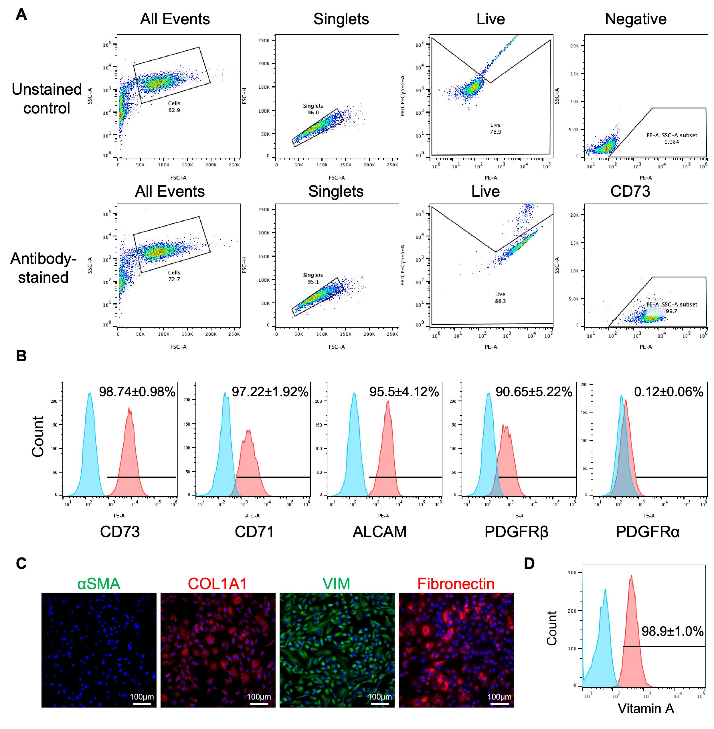
Figure 4. Characteristics of human induced pluripotent stem cell (hiPSC)–hepatic stellate cells (HSCs). (A) Representative flow cytometry gating strategy for the identification of hiPSC-HSCs. (B) Flow cytometry analysis of CD73, CD71, ALCAM, PDGFRβ, and PDGFRα on hiPSC-HSCs (n = 6). (C) Images of immunofluorescence of αSMA, COL1A1, Vimentin (VIM), and Fibronectin in hiPSC-HSCs. (D) Flow cytometry analysis of vitamin A signals in hiPSC-HSCs (n = 3). Data are presented as mean ± SD. Reprinted and adapted with permission from Yang et al. [7].
E2. Vitamin A storage assay
1. Seed 5.2 × 10³ hiPSC-HSCs/cm2 onto an iMatrix-coated 6-well plate and culture for two days (see section B).
2. Prewarm the HSC induction and expansion medium to room temperature. Then, replace the medium with HSC induction and expansion medium supplemented with retinol to a final concentration of 2 μM. Culture for 48 h.
3. Include a control group cultured in medium without retinol.
4. Harvest cells as a single-cell suspension into 1.5 mL Eppendorf tubes (see section B).
5. Wash cells with 0.5 mL of D-PBS, centrifuge using a Tomy Capsulfuge Centrifuge for 1–3 min, and carefully remove the supernatant.
6. Repeat step E2.5.
7. Resuspend cells in 0.5 mL of D-PBS and transfer to FACS tubes. Keep samples on ice and protected from light.
8. Use the Vitamin A blank control to establish the background fluorescence and define the fluorescence threshold for Vitamin A-associated autofluorescence.
9. Acquire 10,000 events for each sample.
10. Gate the main cell population based on forward scatter (FSC-A) and side scatter (SSC-A) to exclude debris and non-cellular events. Perform sequential singlet discrimination using FSC-H vs. FSC-W to exclude cell aggregates and doublets. Assess cellular autofluorescence using the BUV496-A channel within the gated single-cell population. Apply the same fluorescence gate established using the Vitamin A blank control to the assay samples. Cells exhibiting BUV496-A fluorescence above the defined threshold are considered Vitamin A–positive.
11. Analyze data using FlowJo software. Apply the established gating strategy to exclude debris and doublets and quantify the percentage of Vitamin A–positive cells related to the corresponding parent population. About 98% of cells should uptake vitamin A (Figure 4D).
E3. Immunofluorescence staining of cells
1. Seed 1.32 × 10⁴ hiPSC-HSCs/cm2 onto iMatrix-coated 24-well plates and culture for 24 h (see section B).
2. Remove culture medium.
3. Wash cells once with 0.5 mL of D-PBS.
4. Fix cells with 4% PFA for 10 min at room temperature.
5. Wash cells three times with 0.5 mL of D-PBS.
6. Block cells with 0.3 mL of blocking buffer for 60 min at room temperature.
7. Wash cells three times with 0.5 mL of D-PBS.
8. Prepare primary antibodies (αSMA, COL1A1, Vimentin, and Fibronectin; see Reagents for working dilutions) in blocking buffer (see Recipe 11).
9. Incubate cells with 200 μL of primary antibody working solution per well overnight at 4 °C.
10. Wash cells three times with 0.5 mL of D-PBS.
11. Prepare secondary antibodies at a 1:500 dilution in blocking buffer.
12. Incubate cells with 200 μL of secondary antibody solution per well for 60 min at room temperature in the dark.
13. Wash cells three times with 0.5 mL of D-PBS.
14. Counterstain nuclei with 200 μL of DAPI working solution (see Recipe 12) for 5–10 min at room temperature in the dark prior to imaging.
15. Acquire fluorescence images using a Leica fluorescence microscope. Representative images are shown in Figure 4C.
Validation of protocol
This protocol (or parts of it) has been used and validated in the following research article:
Yang et al. [7]. Human pluripotent stem cell-derived fetal hepatic stellate cells promote vascularization and maturation in liver organoids. Developmental Cell.
Specifically, the protocol was applied to the generation, characterization, and expansion of hiPSC-HSCs, as demonstrated by the following data in the referenced article: Figures 1 and S1 demonstrate the highly efficient generation of HSCs from hiPSCs; Figures 2 and S2 show the characteristics of hiPSC-HSCs; and Figures 3 and S3 demonstrate that hiPSC-HSCs have remarkable expandability in vitro.
General notes and troubleshooting
General notes
1. Prepare iMatrix-coated culture vessels as described below
a. Add the recommended volume of plate coating solution (see Recipe 1) to each vessel. For hiPSC-STM or hiPSC-HSC passaging, use 0.5, 1, 2, or 10 mL of coating solution per well/dish for 24-well plates, 12-well plates, 6-well plates, or 10-cm dishes, respectively.
b. Incubate the vessels at 37 °C in a 5% CO2 incubator for 1 h without agitation.
c. Aspirate the coating solution and add an equal volume of fresh D-PBS.
d. Use the coated vessels on the same day. Before use, keep them either in a 37 °C, 5% CO2 incubator or on a cell culture clean bench at room temperature (20–25 °C). Alternatively, coated vessels may be stored at 4 °C for up to 1 week.
2. Recommended volume of dissociation solution for hiPSC-STM and hiPSC-HSC dissociation
a. Use 0.2, 0.3, 0.5, or 2 mL of TrypLETM Express Enzyme (1×) per well/dish for 24-well plates, 12-well plates, 6-well plates, and 10-cm dishes, respectively.
b. After dissociation, terminate the enzymatic reaction by adding an equal volume of quench medium (see Recipe 9).
3. Recommended volume of medium for cell culture and differentiation
Use 0.5, 1, 2, or 10 mL of the appropriate culture medium per well/dish for 24-well plates, 12-well plates, 6-well plates, and 10-cm dishes, respectively.
4. Recommended cell number for hiPSC-STM passage and hiPSC-HSC expansion
When using 24-wells, 12-wells, 6-well plates, or 10-cm dishes for passaging, seed 1 × 10⁴, 3 × 10⁴, 5 × 10⁴, or 2.5 × 10⁵ of cells per well/dish, respectively.
Troubleshooting
Problem 1: Cells at the periphery of the well begin to contract and aggregate toward the center of the well on day 7 (i.e., show signs of edge detachment).
Possible cause: The initial cell seeding density is too high.
Solution: Perform cell passaging on day 7 as described in section B. Culture the passaged cells in STM induction medium supplemented with 10 μM Y-27632 dihydrochloride. On day 8, replace the medium with HSC induction and expansion medium.
Problem 2: Cell confluence is below 50% on day 4 of differentiation.
Possible cause: The initial cell seeding number is too low.
Solution: Increase the initial cell seeding number in subsequent differentiation experiments. In each induction experiment, it is recommended to test four different initial cell seeding densities (see section A) in parallel to determine the optimal seeding condition.
Problem 3: Low percentage of HSC-positive cells on day 12.
Possible causes: Poor quality or unhealthy starting hiPSCs; reduced activity of differentiation reagents.
Solution: Use healthy, well-characterized hiPSCs with compact and well-defined colonies. Ensure proper storage and handling of differentiation reagents. Prepare fresh differentiation medium before use and use the prepared medium within one week.
Problem 4: Poor initial cell attachment after seeding.
Possible cause: Inadequate coating or improper handling of the cell adhesion substrate.
Solution: Ensure that iMatrix-511 is prepared and used according to the manufacturer's instructions and that the culture surface is adequately coated before cell seeding. Avoid excessive mechanical stress during cell handling and seeding.
Acknowledgments
This work was supported by the National Key Research and Development Program of China (Grant No. 2023YFC2308200), National Natural Science Foundation of China (Grant No. 82302396), Japan Agency for Medical Research and Development (JP19lm0203005, JP20fk0210073, 22bm0304002h0010, 22fk0210073, 22bm0804004, 22bk0104102, 23fk0210129, JP23bm1223007, JP23bm1523004, JP23bm1523006, JP24bk0104174) and (JP24ym0126803), Grant-in-Aid for Research A (JP21H04830 and JP24H00641), Grant-in-Aid for Research Activity Start-up (20K22946), Grant-in-Aid for Young Scientists (21K16377 and 23K08045), International Joint Research Project of the Institute of Medical Science, the University of Tokyo (K22-1040), and a donation fund from an individual to Tokyo University. We acknowledge the IMSUT FACS core laboratory for assistance with flow cytometry analysis. We also acknowledge our previous work published in Developmental Cell (2026), DOI: 10.1016/j.devcel.2025.09.002 [7] on which the current protocol is based. Finally, the Graphical overview was created with https://chatgpt.com/.
Author contributions
Conceptualization, X.Y., H.T., and Y.-Z.N.; Investigation, X.Y.; Writing—Original Draft, X.Y.; Writing—Review & Editing, Y.-Z.N.; Funding acquisition, H.T., and Y.-Z.N.; Supervision, H.T., and Y.-Z.N.
Competing interests
X.Y., Y.-Z.N., and H.T. have a patent application related to this work.
References
- Aimaiti, Y., Jin, X., Shao, Y., Wang, W. and Li, D. (2018). Hepatic stellate cells regulate hepatic progenitor cells differentiation via the TGF‐β1/Jagged1 signaling axis. J Cell Physiol. 234(6): 9283–9296. https://doi.org/10.1002/jcp.27609
- Lee, Y., Leslie, J., Yang, Y. and Ding, L. (2020). Hepatic stellate and endothelial cells maintain hematopoietic stem cells in the developing liver. J Exp Med. 218(3): e20200882. https://doi.org/10.1084/jem.20200882
- Camp, J. G., Sekine, K., Gerber, T., Loeffler-Wirth, H., Binder, H., Gac, M., Kanton, S., Kageyama, J., Damm, G., Seehofer, D., et al. (2017). Multilineage communication regulates human liver bud development from pluripotency. Nature. 546(7659): 533–538. https://doi.org/10.1038/nature22796
- Coll, M., Perea, L., Boon, R., Leite, S. B., Vallverdú, J., Mannaerts, I., Smout, A., El Taghdouini, A., Blaya, D., Rodrigo-Torres, D., et al. (2018). Generation of Hepatic Stellate Cells from Human Pluripotent Stem Cells Enables In Vitro Modeling of Liver Fibrosis. Cell Stem Cell. 23(1): 101–113.e7. https://doi.org/10.1016/j.stem.2018.05.027
- Koui, Y., Kido, T., Ito, T., Oyama, H., Chen, S. W., Katou, Y., Shirahige, K. and Miyajima, A. (2017). An In Vitro Human Liver Model by iPSC-Derived Parenchymal and Non-parenchymal Cells. Stem Cell Rep. 9(2): 490–498. https://doi.org/10.1016/j.stemcr.2017.06.010
- Koui, Y., Himeno, M., Mori, Y., Nakano, Y., Saijou, E., Tanimizu, N., Kamiya, Y., Anzai, H., Maeda, N., Wang, L., et al. (2021). Development of human iPSC-derived quiescent hepatic stellate cell-like cells for drug discovery and in vitro disease modeling. Stem Cell Rep. 16(12): 3050–3063. https://doi.org/10.1016/j.stemcr.2021.11.002
- Yang, X., Nie, Y. Z., Lu, C., Li, Y., Hayashi, Y., Plummer, R., Luo, N., Li, Q., Kasai, T., Okumura, T., et al. (2026). Human pluripotent stem cell-derived fetal hepatic stellate cells promote vascularization and maturation in liver organoids. Dev Cell. 61(1): 208–223.e8. https://doi.org/10.1016/j.devcel.2025.09.002
- Takeuchi, K., Tabe, S., Yamamoto, Y., Takahashi, K., Matsuo, M., Ueno, Y., Ohtsuka, M., Morinaga, S., Miyagi, Y., Yamaguchi, T., et al. (2025). Protocol for generating a pancreatic cancer organoid associated with heterogeneous tumor microenvironment. STAR Protoc. 6(1): 103539. https://doi.org/10.1016/j.xpro.2024.103539
- Li, Y., Nie, Y. and Taniguchi, H. (2025). Protocol for generating liver organoids containing Kupffer cells using human iPSCs. STAR Protoc. 6(4): 104138. https://doi.org/10.1016/j.xpro.2025.104138
Article Information
Publication history
Received: Jul 20, 2026
Accepted: Aug 27, 2026
Available online: Sep 16, 2026
Published: Oct 5, 2026
Copyright
© 2026 The Author(s); This is an open access article under the CC BY license (https://creativecommons.org/licenses/by/4.0/).
How to cite
Yang, X., Taniguchi, H. and Nie, Y. Z. (2026). Efficient Generation of Fetal Hepatic Stellate Cells from hiPSC. Bio-protocol 16(19): e5827. DOI: 10.21769/BioProtoc.5827.

 Download Citation in RIS Format
Do you have any questions about this protocol?
Post your question to gather feedback from the community. We will also invite the authors of this article to respond.
 Post a Question


