


- Protocols

- Articles and Issues
- For Authors
- About
- Become a Reviewer


Immune-Complex-Based In Vitro Deubiquitination Assay

Published: Vol 16, Iss 19, Oct 5, 2026 DOI: 10.21769/BioProtoc.5824 Views: 550
Reviewed by: Ágnes Judit JuhászManasi BarathAnonymous reviewer(s)
 PDF
PDF  Ask a question
Original research article
The authors used this protocol in:

Feb 2026

Protocol Collections
Comprehensive collections of detailed, peer-reviewed protocols focusing on specific topics




Abstract
Deubiquitinases (DUBs) are attractive therapeutic targets within the ubiquitin-proteasome system, in part because four of the five DUB subfamilies are cysteine proteases amenable to the development of potent, selective inhibitors, as recently demonstrated for USP7. Identifying DUBs that deubiquitylate and stabilize specific human oncogenic proteins is therefore a promising approach to discovering new mechanism-based targets for cancer therapy. Several complementary experimental strategies are typically required to identify bona fide DUB–substrate pairs. Here, we present an efficient, straightforward in vitro immune-complex protocol to validate USP17-mediated deubiquitylation of the transcriptional co-activator β-catenin. In this assay, both β-catenin and USP17 are immunopurified from transiently transfected 293T cells and then combined to assess USP17 enzymatic activity. The protocol describes the in vitro enzymatic assay performed on immunopurified complexes and the immunoblot-based readout. It can be readily adapted to other DUBs and substrates for mechanistic studies.
Key features
• Requires prior experience with cell culture, immunoprecipitation, and immunoblot analysis.
• Uses plasmids encoding epitope-tagged DUB (here, USP17), the substrate of the DUB of interest (here, β-catenin), and ubiquitin, which are transiently expressed in 293T cells.
• Expression in mammalian cells preserves DUB (here, USP17) essential post-translational modifications.
• Adaptable to other DUB–substrate pairs.
Keywords: USP17 β-catenin
Deubiquitinating enzymes
Immunoprecipitation
In vitro deubiquitinase assay
β-catenin
Deubiquitinating enzymes
Immunoprecipitation
In vitro deubiquitinase assay
Graphical overview
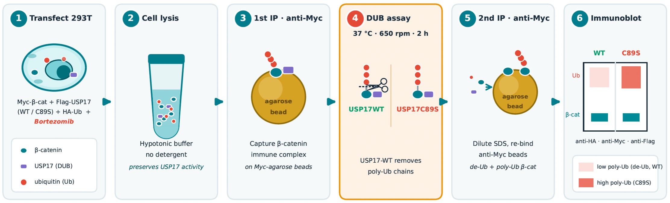
Background
Ubiquitination is a reversible post-translational modification that regulates protein turnover, subcellular localization, enzymatic activity, and protein–protein interactions [1]. The covalent attachment of ubiquitin is reversed by deubiquitinating enzymes (DUBs), also known as deubiquitinases, a functionally diverse class of proteases that remove ubiquitin from mono- and polyubiquitinated proteins [2]. This activity thereby modulates the stability and function of signaling properties and fine-tunes numerous cellular pathways. The human genome encodes approximately 100 DUBs, and dysregulation of their expression has been implicated in many diseases, including cancer and neurodegeneration [2].
Oncogenic transcription factors and co-activators, including MYC, KLF4, SNAIL, ELK1, and β-catenin, are often stabilized by reduced polyubiquitination-mediated proteasomal degradation [3–8]. Therefore, increasing their polyubiquitination by inhibiting DUBs is an attractive strategy to reduce their expression. DUBs are broadly classified into two principal groups based on their catalytic mechanisms: cysteine proteases and metalloproteases. The cysteine protease group includes ubiquitin-specific proteases (USPs), ubiquitin C-terminal hydrolases (UCHs), ovarian tumor domain proteases (OTUs), and Machado–Josephin domain-containing proteins (MJDs). Metalloprotease DUBs are generally referred to as JAMM proteases [2].
Using a genome-wide loss-of-function RNAi screen targeting 99 human DUBs, we recently identified USP17 as a key regulator of β-catenin stability and oncogenic growth in colorectal cancer (CRC) cells [8]. For enzymes such as USP17, target validation typically relies on gain- and loss-of-function approaches in cultured cells and animal models. Although correlations between USP17 expression and functional readouts, such as β-catenin protein levels, transcriptional activity, CRC cell growth, and tumor growth, are essential, additional in vitro biochemical approaches are necessary to establish a direct enzyme–substrate relationship. Therefore, biochemical evidence, including co-immunoprecipitation experiments, co-localization studies, and in vitro deubiquitination assays, is required to directly test whether the candidate DUB can deubiquitinate the putative substrate.
In vitro deubiquitination assays for USP17 have been reported using recombinant USP17 protein purified from bacterial expression systems [9]. However, USP17 requires phosphorylation by cyclin-dependent kinases in mammalian cells to become fully active [4], a requirement that is not recapitulated in bacterial expression systems. Here, we describe a method to directly measure USP17 deubiquitinase activity by immunopurifying both USP17 and its substrate, β-catenin, from mammalian cells. The assay is technically simple and adaptable to other DUBs and substrates.
Materials and reagents
Biological materials
1. HEK 293T/17 (American Type Culture Collection, CRL-11268)
Antibodies and plasmids
1. Pierce Anti-c-Myc agarose: agarose-conjugated anti-c-Myc (9E10) mouse monoclonal antibody (Pierce, catalog number 20168)
2. Purified anti-HA.11 Epitope Tag Antibody (clone 16B12) (BioLegend/Revvity, catalog number: 901502) (previously Covance, catalog number: MMS-101P)
3. Rabbit polyclonal Anti-c-Myc (A-14) antibody, 100 μg/mL (Santa Cruz, catalog number: Sc-789)
4. Rabbit polyclonal Anti-Flag antibody (Sigma, catalog number: F7425)
5. Goat anti-rabbit IgG (H + L), HRP-conjugated (KPL, catalog number: 074-1506)
6. Goat anti-mouse IgG (H + L), HRP-conjugated (KPL, catalog number: 074-1806)
7. pCDNA3.1-Myc-β-catenin, pCDNA3.1-Flag-USP17WT, and pCDNA3.1-Flag-USP17C89S, recently described [8]
8. pCDNA3.1-HA-ubiquitin plasmid, described in [10]
Reagents
1. DMEM Hi-glucose, L-glutamine, Na pyruvate (Wisent, catalog number: 319-005)
2. Fetal bovine serum (FBS), Premium, low endotoxin (Wisent, catalog number: 090-150, lot number: 112733)
3. Trypsine-EDTA (0.05%) (Wisent, catalog number: 325-542-CL)
4. Trypan Blue 0.4% solution (Wisent, catalog number: 609-130-EL)
5. Extreme MEM 1× with Earle’s salts, L-glutamine, and hormones (Wisent, catalog number: 390-005-CL)
6. Methanol 20 L (VWR, catalog number: CAMX0485-5)
7. Hydrochloric acid (HCl) concentrate, 10N ACS (certified) (Fisher, catalog number: SA49_6×100mL)
8. Sodium hydroxide (NaOH) solution (10N certified) (Fisher, catalog number: SS2551)
9. Acetic acid glacial (Fisher, catalog number: 351271500)
10. Anhydrous ethyl alcohol (Greenfield Global, catalog number: P016EAAN)
11. ContecTM PREemptTM concentrate disinfectant (Fisher Scientific, catalog number: 296366215)
12. Polyethylenimine, linear, MW 25000, transfection grade (PEI 25KTM) (Polysciences, catalog number: 23966)
13. Glycerol (Wisent, catalog number: 800-040-LL)
14. Glycine (Wisent, catalog number: 800-045-IK)
15. Tris (hydroxymethyl) aminomethane (Wisent, catalog number: 600-125-IK)
16. Sodium chloride anhydrous (NaCl) (Wisent, catalog number: 600-082 IK)
17. Potassium chloride (KCl) (molecular biology, ≥99.0%) (Sigma, catalog number: P9541)
18. Sodium phosphate dibasic heptahydrate (Na2HPO4·7H2O) (Sigma, catalog number: S9390)
19. Potassium phosphate monobasic (KH2PO4) (molecular biology, anhydrous) (Sigma, catalog number: 60218)
20. Magnesium chloride hexahydrate (MgCl2·6H2O) (Sigma, catalog number: M2670)
21. Sodium fluoride (NaF) (Sigma, catalog number: S7920)
22. β-Glycerophosphate disodium salt hydrate (Sigma, catalog number: G6251)
23. Aprotinin from bovine lung (Roche, catalog number: 10236624001)
24. Pepstatin A from Streptomyces species (Roche, catalog number: 11359053001)
25. Sodium orthovanadate (Na3VO4) (Sigma, catalog number: 567540)
26. DL-Dithiothreitol (DTT) (Sigma, catalog number: D9779)
27. SDS 10% solution (Wisent, catalog number: 880-550-CL)
28. EDTA 0.5 M (pH 8.0) (Wisent, catalog number: 809-163 EL)
29. BSA standard 2 mg/mL (Thermo ScientificTM PierceTM, catalog number: 23209)
30. Triton X-100 (Sigma, catalog number: X100-1L)
31. Tween-20 (Sigma, catalog number: P9416)
32. SDS (sodium dodecyl sulfate) (Wisent, catalog number: 800-100 CG)
33. Ponceau S (Sigma, catalog number: P3504-10G)
34. Sodium azide (NaN3) (EM Science, MilliporeSigma, catalog number: SX0299-1)
35. Dry milk (Carnation, Skim Milk Fat-Free, 500 g)
36. Ammonium persulfate (APS) (Sigma, catalog number: A3678-100G)
37. Bromophenol blue (Bio-Rad, catalog number: 1610404)
38. TEMED (Sigma, catalog number: T9281-25ML)
39. DMSO (Sigma, catalog number: D8418)
40. Bortezomib (Sigma, catalog number: SKU 503140001)
41. Protein assay dye reagent concentrate (Bio-Rad, catalog number: 5000006)
42. 30% Acrylamide/Bis solution 37.5:1 (Bio-Rad, catalog number: 1610158)
43. Western Lightning ECL Pro 340 mL (Revvity; formerly, PerkinElmer’s Life Sciences and Diagnostics; catalog number: NEL121001EA)
44. Super Signal West Femto Maximum Sensitivity Substrate (Pierce, catalog number: 34095)
45. ProtranTM Premium Western blotting membrane, nitrocellulose, 0.2 μm (AmershamTM, catalog number: GE10600001)
46. Parafilm® M sealing film (VWR, catalog number: 52858-000)
47. Plastic film (Saran Wrap)
48. BLUelf Prestained Protein Ladder (Froggabio, catalog number: PM008-0500)
Note: For reagent storage conditions and expected shelf life, please refer to Supplementary File 1.
Solutions
1. 1× PBS buffer (see Recipes)
2. PEI (1 mg/mL) (see Recipes)
3. Bortezomib 10 mM (see Recipes)
4. NaCl 5 M (see Recipes)
5. MgCl2 2 M (see Recipes)
6. NaF 0.5 M (see Recipes)
7. β-Glycerophosphate 1 M (see Recipes)
8. Aprotinin 2 mg/mL (see Recipes)
9. Pepstatin A 1 mg/mL (see Recipes)
10. Na3VO4 0.1 M (see Recipes)
11. DTT 1 M (see Recipes)
12. 10% Triton X-100 (see Recipes)
13. 1 M Tris-HCl pH 7.5 (see Recipes)
14. 1 M Tris-HCl pH 7.4 (see Recipes)
15. Hypotonic buffer + protease inhibitor cocktail (PIC) (see Recipes)
16. Lysis buffer (see Recipes)
17. DUBS assay buffer 5× (see Recipes)
18. 2% SDS in DUBS assay buffer (see Recipes)
19. DUBS assay buffer 1× (see Recipes)
20. 1.5 M Tris pH 8.8 (see Recipes)
21. 2 M Tris pH 6.8 (see Recipes)
22. 0.5 M Tris pH 6.8 (see Recipes)
23. 10% APS (see Recipes)
24. 30% Acrylamide 37.5:1 (see Recipes)
25. 10% SDS-PAGE (see Recipes)
26. 4% SDS-PAGE stacking gel (see Recipes)
27. Sample buffer 5× (see Recipes)
28. 10× Tris-glycine running buffer (see Recipes)
29. 10× transfer buffer (see Recipes)
30. Ponceau S solution (see Recipes)
31. 10% NaN3 (see Recipes)
32. 10× TBS (see Recipes)
33. Stripping solution (see Recipes)
Recipes
1. 10× PBS buffer
| Reagent | Final concentration | Quantity |
|---|---|---|
| NaCl | 1.37 M | 80 g |
| KCl | 26.8 mM | 2 g |
| Na2HPO4·7H2O | 43 mM | 11.5 g |
| KH2PO4 | 14.7 mM | 2 g |
| Milli-Q H2O | Complete to 1 L |
Complete to 1 L with Milli-Q water to make 10×.
2. PEI (1 mg/mL)
| Reagent | Final concentration | Quantity |
|---|---|---|
| PEI | 1 mg/mL | 50 mg |
| Milli-Q H2O | Complete to 50 mL |
Start with 50 mg of PEI + 45 mL of H2O + 12 μL of HCl 10N. Dissolve completely, adding HCl if necessary, but do not drop the pH below 7.0. The final pH should be between 6.5 and 7.5. Adjust the volume to 50 mL. Once the PEI is dissolved and the volume is adjusted, filter-sterilize the solution (0.22 μm), aliquot, and freeze at -20 °C. Stable at 4 °C for 1 month; it may be freeze-thawed five times.
3. Bortezomib 10 mM
| Reagent | Final concentration | Quantity |
|---|---|---|
| Bortezomib | 10 mM | 2.5 mg |
| DMSO | 651 μL |
To prepare a 10 mM stock solution from a 2.5 mg vial of bortezomib (molecular weight: 384.24 g/mol), add 651 μL of DMSO. Aliquot the stock solution into single-use vials and store at -20 °C for up to 6 months.
4. NaCl 5 M
| Reagent | Final concentration | Quantity |
|---|---|---|
| NaCl (54.88 MW) | 5 M | 29.22 g |
| Milli-Q H2O | Complete to 100 mL |
Dissolve completely in 75 mL of Milli-Q water, then dilute to a final volume of 100 mL.
5. MgCl2 2 M
| Reagent | Final concentration | Quantity |
|---|---|---|
| MgCl2·6H2O (203.30 MW) | 2 M | 40.66 g |
| Milli-Q H2O | Complete to 100 mL |
Dissolve completely in 80 mL of Milli-Q water, then dilute to a final volume of 100 mL. Autoclave or filter through 0.22 μm.
6. NaF 0.5 M
| Reagent | Final concentration | Quantity |
|---|---|---|
| NaF (41.99 MW) | 0.5 M | 1.05 g |
| Milli-Q H2O | Complete to 50 mL |
Dissolve completely in 40 mL of Milli-Q water, then dilute to a final volume of 50 mL. Keep at 4 °C.
7. β-Glycerophosphate 1 M
| Reagent | Final concentration | Quantity |
|---|---|---|
| β-Glycerophosphate disodium salt hydrate (216.04 MW) | 1 M | 21.6 g |
| Milli-Q H2O | Complete to 100 mL |
Dissolve completely in 80 mL of Milli-Q water, then dilute to a final volume of 100 mL.
8. Aprotinin 2 mg/mL
| Reagent | Final concentration | Quantity |
|---|---|---|
| Aprotinin | 2 mg/mL | 10 mg |
| Milli-Q H2O | Complete to 5 mL |
Dissolve the contents of the vial in 4 mL of Milli-Q water, then bring the final volume to 5 mL. Divide into aliquots of 75 μL and use at a 1/1,000 dilution (2 μg/mL). Store stocks at -80 °C.
9. Pepstatin A 1 mg/mL
| Reagent | Final concentration | Quantity |
|---|---|---|
| Pepstatin A | 1 mg/mL | 10 mg |
| 100% ethanol | 10 mL |
Dissolve the contents of the vial in 10 mL of 100% ethanol and rotate overnight at 4 °C. Then, prepare 75 μL aliquots and use at a 1/1,000 dilution (1 μg/mL). Store aliquots at -80 °C.
10. Na3VO4 0.1 M
| Reagent | Final concentration | Quantity |
|---|---|---|
| Na3VO4 (183.91 MW) | 0.1 M | 0.92 g |
| Milli-Q H2O | Complete to 50 mL |
a. Dissolve 0.92 g of sodium orthovanadate in 20 mL of Milli-Q water in a 50 mL tube.
b. Adjust the pH to 10 (the solution will turn yellow).
c. Boil the solution in a water bath for 5–10 min (until the solution becomes colorless).
d. Let the solution sit until it cools down to room temperature (RT).
e. Readjust pH to 10.0.
f. Repeat steps c–e until the pH is stable at 10.0 and the solution is colorless.
g. Fill up to 50 mL with Milli-Q H2O to obtain 0.1 M.
h. Prepare 750 μL aliquots and freeze stocks at -80 °C. Refreeze tubes at -20 °C for routine assay.
11. DTT 1 M
| Reagent | Final concentration | Quantity |
|---|---|---|
| DTT (124.25 MW) | 1 M | 248.5 mg |
| Milli-Q H2O | Complete to 2 mL |
Dissolve 248.5 mg of DTT powder into 1.5 mL of Milli-Q water. Once completely dissolved, add water to bring the final volume to 2 mL. Prepare aliquots of 100 μL. Keep at -80 °C. Once thawed, refreeze at -20 °C 3–4 times.
12. 10% Triton X-100
| Reagent | Final concentration | Quantity |
|---|---|---|
| 100% Triton-X-100 | 10% | 1 mL |
| Milli-Q H2O | 9 mL |
With a P1000 tip, take 1 mL of Triton X-100 by releasing the push button of the pipettor very slowly. Wipe the excess viscous solution from the tip with a Kimtech lint-free wipe, add to the water, and rinse the contents of the tip with a few up/down strokes as needed. Store at 4 °C.
13. 1 M Tris pH 7.4
| Reagent | Final concentration | Quantity or volume |
|---|---|---|
| Tris (hydroxymethyl) aminomethane | 1 M | 121.10 g |
| HCl 10N | Up to pH 7.4 | |
| Milli-Q H2O | Complete to 1 L |
Dissolve Tris in approximately 800 mL of MilliQ water. Adjust pH to 7.4 using 10N HCl. Store at 4 °C to cool the solution. Once the solution reaches 4 °C, readjust the pH to 7.4, then fill to 1 L and store at 4 °C.
14. 1 M Tris pH 7.5
| Reagent | Final concentration | Quantity or volume |
|---|---|---|
| Tris (hydroxymethyl) aminomethane | 1 M | 121.10 g |
| HCl 10N | Up to pH 7.5 | |
| Milli-Q H2O | Complete to 1 L |
Dissolve Tris in approximately 800 mL of Milli-Q water. Adjust pH to 7.5 using 10N HCl. Store at 4 °C to cool the solution. Once the solution reaches 4 °C, readjust the pH to 7.5, then fill to 1 L and store at 4 °C.
15. Hypotonic buffer + PIC
| Reagent | Final concentration | Quantity or volume |
|---|---|---|
| 1 M Tris-HCl pH 7.5 (4 °C) | 10 mM | 0.5 mL |
| 5 M NaCl | 10 mM | 0.1 mL |
| 2 M MgCl2 | 1.5 mM | 0,0375 mL |
| 0.5 M NaF | 10 mM | 1 mL |
| 1 M beta-Glycerol-2-phosphate | 10 mM | 0.5 mL |
| 2 mg/mL Aprotinin | 2 μg/mL (1/1,000) | 50 μL |
| 1 mg/mL Pepstatin A | 1 μg/mL (1/1,000) | 50 μL |
| 0.1 M Sodium ortho-vanadate | 100 nM (1/100) | 500 μL |
| Milli-Q H2O | 47.2625 mL (Vt = 50 mL) |
Prepare the buffer freshly as indicated and keep it on ice. The buffer may be frozen.
16. Lysis buffer
| Reagent | Final concentration | Quantity or volume |
|---|---|---|
| 1 M Tris-HCl, pH 7.4 (4 °C) | 50 mM | 2.5 mL |
| 5 M NaCl | 120 mM | 1.2 mL |
| 0.5 M EDTA pH 8 | 1 mM | 0.1 mL |
| 10% Triton-X-100 | 1% | 5 mL |
| 0.5 M NaF | 10 mM | 1.0 mL |
| 1 M β-Glycerophosphate | 10 mM | 0.5 mL |
| Milli-Q H2O | 39.7 mL (Vt = 50 mL) |
Add all the reagents to the Milli-Q water, mix, and keep on ice; no protease inhibitors are added.
17. DUBS assay buffer 5×
| Reagent | Final concentration | Quantity or volume |
|---|---|---|
| 1 M Tris-HCl, pH 7.4 (4 °C) | 750 mM | 2.250 mL |
| 5 M NaCl | 250 mM | 150 μL |
| 0.5 M EDTA | 5 mM | 30 μL |
| 1 M DTT | 25 mM | 75 μL |
| Milli-Q H2O | 495 μL (Vt = 3 mL) |
Add all the reagents to the Milli-Q water, mix, and keep on ice. Freeze the stock at -20 °C.
18. 2% SDS in DUBS assay buffer
| Reagent | Final concentration | Quantity or volume |
|---|---|---|
| 5× DUBS | 1× | 300 μL |
| 10% SDS | 2% | 300 μL |
| Milli-Q H2O | 900 μL (Vt = 1.5 mL) |
Dilute the 5× DUBS assay buffer with water to 1×. Add the SDS, prepare freshly, and use at RT to avoid SDS precipitation.
19. DUBS assay buffer 1×
| Reagent | Final concentration | Quantity or volume |
|---|---|---|
| 1 M Tris pH 7.5 (4 °C) | 150 mM | 2.250 mL |
| 5 M NaCl | 50 mM | 150 μL |
| 0.5 M EDTA | 1 mM | 30 μL |
| 1 M DTT | 5 mM | 75 μL |
| Milli-Q H2O | 12,495 mL (Vt = 15 mL) |
Add all the reagents to the Milli-Q water, mix, and keep on ice. No protease inhibitor is added.
20. 1.5 M Tris pH 8.8
| Reagent | Final concentration | Quantity or volume |
|---|---|---|
| Tris (hydroxymethyl) aminomethane | 1.5 M | 181.7 g |
| HCl 10N | Up to pH 8.8 | |
| Milli-Q H2O | Complete to 1 L |
Dissolve 181.7 g of Tris base in 850 mL of Milli-Q water. Adjust the pH to 8.8 using concentrated HCl (approximately 28 mL). Readjust the pH to 8.8 at room temperature. Add H2O to 1 L. Store the buffer at 4 °C.
21. 2 M Tris pH 6.8
| Reagent | Final concentration | Quantity or volume |
|---|---|---|
| Tris (hydroxymethyl) aminomethane | 2 M | 121.1 g |
| HCl 10N | Up to pH 6.8 | |
| Milli-Q H2O | Complete to 500 mL |
Dissolve 121.1 g of Tris in 400 mL of Milli-Q water. Adjust the pH to 6.8 using concentrated HCl (approximately 40–45 mL). Readjust the pH to 6.8 at room temperature. Add Milli-Q H2O to 500 mL. Store the buffer at 4 °C.
22. 0.5 M Tris pH 6.8
| Reagent | Final concentration | Quantity or volume |
|---|---|---|
| Tris (hydroxymethyl) aminomethane | 0.5 M | 60.55 g |
| HCl 10N | Up to pH 6.8 | |
| Milli-Q H2O | Complete to 1 L |
Dissolve 60.5 g of Tris base in 850 mL of Milli-Q water. Adjust the pH to 6.8 using concentrated HCl (approximately 40– 45 mL). Readjust the pH to 6.8 at room temperature. Add Milli-Q H2O to 1,000 mL. Store the buffer at 4 °C.
23. 10% APS (w/v)
| Reagent | Final concentration | Quantity or volume |
|---|---|---|
| APS | 10% | 1 g |
| Milli-Q H2O | 10 mL |
Dissolve 1 g of APS in 10 mL of Milli-Q H2O. Store at 4 °C. Stable for three months.
24. 30% Acrylamide 37.5:1
| Reagent | Final concentration | Quantity or volume |
|---|---|---|
| Acrylamide/Bis 37.5:1 premixed powder | 30% | 150 g |
| Milli-Q H2O | Add 362 mL |
Add 362 mL of Milli-Q water directly into the bottle, then add a magnetic stirrer (small to medium size) and mix on the magnetic plate for 30 min on medium speed. Once dissolved completely, filter through a 0.45 μm filter unit.
Note: To minimize contact with acrylamide powder or solution, please follow the company protocol. The recommended shelf life of the solution is 1 month at 4 °C in a dark bottle.
Acrylamide, whether in powder or solution, is toxic. The proper use of gloves and masks is highly recommended.
25. 10% SDS-PAGE
| Reagent | Final concentration | Quantity or volume |
|---|---|---|
| 1 M Tris pH 8.8 | 375 mM | 2.5 mL |
| 10% SDS | 0.1% | 100 μL |
| 30% Acrylamide (37.5:1) | 10% | 3.33 mL |
| Milli-Q H2O | 3.96 mL (Vt = 10 mL) | |
| TEMED | 1/1,000 | 10 μL |
| 10% APS | 1/100 | 100 μL |
In a clean tube, add all the solutions except TEMED and APS. At the last minute, when the glass setup is ready, add these last two reagents. Mix with a pipette controller without any bubbles; up/down movements may be performed. Then, disperse onto the casting setup, moving from one side to the other (left to right). Gently add 500 μL of Milli-Q water on top of the acrylamide; there is no need to use any saturated solvent to delimit the top of the gel. Let it polymerize for at least 1 h at RT.
26. 4% SDS-PAGE stacking gel
| Reagent | Final concentration | Quantity or volume |
|---|---|---|
| 0.5 M Tris pH 6.8 | 125 mM | 625 μL |
| 10% SDS | 0.1% | 25 μL |
| 30% Acrylamide (37.5:1) | 4% | 325 μL |
| Milli-Q H2O | 1.5 mL (Vt = 2.5 mL) | |
| TEMED | 1/1,000 | 2.5 μL |
| 10% APS | 1/100 | 25 μL |
Proceed as for Recipe 25; let it polymerize for at least 30 min.
27. Sample buffer 5×
| Reagent | Final concentration | Quantity or volume |
|---|---|---|
| 2 M Tris pH 6.8 | 312.5 mM | 7.8 mL |
| 100% Glycerol | 50% | 25 mL |
| SDS | 10% | 5 g |
| β-Mercaptoethanol* | 25% | 12.5 mL |
| Bromophenol Blue (BPB) | 0.05% | 0.025 g |
| Milli-Q H2O | 4.3 mL |
*Manipulate the solution under the chemical hood when adding β-mercaptoethanol and after.
In a 50 mL conical tube, add Tris, glycerol, SDS, β-mercaptoethanol, and Milli-Q water, then slowly agitate (rocker) until the solution is clear at RT (overnight).
Weigh the BPB and add it to the 50 mL tube; close tightly. Incubate in a closed container filled with warm tap water. Leave the tube on the rocker until the solution is homogeneous. Aliquot in 500 μL and keep stock at -20 °C.
28. 10× Tris-glycine running buffer
| Reagent | Final concentration | Quantity or volume |
|---|---|---|
| Tris (hydroxymethyl) aminomethane | 250 mM | 30 g |
| Glycine | 1,920 mM | 144 g |
| SDS | 1% | 10 g |
| Milli-Q H2O | To 1 L |
Wear a mask and gloves, as SDS is volatile and harmful. In a large beaker, add 600 mL of Milli-Q water, Tris base, glycine, and SDS slowly. Protect with plastic film and agitate on a magnetic stirrer overnight or until dissolved at RT. Complete to 1 L by adding water without creating bubbles.
Note: Mixing the solution produces a lot of foam. Pour gently into the stock bottle.
Prepare 400 mL of 1× SDS running buffer (25 mM Tris, 192 mM Glycine, 0.1% SDS) per montage when ready for electrophoresis. Dilute 40 mL of 10× with 360 mL of Milli-Q water; mix thoroughly.
29. 10× Transfer buffer 1 L
| Reagent | Final concentration | Quantity or volume |
|---|---|---|
| Tris (hydroxymethyl) aminomethane | 250 mM | 30.25 g |
| Glycine | 192 mM | 144 g |
| Milli-Q H2O | To 1 L |
In a 1 L beaker, add Tris and glycine to 350 mL of Milli-Q water. Protect with a plastic film; agitate at RT on a magnetic stirrer until dissolved. Complete to 1 L by adding water. Store at 4 °C.
When ready to transfer, dilute the transfer buffer by adding 100 mL of 10× transfer buffer to 700 mL of cold water, then add 200 mL of methanol (under the chemical hood), mix, and keep at 4 °C. The buffer may be reused once.
30. Ponceau S solution
| Reagent | Final concentration | Quantity or volume |
|---|---|---|
| Ponceau S | 0.1% | 0.5 g |
| Acetic acid glacial | 5% | 15 mL |
| Milli-Q H2O | 475 mL |
In a 475 mL bottle of Milli-Q water, add 0.5 g of Ponceau S and agitate on a magnetic stirrer until fully dissolved. Finally, add 25 mL of acetic acid slowly, close the bottle, keep at RT, and reuse this solution until the staining becomes faint.
31. 10% NaN3 (w/v)
| Reagent | Final concentration | Quantity or volume |
|---|---|---|
| NaN3 | 10% | 1 g |
| 1× PBS | 10 mL |
Dissolve 1 g of sodium azide in 10 mL of 1× PBS. Store at 4 °C. Use at 0.02% final concentration (20 μL of NaN3 in 10 mL of first-antibody dilution).
32. 10× TBS 2 L
| Reagent | Final concentration | Quantity or volume |
|---|---|---|
| Tris (hydroxymethyl) aminomethane | 200 mM | 48.4 g |
| NaCl | 1.37 M | 160.1 g |
| Milli-Q H2O | To 1.5 L | |
| Adjust to pH 8.0 | ||
| Milli-Q H2O | To 2 L |
Before filling to 2 L with Milli-Q water, adjust pH to 8.0. This requires approximately 22 mL of 10N HCl. When pH has been adjusted to 8.0, wait 30 min and take another measurement. If the pH is still at 8.0, fill the solution to 2 L with Milli-Q water. If not, adjust it to 8.0 and wait another 30 min to make sure the pH is stable.
Prepare 1× TBS + Tween-20 (0.1%) (TBST) for all western blot experiments. Dilute 200 mL of 10× TBS in 1,798 mL of Milli-Q water and mix on a magnetic stirrer. With a 5 mL pipette, very slowly retrieve 2 mL of Tween-20 from the 100% solution; wait until the pipettor stops aspirating to the 2 mL volume. With a clean wipe tissue (Kimtech), remove any excess Tween-20 from the outside of the 5 mL pipette, then dispense the 2 mL volume into the solution with many up-and-down strokes until none remains. Continue mixing for at least 15 min.
33. Stripping buffer
| Reagent | Final concentration | Quantity or volume |
|---|---|---|
| 62.5 mM Tris pH 6.8 | 62.5 mM | 1.25 mL |
| 10% SDS | 2% | 2 mL |
| 14.3 M β-mercaptoethanol | 100 mM | 70 μL |
| Milli-Q H2O | 6.68 mL |
Prepare freshly. Add the mercaptoethanol at the last minute, under the chemical hood. Dispose of the waste accordingly.
Laboratory supplies
1. Cell scraper (M) (Sarstedt, catalog number: 83.3951)
2. Four-channel alarm timer with clock (VWR, catalog number: 62344-641)
3. Petri dishes, 10 and 15 cm (Sarstedt, catalog numbers: 83.3902, 83.3903)
4. Pipettes, 5, 10, and 25 mL (Sarstedt, catalog numbers: 86.1253.001, 86.1254.001, 86.1685.001)
5. Tips, 10, 200, 1,000 μL (Ultident, catalog numbers: 87-A10, 87-A200C, 87-A1000B)
6. Glass Pasteur pipette 9 po (VWR, catalog number: 14673-043)
7. Blot paper (Bio-Rad, catalog number: 1650921)
8. Nitrocellulose membrane, 0.45 μm (Bio-Rad, catalog number: 1620115)
9. Kimtech lint-free wipe (Ultident, catalog number: 14-05511)
10. Microplate, clear 96-well, flat-bottom, with cover (Corning, catalog number: 3370)
11. 13 mL × 100 mm borosilicate tubes (Fisherbrand, catalog number: 14-961-27)
12. Microcentrifuge tubes 1.7 mL (Froggabio, catalog number: LMCT1.7B10)
13. Microcentrifuge tubes 5 mL snap cap (Froggabio, catalog number: LMCT5.OB10)
14. Conical centrifuge tubes, 15 and 50 mL (Froggabio, catalog numbers: TB15-500, TB50-500)
15. Filtration unit (500 mL), 0.45 and 0.22 μm (Sarstedt, catalog numbers: 83.3941, 83.3941.001)
16. Polystyrene rectangular dish with four compartments (Thermo Fisher Scientific, catalog number: 267061)
17. Antibody incubation boxes (Servicebio, catalog numbers: G6026-4, G6025-2, and G6020-9)
18. Filter forceps (Sigma, catalog number: XX6200006P)
19. Multiflex gel loading tip (ESBE, catalog number: SRS-2848)
Equipment
1. Hemacytometer (Sigma, model: Bright-Line)
2. Benchtop centrifuge (Sorvall, model: Legend RT)
3. Pipette controller (BrandTech, model: Accu-jet Pro)
4. Pipetman (Gilson, model: P2, P20, P200, P1000)
5. Microplate reader (BioTek, model: SynergyH1)
6. Mini centrifuge with 2 rotors (Benchmark Scientific, catalog number: Z742582)
7. Vortex (MBI-Scientific Industries SI, model: Genie 2)
8. Antibody incubation box (Servicebio Inc, catalog numbers: G6026-4, G6025-2, G6020-9)
9. Rectangular 4-well plate with cover (ThermoFisher, catalog number: 267061)
10. pH meter (Accumet, model: AB150)
11. Isotemp Magnetic Hotplate (Fisher Scientific, model: 11-600-100SH)
12. Dry Bath Incubator BenchMark (Sigma, catalog number: Z742509)
13. Refrigerated centrifuge (Eppendorf, model: 5417R)
14. Tube rotator (Fisher Scientific, model: 346 Chemistry Hematology Mixer test Tube rotator)
15. Hood Class II Type A2 (ESCO, model: LA2-6A2)
16. Cell incubator (Heraeus HERAcell 150i, catalog number: SVH-51022394)
17. Inverted microscope (Hund, model: Wilovert S)
18. Vacusafe (aspiration system) (Mandel, catalog number: TM-158310)
19. Molecular Image ChemiDoc with Universal Hood II (Bio-Rad, model: XRS+)
20. Refrigerated Incubator Max-Q 6000-7 (Barnstead, catalog number: BDI-SHKE6000-7)
21. Freezer -80 °C (REVCO, model: ULTIMA II ULT2586-9S)
22. Freezer -20 °C, 18 cu. ft. refrigerator (Frigidaire, model: FFTR1817LW)
23. Mini-PROTEAN Tetra Electrophoresis system (Bio-Rad, catalog number: 1658006)
24. CriterionTM Blotter with plate electrode (Bio-Rad, catalog number: 1704070)
25. Power supply (Bio-Rad, model: Power PAC 200)
26. Eppendorf Thermomixer Compact (Sigma, catalog number: T1317)
27. Precision reciprocating digital water bath (Thermo Scientific, catalog number: 2870)
28. Rocker dual platform (Bio-Rad, catalog number: 1660709)
29. Digital dry bath (Sigma/Milipore, catalog number: Z742505)
30. Balances (Mettler Toledo, model: PG2002-S and AG204 Delta Range)
31. Spectrophotometer UV visible (GE Amersham, model: Ultrospec 2100 pro)
Software and datasets
1. Image Lab, version 6.1 (Bio-Rad)
Procedure
A. Maintenance of cells
Note: All work should be performed in the ESB2 cabinet, and all components should be at 37 °C or at least at RT before use.
1. Passage 293T/17 cells in complete DMEM containing 10% FBS (DMEM-10% FBS) every 3–4 days and incubate at 37 °C with 5% CO2. Use dilutions of detached cells ranging from 1/10 to 1/15, aiming for a maximum confluence of 90%.
Note: 293T cells are prone to detachment if too confluent or left at RT. They should ideally be used between passage 4 (minimum after thaw) and passage 16 (maximum).
2. Sub-culturing:
a. Rinse with 1× PBS, then detach the cells with 0.05% trypsin-EDTA (0.01 mL/cm2) for 1–2 min at RT.
b. Inactivate with 8–10 volumes of DMEM-10% FBS.
c. Firmly hold the dish and apply a firm knock to the opposite side of the dish with the other hand. Repeat 1–2×.
d. Collect and pool the cells in a 15 or 50 mL tube.
e. Centrifuge for 5 min at 216× g to remove inactivated trypsin.
Note: This step is mandatory when plating counted cells for an experiment or prior to reverse transfection. Otherwise, only passage the cells by diluting the trypsinized cells into media 1/10 to 1/15.
f. After centrifugation, aspirate and firmly snap the end of the tube 3–5 times before adding DMEM-10% FBS (15 mL/15 cm).
Note: This step enables a better resuspension of the pellet.
g. Flush the cell suspension 3–4× with up-and-down circular motions to ensure an homogeneous and complete recovery of cells.
h. Count an aliquot in Trypan Blue (1:1) using a hemacytometer. A concentration of 1.2–2 million cells/mL should be obtained.
3. Upscale the 293T/17 by plating 3 × 15 cm Petri dishes at 3 million cells per dish three days before the experiment.
B. PEI reverse-transfection of 293T/17
Notes:
1. Goal: 60 million freshly resuspended 293T/17 cells are reverse co-transfected at a PEI-to-DNA ratio of 1:2.5 for 24 h, then treated with 20 nM Bortezomib for the final 16 h.
2. All work should be performed in the ESB2 cabinet, and all components should be at 37 °C or at least RT before use.
1. In 5-mL properly labeled tubes, dilute and prepare the DNA mixes (#1ctr, #2WT, #3C89S) in Extreme-MEM, according to Table 1. Then, mix gently up and down 2–3×. This will generate three tubes containing 1,000 μL of DNA mixture. Leave at RT for later use.
Table 1. DNA dilution content of each condition
| DNA quantity (μg) | ||||||
| Conditions (40 μg of DNA total) | pCDNA (Flag) | Bcat (Myc) | Ubi (Ha) | USP17L2+WT (Flag) | USP17L2+C89S (Flag) | Extreme-MEM |
| #1 Bcat+Ubi_CTRL | 20 | 10 | 10 | 0 | 0 | 960 μL |
| #2 Bcat+Ubi+USP17WT | 10 | 10 | 10 | 10 | 0 | |
| #3 Bcat+Ubi+USP17C89S | 0 | 10 | 10 | 0 | 20 | |
Note: Twice the amount of the inactive mutant C89S, compared with the WT, has been tested previously and is required for this assay.
2. Prepare a PEI master mix for the number of conditions to be transfected, adding an extra 0.5× (for a final concentration of 3.5×).
a. Vortex the PEI 1 mg/mL stock for 5 s at medium speed, then add 350 μL of PEI to 3,150 μL of Extreme-MEM in a 5 mL tube for a final volume of 3.5 mL of diluted PEI (now at 0.1 mg/mL).
b. Vortex for 5 s and then incubate at RT for 5 min.
3. Proceed to complex DNA:PEI formation:
a. After the incubation (step B2b), mix twice (up and down) the diluted PEI with a 1,000 μL pipette.
b. Distribute 1,000 μL of PEI to each 1,000 μL of DNA mixture for a total of 2 mL per condition.
c. Vortex samples for 10 s to create a vortex effect. We vortex 2–3 samples simultaneously.
d. Incubate for 20 min at RT.
Note: A 10-s delay between conditions can be implemented when more than 10 conditions are tested.
4. During this incubation, harvest the subconfluent 293T/17 cells.
a. Rinse each Petri dish once with 10 mL of 1× PBS. Disperse the volume without disturbing the cells, then aspirate.
b. Add 1.5 mL of 0.05% trypsin-EDTA. Spread the solution with back-and-forth movements, then rotate the dish to cover the entire surface. Wait 1–2 min.
c. Firmly hold the dish and apply a firm knock to the opposite side of the dish with the other hand. Repeat 1–2×.
d. Inactivate the trypsin by adding 13.5 mL of DMEM-10% FBS (using a 10 mL pipette).
e. Collect and pool the cells in a 50 mL tube by flushing the cell suspension 3–4× with up-and-down circular motions to ensure homogeneous and complete recovery of cells.
f. Count a diluted aliquot in Trypan Blue using a hemacytometer, then centrifuge 70 million viable cells (3.5 conditions × 20 million) at 216× g for 5 min at 4 °C.
5. Label each 15 cm Petri dish appropriately for each reverse-transfection condition:
a. Add 13 mL of DMEM-10% FBS to each Petri dish.
6. Aspirate the supernatant from the centrifuged cells.
a. Gently flick the end of the 50 mL tube 3–5× to disperse the pellet, resulting in better resuspension of the pelleted cells.
b. Dilute the 70 million cells to 4 million cells/mL. To do so, add 5 mL of DMEM-10% FBS, gently swirl the tube 3–4× to resuspend the pellet, then add the remaining 12.5 mL of media and mix gently. Keep at RT.
c. Cells are ready to be plated once the incubation time for the DNA:PEI is complete (step B3).
7. Align the 15 cm Petri dishes, already containing media, from left to right:
a. Half-open the dishes.
b. Invert the diluted cell suspension three times, then gently flush with a 10 mL pipette using circular movements.
c. Dispense 5 mL of the resuspended cells into each 15 cm Petri dish containing media, for a total volume of 18 mL per dish.
8. To evenly distribute the cells in the 15 cm Petri dishes, perform circular rotations (2×), followed by small back-and-forth movements (4×), and finally cross-side movements (4×). Verify the distribution of the cell suspension under a phase-contrast microscope; correct if needed.
9. Proceed to add the DNA:PEI polyplex (from step B3) to the appropriately labeled Petri dish, one at a time.
10. Mix (2×) by inversion the tube containing the 2 mL DNA:PEI polyplex; recover any remaining solution from the cap.
11. Then, remove the Petri dish’s cover and slowly distribute the 2 mL of the DNA:PEI polyplex dropwise over the surface of the dish.
12. Replace the cover and gently disperse the PEI polyplex mixture using the same rotation sequence as described in step B8.
13. Repeat steps B10–12 for the two other conditions.
14. Incubate cells at 37 °C and 5% CO2 for 24 h.
C. Bortezomib treatment to inhibit proteasomal degradation
Note: Bortezomib is used to enrich for ubiquitinated β-catenin by inhibiting proteasomal degradation.
1. Eight hours after reverse-transfection, begin the 20 nM bortezomib treatment for the final 16 h.
2. Prepare a master mix for 3.5× reactions by adding 14.35 μL of 0.1 mM Bortezomib to 1735.65 μL of DMEM-10% FBS. Mix by inversion and vortex, then dispense 500 μL dropwise onto each Petri dish to achieve a final concentration of 20 nM Bortezomib in 20.5 mL of DMEM-10% FBS. Dispense slowly, following the movements described in step B8.
3. Incubate at 37 °C, 5% CO2 for 16 h (24 h post-reverse-transfection); then, proceed directly to cell lysis (preparation of whole-cell extracts).
D. Fresh whole-cell extract preparation
Note: From now on, all work is performed on a lab bench. All solutions, materials, and equipment are kept on ice. A hypotonic buffer is used to preserve the native protein structure and activity; it does not contain any detergent that could interfere with the enzymatic reaction.
1. At a 30° angle, aspirate the media slowly from the 15-cm Petri dishes.
a. Gently deliver 10 mL of ice-cold 1× PBS at the junction of the bottom and the side of each Petri dish.
b. Gently place the Petri dish on an ice bed (well-crushed, compacted ice).
2. Disperse the ice-cold 1× PBS evenly using smooth back-and-forth and left-right movements.
a. Aspirate as stated in step D1. Hold still at an angle that allows the liquid to slide to the bottom of the inclined Petri dish. Wait for 1 min and then carefully remove all PBS.
3. Proceed to direct lysis.
a. Add 600 μL of cold hypotonic buffer + PIC (see Recipe 15) to each 15 cm Petri dish.
b. Spread the buffer evenly across the Petri dish. Then, with the cover on, leave the Petri dish on top of well-crushed compact ice. Make sure the buffer covers the entire surface of the dish.
4. With a cell scraper for each condition, spread the lysis buffer and scrape the entire surface of the Petri dish as follows:
a. Remove the cover and hold it to the side of the Petri dish. Scrape in a zigzag motion from top to bottom (Z) or side to side (W), staying within the Petri dish’s inner circle limit.
b. Secure the inclined Petri dish with crushed ice to achieve an angle (~30°) and place the cover on.
c. Incubate for 30 min on ice.
d. Recover the cell lysate in a cold 1.7 mL mini-centrifuge tube and centrifuge at 20,817× g for 15 min at 4 °C.
e. Transfer the supernatant to another 1.7 mL cold mini-centrifuge tube.
5. Determine the total protein concentration of lysates using the Bio-Rad assay dye reagent (Bradford method).
a. In a 13 mL borosilicate tube, add 5 μL of a BSA standard curve (20 μL pipette) ranging from 0 to 1 mg/mL (diluted in hypotonic buffer). For cellular extracts, use 5 μL per condition. Do not forget the blank, which contains only hypotonic buffer.
b. Filter the reagent through a 0.45 μm filter to remove the common precipitate, then vortex for 2 s before use.
c. Using a 1,000 μL pipette, add 1 mL of Bio-Rad reagent to the first tube, then vortex for 2 s. Repeat for the remaining tubes.
d. Incubate for 5 min at RT, then read on the spectrophotometer at 595 nm.
e. Total protein concentration is expected to be around 1 mg/mL, with 600–750 μg of protein.
Caution: Normalize all samples to the same protein concentration before immunoprecipitation.
E. First Myc-β-catenin immunoprecipitation (IP)
Note: All the materials and buffers should be kept cold work with well-crushed compact ice.
1. Prepare a batch of washed c-Myc agarose beads for the entire protocol. The goal is to prepare enough beads to yield ~40 μL of packed beads across the three transfected conditions for two consecutive IP procedures: #1ctrl, #2USP17WT, and #3USP17C89S; thus, 6 × 40 μL.
Note: According to Pierce’s information, the manufacturer reports a binding capacity of 102–144 nmol of protein per mL of settled resin (tested using a 26–29 kDa c-Myc-tagged fusion protein). For Myc-β-catenin fusion protein (~90 kDa), an estimated theoretical binding capacity of 9.18–12.96 μg/μL of resin is therefore expected (a lower real-world recovery of around 5–7 μg/μL due to steric hindrance). Therefore, 40 μL of packed beads should capture approximately ~240 μg of c-Myc protein (Myc-β-catenin in our assay), which exceeds the amount from our 200 μg total protein extract used in the present experiment.
a. Gently rock back and forth in a circular motion to disrupt the c-Myc agarose beads (the product is supplied as a 25% slurry) into a homogeneous slurry, then withdraw 1,000 μL of slurry (~250 μL of packed beads) into a 1.7 mL mini-centrifuge tube.
Note: Use a 1,000 μL tip with a beveled edge to aliquot bead slurry and perform washes; the wider opening is less damaging to the beads.
b. Removal of the preservation buffer is achieved by substituting 1× PBS twice.
i. Pellet the beads by setting the microcentrifuge to 8,000× g; then, start spinning for 5–10 s.
ii. Without removing the tubes from the centrifuge, rotate each tube 180°, then centrifuge for an additional 5 s.
iii. Leave it on the bench for 1–2 min (always using compact ice), then discard the supernatant.
Note: This step levels the bead surface and improves liquid recovery, reducing loss.
iv. Add 1,000 μL of 1× PBS using a beveled-edge 1,000 μL tip, then resuspend the beads with three slow up-and-down motions.
v. Pellet resin as described in steps E1b.i–iii.
vi. Perform a second wash with 1,000 μL of 1× PBS using a beveled-edge 1,000 μL tip; then, resuspend the beads with three slow up-and-down strokes.
vii. Immediately before centrifugation, split the homogeneous bead preparation into two 500 μL aliquots (~125 μL of packed beads) in 1.7 mL tubes.
Note: This enables two bead preparations for the entire experiment, referred to as the first IP and second IP, performed in different buffers: For the first IP hypotonic buffer + PIC (see Recipe 15); for the second IP lysis buffer with 1% Triton X-100 (see Recipe 16).
viii. Leave in PBS 1× at 4 °C until needed for IP.
c. Final preparation of c-Myc agarose beads for first or second IP: For the final step of resin preparation, wash with the appropriate buffer for the IP.
Note: The last buffer change occurs immediately before the IP. For the first IP, proceed with resin using the hypotonic buffer + PIC, and keep the resin for the second IP in PBS 1× at 4 °C until ready to execute.
i. For the first IP-c-Myc, remove 1× PBS by centrifugation see steps E1b.i–iii.
ii. This time, use 750 μL of hypotonic buffer + PIC with a beveled 1,000 μL tip and resuspend the beads with three slow up-and-down movements.
Note: Volume adjustments are for practical convenience when splitting; for three equal volumes per condition, 250 μL of slurry (~40 μL of packed beads) is a more reproducible volume.
iii. Immediately add 250 μL of the prepared slurry to three 1.7 mL tubes.
iv. Pellet resin as described in steps E1b.i–iii.
v. Perform a second wash with 1,000 μL of hypotonic buffer + PIC using a 1,000 μL tip with a beveled edge; then, resuspend the beads with three slow up-and-down strokes.
vi. Pellet resin as described in steps E1b.i–iii; then, carefully discard the liquid using a Multiflex gel loading tip. Leave the tubes on ice and add the protein lysate.
2. Start the first IP for c-Myc-β-catenin across all three conditions, referred to as #1ctrl, #2USP17WT, and #3USP17C89S.
a. Add to each tube of the packed beads (40 μL packed beads) the corresponding volume of 200 μg of total protein, then complete to 600 μL with hypotonic buffer + PIC.
Note: A ratio of 15 volumes of buffer to 1 volume of packed beads is acceptable.
b. Incubate at 4 °C with orbital rotation (5 rpm) for 4 h.
Note: This incubation period allows the two successive IPs to be performed on the same day.
c. Pellet the beads by setting the microcentrifuge to 8,000× g; then, start spinning for 5–10 s.
d. Remove the liquid with a 1,000 μL pipette.
e. Proceed with two cold washes using hypotonic buffer + PIC, followed by two washes with 1× DUBS assay buffer.
i. In a rack that holds the three IP tubes tightly, add 600 μL of cold hypotonic buffer + PIC, then carefully snap the caps.
ii. Then, holding the tubes to the rack, firmly flip the rack to gently but efficiently displace the beads. Wash 5–10 times with front-to-back motion.
iii. Pellet the beads by setting the microcentrifuge to 8,000× g; then, start spinning for 5–10 s.
iv. Without removing the tubes from the centrifuge, rotate each tube 180°, then centrifuge for another 5 s. Leave them on the bench for 1–2 min, then carefully discard the liquid.
v. Repeat with a second 600 μL cold wash, then proceed the same way by repeating steps E2e.ii–iv.
vi. Switch to the next cold buffer (1× DUBS) and wash it twice by repeating steps E2e.ii–iv.
vii. After the resin has fully settled, carefully discard the liquid, then finish with a Multiflex gel loading tip.
Note: This tip enables less loss of resin when removing the liquid.
f. Resuspend in 40 μL of 1× DUBS assay buffer.
F. On-bead in vitro deubiquitination assay
Note: USP17WT, not USP17C89S, will remove the polyubiquitin chains present on ectopically expressed β-catenin.
1. Enzymatic DUBS assay
a. Incubate the bead-bound immune complex at 37 °C in a thermomixer with agitation at 650 rpm for 2 h.
b. To terminate the DUBS assay, add 40 μL of a 2% SDS solution prepared in 1× DUBS assay buffer.
Note: This stepstopsthe enzymatic reaction.
c. Then, apply a heat treatment to the 1% SDS reaction at 90 °C for 5 min in a thermomixer with agitation set to 650 rpm.
Note: This step will disrupt immune-complex interactions, freeing Myc-βcatenin and Flag-USP17 WT and C89S in solution.
Caution: Ensure the caps are tightly closed. If in doubt, place a heavy weight on top of all three tubes.
G. USP17 immunocomplex control
Note: This essential step confirms the presence of Flag-USP17WT or Flag-USP17C89S in Myc-β-catenin immune complexes.
The goal is to reserve 10% (8 μL) of the immunoprecipitated material to verify the presence of USP17 within the β-catenin immune complexes.
1. After the heat treatment, centrifuge for 5 s at 8,000× g. Without removing the tubes from the centrifuge, rotate each tube 180°, then centrifuge for another 5 s. Leave on the bench for 1–2 min.
2. Remove the 80 μL supernatant and transfer it to a new tube. Reserve an 8 μL aliquot.
Note: These will be referred to as the USP17-Immunocomplex control.
3. Analyze this 10% fraction by SDS-PAGE and western blotting using anti-Flag and anti-Myc antibodies to detect Flag-USP17 WT/C89S and Myc-β-catenin, respectively (please refer to sections I, J, and K).
H. Second Myc-β-catenin immunoprecipitation
1. Dilute 90% of the immunoprecipitated material with Triton-X-100 lysis buffer (see Recipe 16) to reduce the SDS concentration from 1% to 0.1%:
a. To the remaining 72 μL, add 720 μL of Triton-X-100 lysis buffer.
Critical: SDS must be diluted before re-immunoprecipitation to ensure efficient antibody binding.
b. Complete preparation of the c-Myc agarose bead for the second IP (refer to step E1b.viii).
i. Prepare as instructed in steps E1c.i–vi), except that the lysis buffer contains 1% Triton X-100 (Recipe 16)
ii. Remove from the ice where it was kept and pellet the beads at 8,000× g for 5–10 s. Without removing the tubes from the centrifuge, rotate each tube 180° and centrifuge for another 5 s. Leave them on the bench for 1–2 min, then carefully discard the liquid using a Multiflex gel loading tip. The c-Myc agarose beads are ready for use.
c. Proceed to the second c-Myc-β-catenin IP:
i. Add the 0.1%-diluted immunoprecipitated material (step H1a) to the 40 μL of packed beads.
ii. Incubate for 16 h at 4 °C with orbital rotation (5 rpm).
d. Wash beads four times with 600 μL of lysis buffer containing 1% Triton X-100. Proceed as before, and for the last wash, carefully discard the liquid and finish with a Multiflex gel loading tip.
e. Recover immune complexes from the beads by adding 20 μL of 3× SDS-PAGE sample buffer.
i. Vortex vigorously 4× with a 1-s pulse (full speed of the vortex).
ii. Microcentrifuge at 20,817× g for 2 min.
f. Heat samples for 5 min at 95 °C.
i. Vortex vigorously 4× with a 1-s pulse (full speed of the vortex).
ii. Microcentrifuge at 20,817× g for 2 min.
Caution: Ensure the caps are tightly closed. If in doubt, place a heavy weight on top of all three tubes.
Notes:
1. Proceed directly to SDS-PAGE electrophoresis.
2. Samples can be frozen at this step at -20 °C.
I. SDS-PAGE electrophoresis
Refer to the Mini-Protean Tetra cell in the Bio-Rad manual.
Note: Samples from the second IP are already ready. We need to prepare the immunocomplex controls (ctrls) for the first IP, corresponding to 10% of the material used for the second IP.
1. Prepare two homemade 1.5-mm-thick 10% SDS-polyacrylamide resolving gels with a 4% polyacrylamide stacking gel.
a. Pour 7 mL of gel for the resolving portion of the 10% SDS-PAGE into the casting gel setup, then slowly add 500 μL of Milli-Q water on top. Let it polymerize for at least 45 min at RT, away from direct sunlight. If not used the same day, change the water and keep it in a plastic bag at 4 °C, with humidity provided by a hand towel soaked with water.
b. Freshly cast the stacking gel (2 mL/gel) on the day of electrophoresis using a 10-well comb. Avoid bubbles and ensure the wells are complete. Add more stacking if you see a lack of acrylamide between wells during polymerization. Wait more than 30 min.
c. Remove the comb with a straight-up lift toward the top, rinse the wells with water to remove all acrylamide residue, and install the gel in the system (see manual).
2. Pre-run the gels for 10 min at 100 V while preparing samples for the two gels.
3. For gel number 1: Prepare samples #1CTLR, #2USP17WT, and #3USP17C89S. USP17 immunocomplex controls are described in step G2).
i. Simply add 3 μL of 5× sample buffer to the already aliquoted 8 μL sample, then complete to 15 μL with 1% Triton-X-100 buffer.
ii. Vortex vigorously 4× with a 1-s pulse (full speed of the vortex).
iii. Microcentrifuge at 20,817× g for 2 min.
iv. Heat the samples for 5 min at 95 °C.
v. Vortex vigorously 4× 1s pulse (full speed of the vortex).
vi. Microcentrifuge at 20,817× g for 2 min.
Caution: Ensure that the caps are tightly closed. If in doubt, place a heavy weight on top of all three tubes.
Note: Proceed directly to SDS-PAGE electrophoresis.
4. For gel number 2: The IP samples are already prepared (refer to step H1f).
5. Replace the upper tank buffer by adding a new migration buffer to displace and create a washing effect in the wells.
6. Load the samples with a 20 μL pipette, then add 5 μL of prestained standard (BLUeye).
7. Run the gel in the cold room at 150 V until the front dye reaches the end of the gel (about 1.5 h).
J. Transfer on nitrocellulose membrane and Red Ponceau staining
Refer to the Criterion Blotter’s Bio-Rad Manual.
Note: The Red Ponceau step is often underestimated. Also, keeping a picture of the stained transferred membrane can be a useful tool for later analysis of stripping, transfer quality, and loading efficiency.
1. Transfer the gel to a 0.45 μm nitrocellulose membrane at 100 V for 45 min. Keep transfer buffer 1× at 4 °C and perform transfer with gentle agitation in an ice-filled container.
Note: Buffer may be reused once.
2. Verify transfer efficiency using Red Ponceau staining.
a. Overlay the nitrocellulose membrane with Ponceau and agitate on a rocker for less than 3 min or until the bands appear.
b. Rinse the membrane with water until the background is clear.
c. On a glass plate, use a scalpel and a clean ruler to cut away the excess membrane.
d. With a pencil, mark the ladder and identify the order of the loaded samples.
e. Only cut the membrane of gel number 1 between markers to allow incubation with different antibodies for the same samples (Figure 1, left panel).
i. Cut a first portion between the 245 KDa molecular weight marker (MWM) and just before the 75 KDa MWM (to reveal Myc-β-catenin, a signal around 90 kDa), and a second portion between 75 and 63 KDa MWMs (to reveal Flag-USP17, a signal around 63 kDa).
f. Take a picture of the transferred membrane, as shown in Figure 1.
g. Remove reversible staining by rinsing twice with 1× TBS. The membranes are ready for immunoblotting.
Note: Membranes can be dried and stored at RT for later immunoblotting. Rehydrate slowly in 1× TBS under mild agitation at RT.
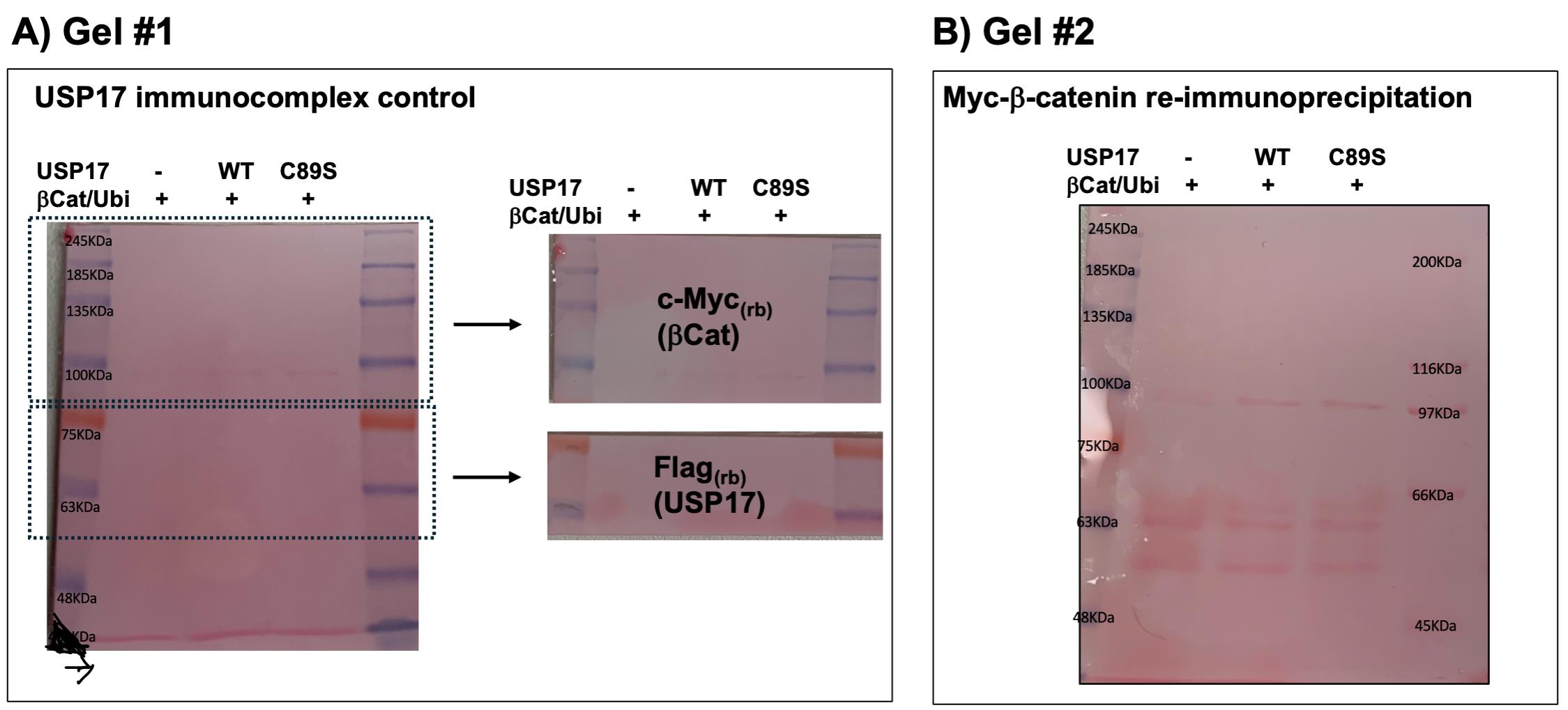
Figure 1. Samples derived from the indicated transfection conditions and subjected to Myc-β-catenin immunoprecipitation (IP) were resolved by SDS-PAGE electrophoresis and transferred onto nitrocellulose membranes as described in sections I and J. Panel A (Gel #1) refers to section G (first IP). Panel B (Gel #2) refers to section H (second IP).
K. Immunoblots and data analysis
1. Block the membranes with 5% milk in TBS-Tween-20 (0.1%) for 1 h at RT on a Bio-Rad UltraRocker set to 50 rpm.
2. Rinse 3× for 5 min with TBS-Tween-20 (0.1%) at 90 rpm.
3. Incubate membranes with first-antibody dilutions, as shown in Table 2, overnight (16 h) at 4 °C with gentle agitation at 40 rpm.
Note: The volume of antibody diluent varies depending on the size of the membrane to be covered. We used 3–10 mL. Incubation is performed in a rectangular 4-well plate with cover or in the antibody incubation box.
Table 2. Primary antibodies used for the western blot analysis
| Gel number 1 | ||||
| Ctrl | Name | Company, catalog number | Dilution | |
| 1st portion | Anti-c-Myc (rabbit) | Santa-Cruz, sc-789 | 1:1,000 | 5% milk + TBS-Tween-20 (0.1%) Add 0.02% NaN3 |
| 2nd portion | Anti-Flag (rabbit) | Sigma, F7425 | 1:1,000 | |
| Gel number 2 | ||||
| IP c-Myc | Name | Company, catalog number | Dilution | |
| Full membrane | Anti-Ha-ubi (mouse) | Covence, Ha.11# MMS-101P | 1:1,000 | |
| Striping and redo second immunoblotting (step K6) | ||||
| Anti-c-Myc (rabbit) | Santa-Cruz, sc-789 | 1:1,000 | ||
4. Rinse 3× for 5 min with TBS-T (0.1%) under the same agitation conditions.
5. Incubate membranes with the second antibody (HRP-conjugated) for 1 h at RT, as outlined in Table 3.
Table 3. Secondary antibodies used for the western blot analysis
| Gel number 1 | ||||
| Ctrl | Name | Company, catalog number | Dilution | 5% milk + TBS-Tween-20 (0.1%) |
| First portion | Goat anti-rabbit | KPL, KPL-074-1506 | 1:10,000 | |
| Second portion | Goat anti-rabbit | KPL, KPL-074-1506 | 1:10,000 | |
| Gel number 2 | ||||
| IP c-Myc | Name | Company, catalog number | Dilution | |
| Full membrane | Goat anti-mouse | KPL, KPL-074-1806 | 1:10,000 | |
| Striping and redo second immunoblotting | ||||
| Goat anti-rabbit | KPL, KPL-074-1506 | 1:10,000 | ||
6. Detection of the chemiluminescence signal
Note: Western Lightning ECL Pro from Perkin-Elmer and Super-Signal West Femto from Pierce were used. Please refer to the proper manual.
a. Prepare the ECLPro or the West Femto by mixing equal parts of the substrate and stable peroxide components. The manufacturer’s suggested volume is more than needed; we usually cut it in half. We prepare a total of 3 mL (1.5 mL + 1.5 mL) in a 5 mL container and protect it from light.
b. After the last wash following the second antibody (HRP-conjugated) incubation, with forceps, place the membrane between two Kimtech wipes, each folded into four layers, and apply gentle pressure to remove the buffer.
c. Quickly transfer the membrane onto a clean, very dry glass plate, protein side up.
d. Add the prepared detection solution slowly, starting in the middle of the membrane, and add enough volume to cover the entire membrane.
i. A “dome effect” is desired, spreading the liquid evenly over the membrane. The tip may be used, but try not to let the detection solution extend beyond the membrane surface.
Caution: No agitation is needed if the dome effect is obtained. Otherwise, turn the membrane upside down so that the membrane protein side touches the liquid on the glass plate, then move the membrane back and forth to ensure the solution contacts the whole surface.
ii. Incubate for 1–3 min, then lift the membrane and drain off the liquid.
iii. Place it on top of a pellicle (like a sheet protector, not too thick). Avoid any bubbles.
vi. Move the pellicle and membrane settings on the Bio-Rad UV transilluminator for Chemiluminescence.
v. Set the Bio-Rad CCD Imager to the automated setting for either strong or faint band detection (Image Lab, version 6.1, Bio-Rad), depending on the used antibody and the signal obtained.
7. Stripping procedure used to reprobe the IP c-Myc (mouse) membrane with the c-Myc (rabbit) antibody:
a. Directly after the ECL detection, soak the membrane in 10 mL of stripping buffer within a leakproof box.
c. Incubate for 15 min at 50 °C in an agitated water bath at 100 rpm.
d. Wash 3× for 5 min with TBS 1× (no Tween-20) on a UltraRocker at 90 rpm.
e. Proceed with the blocking (5% milk + 0.1% Tween-20) and immunoblotting using the conditions described in Table 2.
Data analysis
Briefly, the results of the first Myc-β-catenin IP (gel number 1; before heat denaturation) should show that the DUBs (here, Flag-USP17WT and USP17C89S ~63 kDa chemiluminescent signals) are present in the immunocomplex with the tested substrate (Myc-β-catenin ~90 kDa chemiluminescent signals). Gel number 2 (re-immunoprecipitation of Myc-β-catenin) should show that USP17WT, and not the catalytically inactive mutant C89S, reduces the polyubiquination (a signal observed above 90 kDa) of β-catenin. Please refer to Figure 2 in [8].
Validation of protocol
This protocol (or parts of it) has been used and validated in the following research article:
Acevedo et al. [8]. The deubiquitinase USP17 regulates the expression and activity of the oncogenic driver beta-catenin in colorectal cancer. Oncogene.
General notes and troubleshooting
Troubleshooting
1. Low transfection efficiency: Increase the PEI-to-DNA ratio to 3:1 up to 5:1. Incubate for 48 h.
2. Weak immunoprecipitation signals: Increase the amount of proteins.
3. Weak or absent ubiquitination signals: The polyubiquitination of immunoprecipitated β-catenin should be easy to detect under the described conditions (15–30 s revelation). If the polyubiquitination signal is weak, consider that transfection efficiency might be too low and/or that the Bortezomib treatment was inefficient. The latter can be tested with a highly unstable protein, such as HIF1α, under normoxic conditions. The quantity of the pCDNA3.1-HA-ubiquitin plasmid could also be increased.
4. Weak or absent deubiquitination signals: USP17 is a highly efficient DUB enzyme. In situations where the deubiquitination signal is weak, the polyubiquitination of overexpressed Myc-β-catenin might be too low (see above), making it difficult to assess a reduction in polyubiquitination by immunoblot analysis. Results of the first IP are also important, as they confirm the presence of USP17 in the β-catenin immune complexes. If USP17 is only weakly present in these complexes, this will significantly affect the deubiquitination of β-catenin.
Acknowledgments
This work was supported by a Cancer Research Society (CRS) grant to MJS and a Canadian Institutes for Health Research (CIHR) Grant to MJS and SM. This protocol was used in [8].
Graphical overview was created using Claude Sonnet 5 (language models by Anthropic).
Author contributions
Conceptualization: F.D. and M.S.; Writing—original draft: F.D. and M.S.; Editing: M.S. and F.D. (S.M.; Background and Key features); Protocol optimization: F.D.; Data collection: F.D.; Funding acquisition: M.S. and S.M.; Supervision: M.S. and S.M.
Competing interests
The authors declare no conflicts of interest.
References
- Oh, E., Akopian, D. and Rape, M. (2018). Principles of Ubiquitin-Dependent Signaling. Annu Rev Cell Dev Biol. 34(1): 137–162. https://doi.org/10.1146/annurev-cellbio-100617-062802
- Reyes-Turcu, F. E., Ventii, K. H. and Wilkinson, K. D. (2009). Regulation and Cellular Roles of Ubiquitin-Specific Deubiquitinating Enzymes. Annu Rev Biochem. 78(1): 363–397. https://doi.org/10.1146/annurev.biochem.78.082307.091526
- Nagasaka, M., Inoue, Y., Yoshida, M., Miyajima, C., Morishita, D., Tokugawa, M., Nakamoto, H., Sugano, M., Ohoka, N., Hayashi, H., et al. (2022). The deubiquitinating enzyme USP17 regulates c‐Myc levels and controls cell proliferation and glycolysis. FEBS Lett. 596(4): 465–478. https://doi.org/10.1002/1873-3468.14296
- Liu, T., Yu, J., Deng, M., Yin, Y., Zhang, H., Luo, K., Qin, B., Li, Y., Wu, C., Ren, T., et al. (2017). CDK4/6-dependent activation of DUB3 regulates cancer metastasis through SNAIL1. Nat Commun. 8(1): e1038/ncomms13923. https://doi.org/10.1038/ncomms13923
- Wu, Y., Wang, Y., Lin, Y., Liu, Y., Wang, Y., Jia, J., Singh, P., Chi, Y. I., Wang, C., Dong, C., et al. (2017). Dub3 inhibition suppresses breast cancer invasion and metastasis by promoting Snail1 degradation. Nat Commun. 8(1): e1038/ncomms14228. https://doi.org/10.1038/ncomms14228
- Ducker, C., Chow, L. K. Y., Saxton, J., Handwerger, J., McGregor, A., Strahl, T., Layfield, R. and Shaw, P. E. (2019). De-ubiquitination of ELK-1 by USP17 potentiates mitogenic gene expression and cell proliferation. Nucleic Acids Res. 47(9): 4495–4508. https://doi.org/10.1093/nar/gkz166
- Jia, X., Li, L., Wang, F., Xue, Y., Wu, T., Jia, Q., Li, Y., Wu, C., Chen, Y., Wu, J., et al. (2022). DUB3/KLF4 combats tumor growth and chemoresistance in hepatocellular carcinoma. Cell Death Discov. 8(1): e1038/s41420–022–00988–5. https://doi.org/10.1038/s41420-022-00988-5
- Acevedo, M., Dô, F., El-Mortada, F., Tanguay, P. L., Voisin, L., Houles, T., Lavoie, G., Allard, D., Roux, P. P., Stagg, J., et al. (2026). The deubiquitinase USP17 regulates the expression and activity of the oncogenic driver β-catenin in colorectal cancer. Oncogene. 45(11): 989–998. https://doi.org/10.1038/s41388-026-03695-y
- Hjortland, N. M. and Mesecar, A. D. (2016). Steady-state kinetic studies reveal that the anti-cancer target Ubiquitin-Specific Protease 17 (USP17) is a highly efficient deubiquitinating enzyme. Arch Biochem Biophys. 612: 35–45. https://doi.org/10.1016/j.abb.2016.10.008
- Coulombe, P., Rodier, G., Pelletier, S., Pellerin, J. and Meloche, S. (2003). Rapid Turnover of Extracellular Signal-Regulated Kinase 3 by the Ubiquitin-Proteasome Pathway Defines a Novel Paradigm of Mitogen-Activated Protein Kinase Regulation during Cellular Differentiation. Mol Cell Biol. 23(13): 4542–4558. https://doi.org/10.1128/mcb.23.13.4542-4558.2003
Article Information
Publication history
Received: Jun 30, 2026
Accepted: Aug 19, 2026
Available online: Sep 3, 2026
Published: Oct 5, 2026
Copyright
© 2026 The Author(s); This is an open access article under the CC BY-NC license (https://creativecommons.org/licenses/by-nc/4.0/).
How to cite
Dô, F., Meloche, S. and Servant, M. J. (2026). Immune-Complex-Based In Vitro Deubiquitination Assay. Bio-protocol 16(19): e5824. DOI: 10.21769/BioProtoc.5824.

 Download Citation in RIS Format
Category
Cancer Biology > Proliferative signaling > Biochemical assays > Protein analysis
Biochemistry > Protein > Posttranslational modification
Molecular Biology > Protein > Ubiquitinylation
Do you have any questions about this protocol?
Post your question to gather feedback from the community. We will also invite the authors of this article to respond.
 Post a Question


