


- Protocols

- Articles and Issues
- For Authors
- About
- Become a Reviewer


Isolation of Human Umbilical Cord Blood Hematopoietic Stem Cells and Directed Differentiation Into Megakaryocytes

Published: Vol 16, Iss 18, Sep 20, 2026 DOI: 10.21769/BioProtoc.5822 Views: 338
Reviewed by: Jessica DavisXinyuan ZhangAnonymous reviewer(s)
 PDF
PDF  Ask a question
Original research article
The authors used this protocol in:
Oct 2025

Protocol Collections
Comprehensive collections of detailed, peer-reviewed protocols focusing on specific topics




Related protocols
Abstract
Platelets originate from megakaryocytes, whose generation involves a series of biological processes including directed differentiation, proliferation, polyploidization, and maturation of hematopoietic stem cells. Abnormalities in megakaryocyte development and maturation can lead to quantitative and functional defects in platelets, thereby contributing to hemostatic or thrombotic disorders as well as the development of malignancies. Investigating megakaryocyte development and maturation and platelet production can provide important theoretical foundations for the diagnosis and treatment of thrombocytopenia, thrombotic diseases, and myeloproliferative neoplasms. Currently, there are three main clinical sources of hematopoietic stem cells (HSCs): bone marrow (BM), peripheral blood (PBSC), and umbilical cord blood (UCB). Among these, umbilical cord blood (UCB)-derived HSCs, due to their higher differentiation efficiency and stronger proliferative capacity, are the preferred starting cell source for studying megakaryocyte (MK) development and maturation and the mechanisms of platelet production. This article describes a detailed protocol covering all necessary steps for isolating CD34+ hematopoietic stem cells from umbilical cord blood, followed by in vitro induction culture with stem cell factor (SCF) and thrombopoietin (TPO) to generate mature megakaryocytes that highly express early megakaryocyte markers (CD41a, CD61) and late maturation markers (CD42a, CD42b). This protocol provides an effective tool for studying megakaryocyte development and platelet production and holds potential value for application in research on megakaryocyte-related diseases.
Key features
• Enables efficient isolation of highly pure and viable CD34+ hematopoietic stem cells from human umbilical cord blood using magnetic bead enrichment.
• Establishes a serum-free, chemically defined culture system that minimizes batch-to-batch variability and ensures reproducible megakaryocyte differentiation.
• Uses two key cytokines (SCF and TPO) to effectively induce directed differentiation of CD34+ cells into mature megakaryocytes.
• This protocol is compatible with downstream analyses, including flow cytometry, qPCR, and platelet morphology, and can be used for disease modeling and drug screening.
Keywords: Umbilical cord blood Hematopoietic stem cells
Megakaryocyte differentiation
Thrombopoiesis
CD34+
SCF
TPO
Hematopoietic stem cells
Megakaryocyte differentiation
Thrombopoiesis
CD34+
SCF
TPO
Graphical overview
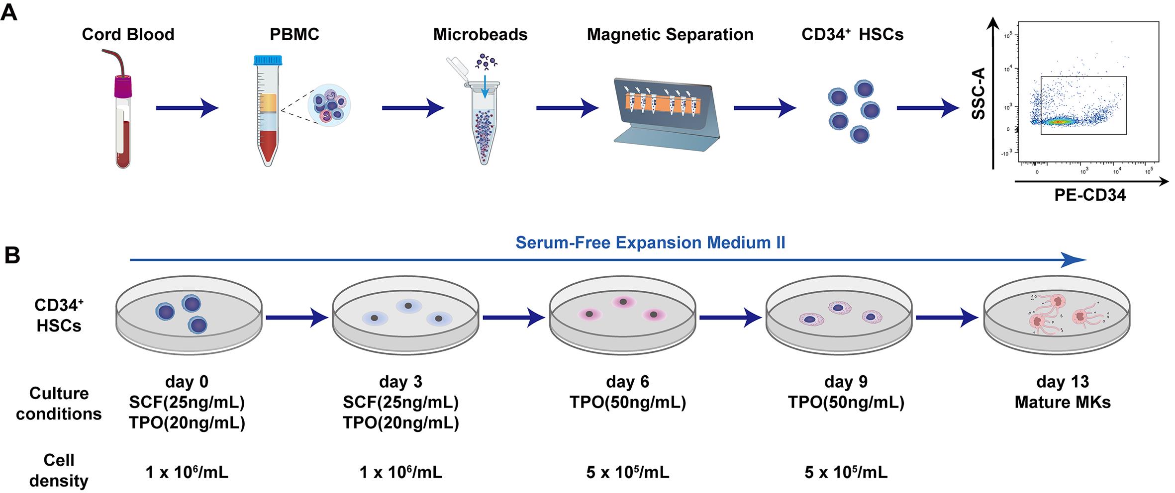
Schematic of human umbilical cord blood–derived CD34+ hematopoietic stem cell (HSC) isolation (A) and directed differentiation into mature megakaryocytes (B)
Background
Megakaryocytes are the precursor cells of platelets. Platelets are indispensable for hemostasis and tissue repair, and their clinical demand is enormous. However, reliance on donor-derived platelets faces challenges such as supply shortages and short storage periods. Therefore, in vitro generation of megakaryocytes and platelets has become a key direction in transfusion medicine and regenerative medicine [1]. Common sources of hematopoietic stem cells include bone marrow, peripheral blood, and umbilical cord blood. Bone marrow is a classic source, but its collection is invasive, and the quality of cells is influenced by donor age and health status. Under homeostatic conditions, only a very small number of stem cells circulate in peripheral blood [2]. Hematopoietic stem cells (HSCs) reside within the bone-marrow niche, where they are regulated by a complex network of cellular components [3]. Meanwhile, hematopoietic stem cells are capable of trafficking between bone marrow and peripheral blood. Accordingly, stem cell mobilization can be achieved via specific pharmacological agents or interventions, which displace hematopoietic stem and progenitor cells (HSPCs) from the bone-marrow niche into the peripheral circulation. Sufficient CD34+ cells can then be collected by leukapheresis [4]. Clinically, the most frequently applied mobilization regimens include granulocyte colony-stimulating factor (G-CSF) monotherapy or its combination with plerixafor (AMD3100) [4]. In addition, cyclophosphamide (CY)-based chemotherapy-assisted mobilization represents a classic strategy: cyclophosphamide is administered on day 1, followed by G-CSF treatment from day 2 to day 11, and leukapheresis is initiated on day 12. Nevertheless, multiple factors may affect CD34+ cell collection efficiency, including donor demographics (age, sex, body weight), baseline blood cell counts, and the selection of the mobilization regimen. In contrast, umbilical cord blood can be collected non-invasively and is rich in CD34+ cells, making it an ideal starting material for in vitro megakaryocytic differentiation [5].
This protocol describes in detail the isolation of CD34+ hematopoietic stem cells from human cord blood using magnetic bead separation technology and the induction of these hematopoietic stem cells into mature megakaryocytes [1,6,7]. Using magnetic bead enrichment, high-purity CD34+ cells are efficiently obtained from cord blood mononuclear cells and then directed toward megakaryocytic differentiation in serum-free medium supplemented with only thrombopoietin (TPO) and stem cell factor (SCF). As core cytokines governing hematopoiesis, SCF and TPO exert complementary and synergistic pivotal functions in the HSC-to-MK induction system. SCF delivers basal signals supporting the proliferation and self-renewal of hematopoietic stem and progenitor cells, whereas TPO drives lineage commitment and maturation toward the megakaryocytic lineage. At the signaling level, their synergistic effect manifests as enhanced and prolonged activation of pathways such as JAK2-STAT5. Functionally, this synergy drives massive expansion of megakaryocyte progenitors for subsequent megakaryocyte differentiation [8–10]. Compared with existing methods, this protocol provides more detailed operational steps and offers compatibility with various downstream analyses, including flow cytometry, qPCR, and platelet morphology. At the application level, this protocol can be used not only for basic research but also to provide an alternative source for platelet transfusion. Clinical trials based on cord blood–derived megakaryocyte progenitors have already shown good tolerability [5,11–12]. Furthermore, it can be used to construct models of inherited platelet disorders or serve as a platform for screening candidate drugs for hematological toxicity.
To address the technical limitations of previously reported megakaryocyte induction systems, we established an optimized two-stage gradient cytokine differentiation protocol modified from existing culture workflows. Published differentiation protocols either employ complex multi-cytokine cocktails that increase experimental batch variability or rely solely on TPO, which restricts early HSPC expansion. Our simplified SCF/TPO dual-factor culture system retains robust megakaryocyte terminal maturation efficiency while minimizing reagent-induced variation. More importantly, we established a standardized double-round MACS enrichment workflow; this purification strategy consistently yields CD34+ cell purity above 90%, which markedly outperforms the 60%–70% purity obtained by single-column separation in reference protocols [13]. Beyond cytokine and enrichment optimizations, continuous multi-time-point sampling for dynamic differentiation tracking and standardized post-staining antibody washing steps are integrated into our complete workflow to resolve the deficiencies of prior protocols, including insufficient CD34 purity control and non-standardized flow cytometry staining procedures [14–16].
Materials and reagents
Biological materials
1. Human umbilical cord blood (UCB): Fresh human umbilical cord blood collected from full-term deliveries
Reagents
1. Ficoll-PaqueTM PLUS (Cytiva, catalog number: 17144002)
2. AutoMACS rinsing solution (Miltenyi Biotec, catalog number: 130-091-222)
3. MACS BSA stock solution (Miltenyi Biotec, catalog number: 130-091-376)
4. CD34 MicroBead kit, human (Miltenyi Biotec, catalog number: 130-046-702)
5. Penicillin/streptomycin (Invitrogen, catalog number: 15140-122)
6. 10 μg recombinant human TPO (Peprotech, catalog number: 300-18)
7. 10 μg recombinant human SCF (Peprotech, catalog number: 300-07)
8. Phosphate-buffered saline (PBS) (without Ca2+ and Mg2+) (VivaCell BIOSCIENCES, catalog number: C3580-0500)
9. Fetal bovine serum (FBS) (Gibco, catalog number: A3161001C)
10. FcR blocking reagent, human (Miltenyi Biotec, catalog number: 130-059-901)
11. Dimethyl sulfoxide (DMSO) (Sigma-Aldrich, catalog number: D2650)
12. DMEM (Gibco, catalog number: C11995500BT)
13. EDTA, 0.5 M, pH 8.0 (Beyotime, catalog number: ST066)
14. APC mouse anti-human CD41a antibody (clone: HIP8) (BD Biosciences, catalog number: 559777)
15. PE mouse anti-human CD61 antibody (clone: VI-PL2) (BD Biosciences, catalog number: 555754)
16. PE mouse anti-human CD42a antibody (clone: ALMA.16) (BD Biosciences, catalog number: 558819)
17. PE mouse anti-human CD42b antibody (clone: HIP1) (BD Biosciences, catalog number: 555473)
18. APC mouse IgG1 κ isotype control (clone: MOPC-21) (BD Biosciences, catalog number: 555751)
19. PE mouse IgG1 κ isotype control (clone: MOPC-21) (BD Biosciences, catalog number: 555749)
20. PE mouse anti-human CD34 antibody (clone: 581) (BD Biosciences, catalog number: 555822)
21. Cell staining buffer (Biolegend, catalog number: 420201)
22. Trypan Blue stain, 0.4% (Gibco, catalog number: 15250-061)
23. 75% ethanol (LIRCON, 500 mL)
24. StemSpanTM SFEM II (Stem Cell Technologies, catalog number: 09605)
Solutions
1. Buffer 1 (see Recipes)
2. Buffer 2 (see Recipes)
3. Cell cryopreservation medium (see Recipes)
4. DMEM + 10% FBS (see Recipes)
5. 25 μg/mL SCF (see Recipes)
6. 20 μg/mL TPO (see Recipes)
7. SFEM (see Recipes)
Recipes
1. Buffer 1
| Reagent | Final concentration | Volume |
|---|---|---|
| PBS | n/a | 488 mL |
| FBS | 2% | 10 mL |
| EDTA | 2 mM | 2 mL |
| Total | n/a | 500 mL |
After buffer 1 is prepared, store it at 4 °C.
2. Buffer 2
| Reagent | Final concentration | Quantity or volume |
|---|---|---|
| AutoMACS rinsing solution | n/a | 100 mL |
| MACS BSA stock solution | n/a | 5 mL |
| Total | n/a | 105 mL |
After Buffer 2 is prepared, filter through a 0.22 μm membrane, store at 4 °C protected from light, and do not warm to room temperature (RT) before use.
3. Cell cryopreservation medium
| Reagent | Final concentration | Quantity or volume |
|---|---|---|
| DMEM | 75% | 750 μL |
| FBS | 15% | 150 μL |
| DMSO | 10% | 100 μL |
| Total | n/a | 1 mL |
For every 5 × 10⁵ cells, use 1 mL of cryopreservation medium (prepare fresh and use immediately).
4. DMEM + 10% FBS
| Reagent | Final concentration | Quantity or volume |
|---|---|---|
| DMEM | 89% | 89 mL |
| FBS | 10% | 10 mL |
| Penicillin-streptomycin | 1% | 1 mL |
| Total | n/a | 100 mL |
Store the prepared complete medium at 4 °C and use within 2–4 weeks. Allow the medium to equilibrate to RT prior to experimental use.
5. 25 μg/mL SCF
| Reagent | Final concentration | Quantity or volume |
|---|---|---|
| SCF | 25 μg/mL | 10 μg |
| ddH2O | n/a | 0.4 mL |
| Total | n/a | 0.4 mL |
Prepare single-use 10 μL aliquots of working stocks. Store at -80 °C, avoid repeated freeze-thaw cycles, and use within 6 months after preparation.
6. 20 μg/mL TPO
| Reagent | Final concentration | Quantity or volume |
|---|---|---|
| TPO | 20 μg/mL | 10 μg |
| ddH2O | n/a | 0.5 mL |
| Total | n/a | 0.5 mL |
Prepare single-use 10 μL aliquots of working stocks. Store at -80 °C, avoid repeated freeze-thaw cycles, and use within 6 months after preparation.
7. SFEM
| Reagent | Final concentration | Quantity or volume |
|---|---|---|
| StemSpanTM SFEM II | 90% | 99 mL |
| Penicillin-streptomycin | 1% | 1 mL |
| Total | n/a | 100 mL |
Store the prepared medium at 4 °C and use within 2–4 weeks. Allow the medium to equilibrate to RT prior to experimental use.
Note: All commercial reagents shall be stored according to the manufacturer’s instructions and used before the manufacturer-indicated expiry date. For opened commercial reagents and laboratory-prepared cytokine working stocks, an additional in-house recommended shelf-life is specified. Do not use any reagent beyond its expiry or recommended usage period.
Laboratory supplies
1. 3 mL Pasteur pipette (NEST, catalog number: 318314)
2. 12-well cell culture plate (NEST, catalog number: 712001)
3. 24-well cell culture plate (NEST, catalog number: 702001)
4. 15-mL centrifuge tube (NEST, catalog number: 601052)
5. 50-mL centrifuge tube (NEST, catalog number: 602052)
6. FACS tubes 5 mL (Falcon, catalog number: 352054)
7. 1.5 mL Eppendorf tubes (Axygen, catalog number: MCT-150-CS)
8. 0.22 μm filter (Millipore, catalog number: 431229)
9. Hemocytometer (Countstar, catalog number: C0010101)
10. 2 mL cryogenic vials (Corning, catalog number: 430659)
11. 25 mL pipette (BeyoGold, catalog number: FPIP125)
12. μ-dish 35 mm, high (ibidi, catalog number: 81156)
13. 10 mL BD Vacutainer® K2EDTA blood collection tubes (BD, catalog number: 367525)
14. MS columns (Miltenyi Biotec, catalog number: 130-042-201)
15. LS columns (Miltenyi Biotec, catalog number: 130-042-401)
16. Cell strainers (BeyoGold, catalog number: FSTR070)
17. RNase-free pipette tips (10 μL, 200 μL, 1 mL) (Axygen, catalog numbers: AXY-TF-300-R-S, TF-200, TF-1000)
Equipment
1. Biosafety cabinet, BL2 level (Haier, model: HR1200-IIA2)
2. CO2 cell incubator (37 °C, 5% CO2) (Thermo Fisher Scientific, model: 3111)
3. Benchtop centrifuge (Xiangyi, model: L5-35R)
4. Automated cell counter (Countstar, model: IC1000)
5. 4 °C refrigerator (Haier, model: HYC-940C)
6. -80 °C laboratory freezer (Thermo Fisher Scientific, model: 994)
7. Liquid nitrogen storage tank (Haier, model: YDS-115-216-FZ)
8. Inverted microscope (Olympus, model: IX73)
9. Flow cytometer (BD, model: FACSCantoII)
10. Motorized pipette filler (Thermo Fisher Scientific, model: S1)
11. Water bath (Bluepard, model: HWS-12)
12. MiniMACS separator (Miltenyi Biotec, catalog number: 130-042-102/120-060-068)
13. Adjustable-volume pipette (1–10 μL, 20–200 μL, 100–1,000 μL) (Thermo ScientificTM FinnpipetteTM F1, model: 4641040N, 4641080N, 4641100N)
14. Cell freezing container (Corning, model: 432002)
Procedure
A. Isolation of human cord blood CD34+ cells
1. Prewarm buffer 1 and Ficoll-PaqueTM PLUS to RT in the biological safety cabinet for 30 min.
2. Collect human umbilical cord blood in 10 mL BD Vacutainer® K2EDTA blood collection tubes. Process the blood sample immediately after collection.
Pause point: If the collected cord blood cannot be processed promptly, store it in a refrigerator at 4 °C for no longer than 24 h.
3. Dilute the cord blood sample with an equal volume (1:1) of buffer 1 and mix gently.
4. Add 7.5 mL of Ficoll-PaqueTM PLUS to the bottom of a 50 mL centrifuge tube. Using a motorized pipette filler, slowly overlay 15 mL of diluted cord blood onto the surface of the Ficoll-PaqueTM PLUS medium along the inner wall of the tube. The combined volume of Ficoll-PaqueTM PLUS and diluted cord blood should reach 22.5 mL. Avoid disturbing the interface between the blood and the density-gradient medium during overlaying (Figure 1A).
Critical: Ensure that all centrifuge tubes are well-balanced before centrifugation. Proper tube balancing is essential to achieve distinct cell-layer stratification after centrifugation.
Caution: When adding the diluted cord blood, pipette gently and slowly to avoid the blood penetrating into the Ficoll-PaqueTM PLUS layer. Avoid shaking when holding the pipette and centrifuge tube; set the ejection speed of the motorized pipette filler to the lowest level.
5. Centrifuge blood at 383× g (1,390 rpm) for 30 min (acceleration 1, brake 0) at 20 °C.
6. After centrifugation, the 50 mL centrifuge tube will contain four layers from top to bottom: plasma layer, mononuclear cell layer (buffy coat), Ficoll-PaqueTM PLUS layer, and erythrocyte layer (Figure 1B). First, use a Pasteur pipette to remove the plasma layer. Then, using a 1 mL pipette, gently rotate along the tube wall to aspirate the buffy coat cells and transfer them to a new 50 mL centrifuge tube. If multiple separation tubes are used, do not pool the buffy coat layers; instead, transfer each to its own new 50 mL centrifuge tube.
Note: Be careful not to aspirate the Ficoll-PaqueTM PLUS. The buffy coat may contain some red blood cells; try to minimize their collection. The total volume of the aspirated buffy coat is approximately 2–3 mL.
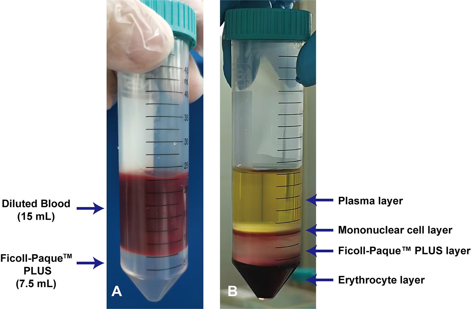
Figure 1. Before and after comparison of mononuclear cell isolation using Ficoll-PaqueTM PLUS. (A) Diluted umbilical cord blood is carefully layered onto Ficoll-PaqueTM PLUS, taking care to avoid mixing. Red blood cell precipitation may be observed upon excessive standing. (B) Distinct layers formed after centrifugation. The buffy-coat layer containing mononuclear cells is harvested for CD34+ cell enrichment. In umbilical cord blood samples, red blood cell contamination within the leukocyte layer, clumpy appearance of the leukocyte layer, or reddish discoloration of the plasma layer caused by erythrocyte lysis are considered normal phenomena.
7. Add buffer 1 to the isolated buffy coat to a final volume of 50 mL, mix thoroughly, and centrifuge at 285× g (1,200 rpm) for 10 min (acceleration 9, brake 9) at 20 °C. Discard the supernatant.
8. Resuspend the cell pellet in each 50 mL tube with 2 mL of buffer 1. Pool all suspensions into a single new 50 mL centrifuge tube. Rinse the original tubes with 1 mL of buffer 1 and transfer the wash to the collection tube. Mix the cells thoroughly and count the cells using an automated cell counter following Trypan Blue staining. Bring the volume to 50 mL with buffer 1, then centrifuge at 190× g (980 rpm) for 10 min (acceleration 9, brake 9) at 20 °C. Discard the supernatant. Resuspend the resulting cell pellet in an appropriate volume of buffer 2 according to the total cell number.
9. Transfer an aliquot containing 1 × 10⁸ cells to a 1.5 mL Eppendorf tube. Centrifuge at 190× g (980 rpm) for 10 min (acceleration 9, brake 9) at 20 °C, discard the supernatant, and resuspend the cell pellet in 300 μL of buffer 2. Add 100 μL of FcR blocking reagent. Mix thoroughly and incubate at 4 °C for 10 min in the dark.
10. Add 100 μL of CD34 MicroBeads, mix well, and incubate at 4 °C for 30 min in the dark.
11. Transfer the cell suspension from step A10 (pooling multiple incubation tubes if applicable) to a new 50 mL centrifuge tube. Bring the volume to 50 mL with buffer 2, gently mix, and centrifuge at 285× g (1,200 rpm) for 10 min (acceleration 9, brake 9) at 4 °C. Discard the supernatant.
12. Place the separation column (choose an LS or MS column based on the total cell number) on the magnetic stand. Place a 50 mL centrifuge tube under the column to collect the flowthrough. Equilibrate the column with 0.5 mL of buffer 2 for MS columns or 1 mL of buffer 2 for LS columns. Ensure the column reservoir is completely empty before proceeding to the next step (Figure 2A).
Note: LS columns can bind up to 1 × 10⁸ magnetically labeled cells and accommodate up to 2 × 10⁹ total cells. MS columns can bind up to 1 × 10⁷ magnetically labeled cells and accommodate up to 2 × 10⁸ total cells.
13. When the buffer in the column from step A12 has just run dry, apply the cell suspension in 1 mL aliquots for LS columns or 0.5 mL aliquots for MS columns for magnetic separation (Figure 2B, C).
14. After the cell suspension has run through, wash the column three times with buffer 2 (0.5 mL × 3 for MS columns; 1 mL × 3 for LS columns) (Figure 2D).
15. Remove the column from the magnet and place it on a new 15 mL centrifuge tube. Add the appropriate volume of buffer 2 (MS: 1 mL; LS: 2 mL) to the column and immediately flush out the magnetically labeled cells by firmly pushing the plunger into the column (Figure 2E).
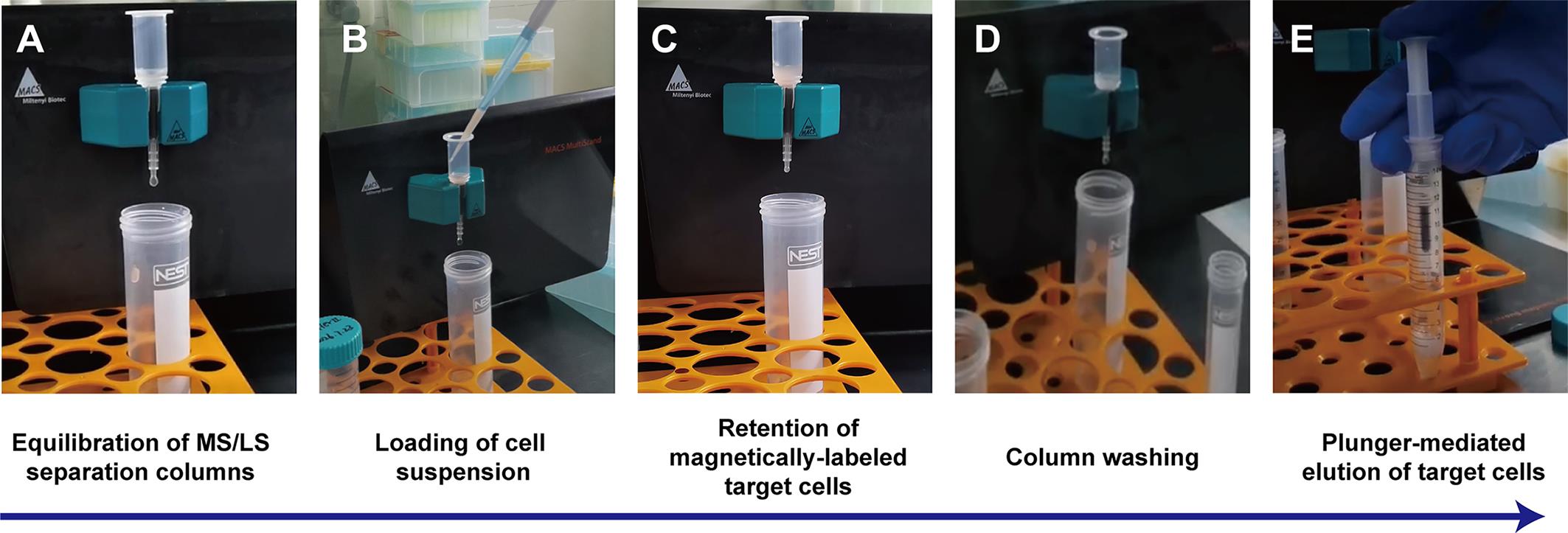
Figure 2. Operational workflow of magnetic cell separation using MS/LS separation columns. (A) Equilibration of MS or LS separation column with buffer 2 fixed on the magnetic stand. (B) Loading of cell suspension onto the preequilibrated separation column by pipette. (C) Magnetically labeled target cells are retained inside the column under a magnetic field while unlabeled cells flow through. (D) Column washing with buffer 2 to thoroughly remove residual non-target cells. (E) The column is detached from the magnet, and target cells are eluted completely by pushing the matched plunger. The procedures shown in Figure 2 are repeated with a brand-new separation column to finish the second round of magnetic enrichment, followed by Trypan Blue staining and cell counting to detect cell concentration and viability.
16. Take a new separation column and equilibrate it with buffer 2. Load the cell suspension obtained from step A15 in 1 mL aliquots for LS columns or 0.5 mL aliquots for MS columns, then wash the column with 3 mL of buffer 2 for LS columns or 1 mL of buffer 2 for MS columns to complete the second round of magnetic separation.
17. Remove the column from the magnet and place it on a new 15 mL centrifuge tube. Add the appropriate volume of buffer 2 (MS: 1 mL; LS: 2 mL) to the column and immediately flush out the magnetically labeled cells by firmly pushing the plunger. Mix 10 μL of cell suspension with 10 μL of Trypan Blue solution, load the mixture into the counting slide, and determine cell concentration and viability using an automated cell counter.
18. Dispense 1 × 10⁵ cells per tube into two 1.5 mL Eppendorf tubes (labeled PE-IgG and PE-CD34+, respectively) for analysis by flow cytometry. Add 0.1 test (one-tenth of the standard “1 test” antibody dose, where 1 test is defined as the amount sufficient to label 1 × 10⁶ cells, matching 1 × 10⁵ cells per tube in this experiment) of pre-diluted antibody (isotype control antibody is used at the same test dosage). Gently mix and incubate at room temperature in the dark for 30 min. Centrifuge the tubes at 198× g (1,000 rpm) for 7 min (acceleration 9, brake 9) and completely discard the supernatant to remove unbound excess antibody. Resuspend cells in 500 μL of PBS. Filter the cell suspension using a cell-strainer accessory and collect the filtrate into a 5 mL FACS tube.
Notes:
1. Isotype control staining was performed in parallel under identical experimental conditions to define background fluorescence levels. All flow cytometric gating thresholds for positive marker expression were strictly determined based on the isotype control signals to exclude nonspecific binding and ensure accurate population identification. Analyze CD34+ cell frequency by flow cytometry to validate magnetic bead enrichment (Figure 3).
2. QC metrics: Post-sort CD34+ cell purity: >90% (flow cytometry quantification) after two-round column-based MACS enrichment; single-column enrichment routinely achieves ~70% CD34+ purity.
3. Typical yield: Starting from a 50 mL diluted human umbilical cord blood sample, the yield of viable CD34+ cells ranges from 1.5 × 10⁶ to 2 × 10⁶ following two-round MACS enrichment.
Critical: Assess CD34+ purity by flow cytometry immediately after cell enrichment before proceeding to HSC differentiation culture. Proceed to downstream culture only when CD34+ cell purity exceeds 90% following two-round MACS enrichment.
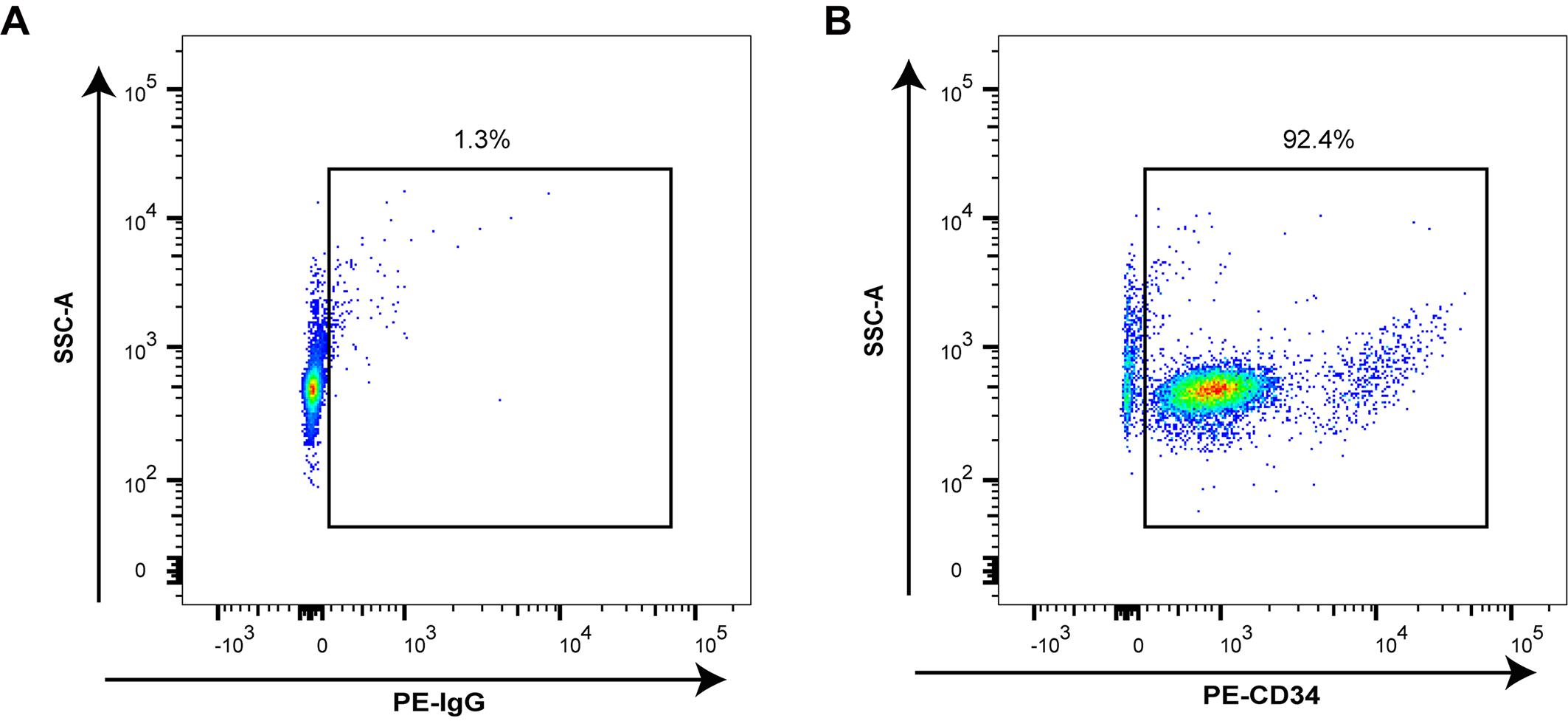
Figure 3. Percentage of CD34+ cells isolated from human umbilical cord blood. (A) Isotype control, PE-IgG-stained cells used to set PE+ cell gate and population. (B) Gated CD34+ cells.
19. Centrifuge the remaining enriched CD34+ cells from step A17 (after removing aliquots for flow cytometry analysis in step A18) at 285× g (1,200 rpm) for 10 min (acceleration 9, brake 9) at 4 °C, then discard the supernatant. The cell pellet is now ready for subsequent culture or cryopreservation.
Pause point: For cryopreservation: Resuspend 1 × 10⁶ cells in 1 mL of cell cryopreservation medium, then transfer the cell suspension into a cryovial. Place the cryovial inside a freezing container and store directly at -80 °C overnight. Transfer to liquid nitrogen for long-term storage after 24 h. The post-thaw cell viability is greater than 90%.
B. CD34+-derived primary MK differentiation
B1. Day 0
For cryopreserved cells, perform the following steps:
1. Rapidly thaw cryopreserved CD34+ cell cryovials by full immersion in a 37 °C water bath with gentle manual shaking until only a tiny trace of ice remains.
2. Remove the cryovials from the water bath and disinfect the surface by spraying with 75% ethanol.
3. Immediately transfer the thawed cell suspension into a 15 mL centrifuge tube pre-loaded with 9 mL of DMEM + 10% FBS.
4. Centrifuge the cell suspension at 285× g (1,200 rpm) for 7 min (acceleration 9, brake 9) at RT. Discard the supernatant after centrifugation.
5. Resuspend 1 × 10⁶ CD34+ hematopoietic stem cells in 1 mL of StemSpanTM SFEM medium containing 1% penicillin-streptomycin, 25 ng/mL SCF, and 20 ng/mL TPO (Table 1).
Table 1. Stage-specific cytokine and medium scheme for HSC megakaryocytic differentiation
| Culture time window | Medium type | Cytokine supplementation | Working concentration | Experimental purpose |
|---|---|---|---|---|
| Days 0–3 | SFEM | SCF + TPO | SCF: 25 ng/mL; TPO: 20 ng/mL | Maintain the survival and proliferation of primary human umbilical cord blood–derived CD34+ HSCs and initiate megakaryocyte lineage commitment |
| Days 3–6 | SFEM | SCF + TPO | SCF: 25 ng/mL; TPO: 20 ng/mL | Sustain continuous differentiation stimulation, support early megakaryocyte proliferation and lineage maturation |
| Days 6–9 | SFEM | TPO | TPO: 50 ng/mL | Promote maturation of megakaryocytes and stabilize the mature megakaryocyte phenotype |
| Days 9–13 | SFEM | TPO | TPO: 50 ng/mL | Support complete terminal maturation of megakaryocytes and facilitate proplatelet formation. |
6. Add 1 mL of the suspension to one well of a 12-well cell culture plate.
7. Culture for three days in a CO2 incubator at 37 °C.
B2. Day 3
1. Collect hematopoietic stem cells after three days of culture and transfer the cell suspension into a 15 mL centrifuge tube.
2. Rinse the 12-well plate with 1 mL of sterile PBS to collect residual cells and transfer all the washing medium into the same 15 mL centrifuge tube.
3. Centrifuge the cell suspension at 198× g (1,000 rpm) for 7 min (acceleration 9, brake 9). Discard the supernatant.
4. Gently resuspend the cell pellet in 1 mL of sterile PBS and count the cells using a hemocytometer.
5. Centrifuge the cell suspension again at 198× g (1,000 rpm) for 7 min (acceleration 9, brake 9) and discard the supernatant.
6. Resuspend 1 × 10⁶ cells in 1 mL of StemSpanTM SFEM medium supplemented with 1% penicillin-streptomycin in a 15 mL centrifuge tube, mix thoroughly, and seed 1 mL of cell suspension per well into a 12-well cell culture plate.
7. Add 1 μL of 25 μg/mL SCF and 1 μL of 20 μg/mL TPO to each well to achieve final concentrations of 25 ng/mL SCF and 20 ng/mL TPO (Table 1). Continuously culture the cells for another 3 days in a 37 °C, 5% CO2 incubator.
B3. Day 6
1. Collect hematopoietic stem cells after six days of culture and transfer the cell suspension into a 15 mL centrifuge tube.
2. Rinse the 12-well plate with 1 mL of sterile PBS to recover residual cells and transfer the washing solution to the same 15 mL tube.
3. Centrifuge at 198× g (1,000 rpm) for 7 min (acceleration 9, brake 9) and discard the supernatant.
4. Gently resuspend the cell pellet in 1 mL of sterile PBS for cell counting.
5. Perform a second centrifugation at 198× g (1,000 rpm) for 7 min (acceleration 9, brake 9), then discard the supernatant.
6. Resuspend 5 × 10⁵ cells in 1 mL of StemSpanTM SFEM containing 1% penicillin-streptomycin, mix well, and seed 1 mL suspension per well in a 12-well plate.
7. Add 2.5 μL of 20 μg/mL TPO to each well to obtain a final TPO concentration of 50 ng/mL (Table 1). Culture the cells for 3 days in a 37 °C, 5% CO2 incubator.
8. Label four 1.5 mL microcentrifuge tubes (1 isotype-control tube and 3 experimental tubes); aliquot 1 × 10⁵ day 6–cultured hematopoietic stem cells into each tube.
9. Centrifuge the tubes at 198× g (1,000 rpm) for 7 min (acceleration 9, brake 9) and aspirate the supernatant completely.
10. Add 100 μL of cell staining buffer to each tube and gently resuspend the cells.
11. To the isotype-control tube, add 1 μL of APC-IgG antibody and 1 μL of PE-IgG antibody and mix well.
12. To each of the three experimental tubes, add 1 μL of APC-CD41a antibody together with 1 μL of PE-CD61 antibody. The antibody combinations of APC-CD41a/PE-CD42a and APC-CD41a/PE-CD42b are prepared in the same manner.
13. Incubate at room temperature in the dark for 30 min. Centrifuge the tubes at 198× g (1,000 rpm) for 7 min (acceleration 9, brake 9) and completely discard the supernatant to remove unbound excess antibody. Resuspend the cell pellet in 500 μL of PBS for flow cytometric analysis (Figure 4).
Note: Expected flow cytometry observations at day 6 of differentiation: At this intermediate time point, the proportion of CD41a+CD61+ double-positive cells is relatively low, indicating successful initiation of megakaryocyte lineage differentiation. Of note, CD42a and CD42b are also expressed at low levels on day 6. These are late-stage maturation markers that are progressively upregulated during subsequent culture phases.
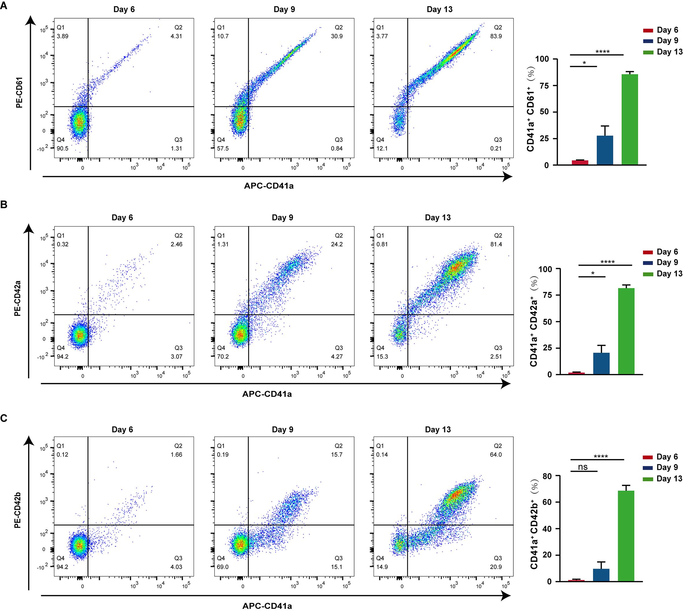
Figure 4. Megakaryocyte cultures generated from human umbilical cord blood CD34+ cells. (A–C) Representative flow cytometry dot plots of cultured cells on days 6, 9, and 13. (A) Dual staining results and statistical analysis of cultured cells with PE-labeled anti-CD61 and APC-labeled anti-CD41a antibodies (n = 3). (B) Dual staining results and statistical analysis of cultured cells with PE-labeled anti-CD42a and APC-labeled anti-CD41a antibodies (n = 3). (C) Dual staining results and statistical analysis of cultured cells with PE-labeled anti-CD42b and APC-labeled anti-CD41a antibodies (n = 3). IgG isotype controls for CD61, CD41a, CD42a, and CD42b were used to determine the background. Statistical analysis for two-group comparisons was carried out using the unpaired two-tailed Student’s t-test. Statistical significance: *P < 0.05, **P < 0.01, ***P < 0.001; ns = not significant.
B4. Day 9
1. Collect hematopoietic stem cells after 9 days of culture and transfer the cell suspension into a 15 mL centrifuge tube.
2. Rinse the 12-well plate with 1 mL of sterile PBS to collect residual cells and combine the rinse with the cell suspension in the same tube.
3. Centrifuge at 198× g (1,000 rpm) for 7 min (acceleration 9, brake 9), discard the supernatant, and resuspend the cells in 1 mL of PBS for cell counting.
4. Centrifuge the cell suspension again under the same conditions and discard the supernatant.
5. Resuspend 5 × 10⁵ cells in 1 mL of StemSpanTM SFEM supplemented with 1% penicillin-streptomycin, mix well, and seed 1 mL per well in a 12-well plate.
6. Add 2.5 μL of 20 μg/mL TPO to each well (final concentration of 50 ng/mL TPO) and culture the cells in a 37 °C, 5% CO2 incubator for another four days (Table 1).
7. Proplatelet observation culture: Collect day 9–induced cells, adjust the cell density to 1 × 10⁵ cells/mL in 500 μL of StemSpanTM SFEM, and seed the cell suspension into the center of a 35 mm high μ-dish. Add 1.25 μL of 20 μg/mL TPO to reach a final concentration of 50 ng/mL TPO and incubate for 4 days in a 37 °C, 5% CO2 incubator.
8. Label four 1.5 mL microcentrifuge tubes and transfer 1 × 10⁵ day 9–cultured cells into each tube.
9. Centrifuge at 198× g (1,000 rpm) for 7 min (acceleration 9, brake 9) and remove the supernatant.
10. Resuspend the cell pellet in 100 μL of cell staining buffer.
11. Add 1 μL of APC-IgG and 1 μL of PE-IgG antibodies for isotype control staining.
12. Add antibody combinations of APC-CD41a/PE-CD61, APC-CD41a/PE-CD42a, and APC-CD41a/PE-CD42b to the corresponding experimental tubes separately.
13. Incubate at room temperature in darkness for 30 min. Centrifuge the tubes at 198× g (1,000 rpm) for 7 min (acceleration 9, brake 9) and discard the supernatant to wash away residual unbound antibody. Resuspend cells in 500 μL of PBS and prepare for flow cytometric analysis (Figure 4).
Note: Expected flow cytometry observations at day 9 of differentiation: At this mid-late differentiation stage, the proportion of CD41a+CD61+ double-positive cells reaches approximately 30%, indicating sustained and enhanced megakaryocyte-lineage differentiation. Importantly, the late-stage maturation markers CD42a and CD42b start to be markedly upregulated on day 9. The percentage of CD41a+CD42a+ double-positive cells is around 20%, whereas CD41a+CD42b+ double-positive cells account for approximately 10%, reflecting progressive maturation of differentiating megakaryocytes.
B5. Day 13
1. Collect hematopoietic stem cells after 13 days of culture and transfer the cell suspension into a 15 mL centrifuge tube.
2. Rinse the culture plate with 1 mL of sterile PBS and combine all residual cells into the same 15 mL tube.
3. Centrifuge at 198× g (1,000 rpm) for 8 min (acceleration 9, brake 0), discard the supernatant, and gently resuspend the cells in 1 mL of PBS for cell counting.
4. Label four 1.5 mL microcentrifuge tubes and dispense 1 × 10⁵ day 13–cultured cells into each tube.
5. Centrifuge the tubes at 198× g (1,000 rpm) for 8 min (acceleration 9, brake 0) and aspirate the supernatant.
6. Resuspend each cell pellet in 100 μL of cell staining buffer.
7. Stain the control tube with 1 μL of APC-IgG and 1 μL of PE-IgG antibodies.
8. Stain experimental tubes with antibody combinations of APC-CD41a/PE-CD61, APC-CD41a/PE-CD42a, and APC-CD41a/PE-CD42b.
9. After 30 min of dark incubation at room temperature, centrifuge the tubes at 198× g (1,000 rpm) for 8 min (acceleration 9, brake 0) and discard the supernatant to remove excess unbound antibody. Resuspend the cell pellet in 500 μL of PBS for flow cytometric detection (Figure 4).
10. Cells are cultured until day 13, and proplatelets derived from day 9–cultured cells in the 35 mm high μ-dish are observed under a microscope (Figure 5).
Note: Expected flow cytometry observations at day 13 of differentiation: At this mid-late differentiation stage, the proportion of CD41a+CD61+ double-positive cells reaches approximately 80%, indicating sustained and enhanced megakaryocyte-lineage differentiation. Importantly, the late-stage maturation markers CD42a and CD42b remain markedly upregulated on day 13. The percentage of CD41a+CD42a+ double-positive cells is around 80%, while CD41a+CD42b+ double-positive cells account for approximately 60%, reflecting the terminal maturation of differentiating megakaryocytes. In addition, proplatelets can be observed under the microscope on day 13. Observation of proplatelets by light microscopy allows users to visually assess the terminal maturation status of megakaryocytes. Proplatelet formation represents a key functional hallmark of mature megakaryocytes capable of platelet release. Mature megakaryocytes and proplatelet-bearing cells are highly fragile and not recommended for cryopreservation, as freezing-thawing procedures severely impair cell viability and proplatelet-forming capacity. At the end of this protocol, harvested cells can be used for multiple downstream assays, including flow cytometric phenotyping, qPCR, immunofluorescence staining, transcriptomic sequencing, in vitro platelet functional assays, and co-culture and in vivo transplantation studies.
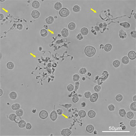
Figure 5. Representative images of proplatelet formation in megakaryocytes (MKs). Yellow arrows indicate MKs with proplatelets. Scale bar, 50 μm.
Data analysis
1. Statistical analysis
All experiments were performed with a minimum of three independent biological replicates to ensure the stability and reproducibility of the results. All flow cytometry data were first subjected to normality and lognormality tests to verify normal distribution. F-tests were performed to examine the homogeneity of variances between groups. Two-group comparisons were carried out using an unpaired two-tailed Student’s t-test: day 6 vs. day 9, and day 6 vs. day 13.
2. Flow cytometry gating strategy
Single-stained compensation tubes (PE-CD42a, APC-CD41a) were measured once to establish a fixed compensation template, which was adopted for all subsequent experimental batches. Isotype-matched IgG controls were applied to define positive-signal thresholds for each surface marker in every experiment. Gate P1 encompassed all acquired cellular events, and debris was not pre-excluded. The fluorescence intensity distribution of isotype-control samples within gate P1 was used to set quadrant boundaries for CD41a, CD61, CD42a, and CD42b; quadrant lines were adjusted so that ≤1% of events fell within positive quadrants for the isotype control. These identical quadrant boundaries were then applied to fully stained experimental samples. Representative dot plots illustrating this gating workflow are shown in Supplementary Figure 1.
3. Interpretation of megakaryocyte lineage marker dynamics
CD41a, CD61, CD42a, and CD42b are pivotal cell-surface markers tracing human megakaryocyte development. CD41a (integrin αIIb) and CD61 (integrin β3) assemble into the fibrinogen-receptor complex GPIIb/IIIa, representing early-stage markers activated upon megakaryocyte lineage commitment. In our two-color flow cytometry analyses, CD41a+CD61+ double-positive cells gradually increase from day 6 to day 13, reflecting the progressive commitment of cord blood–derived CD34+ progenitors toward the megakaryocyte lineage.
CD42a and CD42b are functional late-maturation markers of megakaryocytes. At day 6 of differentiation, CD41a+CD42a+ and CD41a+CD42b+ populations remain scarce, which is a physiological feature of early-phase megakaryocyte priming. Along with differentiation progression toward day 9, CD42a and CD42b are markedly upregulated in subsets of CD41a-positive cells, marking the transition from immature to maturing megakaryocytes. By day 13, CD41a+CD42a+ and CD41a+CD42b+ double-positive proportions rise substantially, accompanied by the appearance of proplatelet-forming structures under microscopy; these observations indicate the acquisition of terminal maturation and platelet-biogenesis potential.
It should be noted that our marker profiling relies exclusively on pairwise two-color flow cytometry. High fractions of CD41a+CD61+, CD41a+CD42a+, and CD41a+CD42b+ cells at day 13 collectively support robust megakaryocyte differentiation within the culture system. Nevertheless, due to the lack of a suitable multicolor antibody panel, we cannot formally verify whether all four markers are simultaneously expressed on individual single cells.
Validation of protocol
This protocol (or parts of it) has been used and validated in the following research article(s):
• Wu et al. [17]. circFUT8 promotes proplatelet formation by interacting with IGF2BP2 and stabilizing TNS1 mRNA in megakaryocytes. Blood (Figure S1A–E).
General notes and troubleshooting
Troubleshooting
Problem 1: The yield of CD34+ cells obtained from fewer than 30 mL of fresh cord blood is not adequate for the subsequent induced differentiation assays. From 30 mL of cord blood, approximately 6–8 × 10⁵ CD34+ cells are generally obtained.
Possible cause: The CD34+ yield varies between individual blood donors.
Solution: Increase the volume of the blood sample.
Problem 2: Low percentage of CD41a+ CD61+ double-positive cells at days 6, 9, or 13 during megakaryocyte differentiation.
Possible causes: Impaired quality of initial CD34+-enriched cells, loss of bioactivity of key cytokines (SCF and TPO), or expired culture medium, which hinders normal megakaryocyte lineage differentiation.
Solutions: Check the purity and viability of starting CD34+-enriched cells before culture; confirm the bioactivity of SCF and TPO working stocks and avoid repeated freeze-thaw cycles of cytokines; verify that the culture medium is within its recommended shelf-life before use.
Problem 3: Poor cell viability after cell antibody staining for flow cytometry detection.
Possible causes: Excessive mechanical shear stress during pipetting, non-low-temperature staining operation, or over-centrifugation during the staining process, resulting in cell damage and death.
Solutions: Reduce mechanical shear stress during cell pipetting and handle cells gently; perform the entire antibody staining workflow on ice to maintain cell activity; avoid over-centrifugation during cell washing and staining steps.
Problem 4: Unexpected extensive cell death occurs at the early stage of differentiation prior to days 6, 9, or 13.
Possible causes: Inappropriate initial cell-seeding density, microbial contamination in the culture system, or inaccurate incubator parameters (temperature and CO2 concentration), leading to abnormal cell growth and massive cell death.
Solutions: Confirm and adjust the optimal initial cell seeding density strictly according to the protocol; routinely screen for microbial contamination of culture medium and cells before and during culture; calibrate incubator temperature and CO2 concentration settings regularly to ensure stable culture conditions.
Problem 5: Near-undetectable CD41a+CD42a+ and CD41a+CD42b+ cell populations at day 6 of differentiation.
Possible cause: This phenomenon is a normal physiological characteristic of early-stage megakaryocyte differentiation, rather than improper experimental operation or experimental failure. Late maturation markers of megakaryocytes are not yet expressed at the early differentiation stage.
Solution: Continue the cell culture following the standard protocol until day 9 or day 13. The robust expression of late-stage megakaryocyte markers (CD42a and CD42b) and mature cell populations can be observed with the progression of differentiation.
Supplementary information
The following supporting information can be downloaded here:
1. Supplementary Figure 1. Representative flow cytometry gating strategy for CD41a and CD42a detection.
Acknowledgments
Funding sources that supported the work: New Chongqing Young Innovative Talents Project (CSTB2025YITP-QCRCX0040). The authors thank Xiuhui Zheng (Department of Gynecology and Obstetrics, Daping Hospital, Army Medical University) for technical assistance with human cord blood–derived CD34+ cell isolation. This protocol was used in [17]. The protocol for isolation of human cord blood CD34+ cells was adapted from [18].
Author contributions
Specific contributions of each author: Experiment design and data analysis, Huang Wu, Yahan Fan; Writing—Original Draft, Wenjun Xia; Writing—Review & Editing, Huang Wu; Experiments and data curation, Wenjun Xia, Zeqing Miao, Weiwei Zhang, Zhixia Liu.
Competing interests
The authors declare no competing financial interests.
Ethical considerations
The study was approved by the Ethics Committee of Daping Hospital, Army Medical University (Chongqing, China) [Approval No. Yiyanlun Shen (2021)-71]. All UCB samples were collected from eligible pregnant women who met the inclusion criteria and provided written informed consent. Sample acquisition, transportation, and experimental usage were performed in strict accordance with institutional guidelines.
References
- Bhatlekar, S., Manne, B. K., Basak, I., Edelstein, L. C., Tugolukova, E., Stoller, M. L., Cody, M. J., Morley, S. C., Nagalla, S., Weyrich, A. S., et al. (2020). miR-125a-5p regulates megakaryocyte proplatelet formation via the actin-bundling protein L-plastin. Blood. 136(15): 1760–1772. https://doi.org/10.1182/blood.2020005230
- Klein, G., Schmal, O. and Aicher, W. K. (2015). Matrix metalloproteinases in stem cell mobilization. Matrix Biol. 44–46: 175–183. https://doi.org/10.1016/j.matbio.2015.01.011
- Gao, X., Zhang, D., Xu, C., Li, H., Caron, K. M. and Frenette, P. S. (2020). Nociceptive nerves regulate haematopoietic stem cell mobilization. Nature. 589(7843): 591–596. https://doi.org/10.1038/s41586-020-03057-y
- Sun, D. S., Chang, H. H. and Liou, Y. S. (2021). Hematopoietic stem cell mobilization. Tzu Chi Med J. 34(3): 270–275. https://doi.org/10.4103/tcmj.tcmj_98_21
- He, Y., Liang, Y., He, J., Shen, Y., Wu, Z., Pei, S. and Zhu, F. (2026). Generation and single-cell characterization of functional megakaryocytes derived from umbilical cord blood. Stem Cell Res Ther. 17(1): e1186/s13287–026–05047–9. https://doi.org/10.1186/s13287-026-05047-9
- Montenont, E., Bhatlekar, S., Jacob, S., Kosaka, Y., Manne, B. K., Lee, O., Parra-Izquierdo, I., Tugolukova, E., Tolley, N. D., Rondina, M. T., et al. (2021). CRISPR-edited megakaryocytes for rapid screening of platelet gene functions. Blood Adv. 5(9): 2362–2374. https://doi.org/10.1182/bloodadvances.2020004112
- Bhatlekar, S., Basak, I., Edelstein, L. C., Campbell, R. A., Lindsey, C. R., Italiano, J. E., Weyrich, A. S., Rowley, J. W., Rondina, M. T., Sola-Visner, M., et al. (2019). Anti-apoptotic BCL2L2 increases megakaryocyte proplatelet formation in cultures of human cord blood. Haematologica. 104(10): 2075–2083. https://doi.org/10.3324/haematol.2018.204685
- Morino-Koga, S., Tsuruda, M., Zhao, X., Oshiro, S., Yokomizo, T., Yamane, M., Tanigawa, S., Miike, K., Usuki, S., Yasunaga, K. I., et al. (2024). Transition of signal requirement in hematopoietic stem cell development from hemogenic endothelial cells. Proc Natl Acad Sci USA. 121(31): e2404193121. https://doi.org/10.1073/pnas.2404193121
- Kaushansky, K. (2024). Thrombopoietin, the Primary Regulator of Platelet Production: From Mythos to Logos, a Thirty-Year Journey. Biomolecules. 14(4): 489. https://doi.org/10.3390/biom14040489
- Drayer, A. L., Boer, A., Los, E. L., Esselink, M. T. and Vellenga, E. (2005). Stem Cell Factor Synergistically Enhances Thrombopoietin‐Induced STAT5 Signaling in Megakaryocyte Progenitors through JAK2 and Src Kinase. Stem Cells. 23(2): 240–251. https://doi.org/10.1634/stemcells.2004-0153
- Xi, J., Zhu, H., Liu, D., Nan, X., Zheng, W., Liu, K., Shi, W., Chen, L., Lv, Y., Yan, F., et al. (2013). Infusion of Megakaryocytic Progenitor Products Generated from Cord Blood Hematopoietic Stem/Progenitor Cells: Results of the Phase 1 Study. PLoS One. 8(2): e54941. https://doi.org/10.1371/journal.pone.0054941
- Mohapatra, D., Bakhshi, S., Sahoo, R. K., Batra, A., Sharawat, S. K. and Pushpam, D. (2025). Outcome of Unrelated Umbilical Cord Blood Transplantation in India: A Less Explored Horizon. Indian Pediatr. 62(12): 881–885. https://doi.org/10.1007/s13312-025-00143-0
- Mazharian, A. (2011). Assessment of Megakaryocyte Migration and Chemotaxis. Methods Mol Biol. 788: 275–288. https://doi.org/10.1007/978-1-61779-307-3_19
- Cortin, V., Pineault, N. and Garnier, A. (2009). Ex Vivo Megakaryocyte Expansion and Platelet Production from Human Cord Blood Stem Cells. Methods Mol Biol. 482: 109–126. https://doi.org/10.1007/978-1-59745-060-7_7
- Perdomo, J., Yan, F., Leung, H. H. and Chong, B. H. (2017). Megakaryocyte Differentiation and Platelet Formation from Human Cord Blood-derived CD34+ Cells. J Visualized Exp. 130: e3791/56420. https://doi.org/10.3791/56420
- Zhong, Z., Chen, C., Wang, N., Qiu, Y., Li, X., Liu, S., Wu, H., Tang, X., Fu, Y., Chen, Q., et al. (2024). Generation of Functioning Platelets from Mature Megakaryocytes Derived from CD34+ Umbilical Cord Blood Cells. Stem Cells Dev. 33: 677–691. https://doi.org/10.1089/scd.2024.0095
- Wu, H., Lu, Y., Sun, D., Miao, Z., Chen, S., Xia, W., Chen, Y., Yu, Y., Zhang, W., Chen, W., et al. (2025). circFUT8 promotes proplatelet formation by interacting with IGF2BP2 and stabilizing TNS1 mRNA in megakaryocytes. Blood. 146(17): 2119–2132. https://doi.org/10.1182/blood.2025028527
- Chen, Q. and Chen, J. (2013). Isolation of CD34+ Cells from Human Fetal Liver and Cord Blood. Bio Protoc. 3(23): e991. https://doi.org/10.21769/bioprotoc.991
Article Information
Publication history
Received: Jul 2, 2026
Accepted: Aug 18, 2026
Available online: Aug 31, 2026
Published: Sep 20, 2026
Copyright
© 2026 The Author(s); This is an open access article under the CC BY-NC license (https://creativecommons.org/licenses/by-nc/4.0/).
How to cite
Xia, W., Miao, Z., Zhang, W., Liu, Z., Wu, H. and Fan, Y. (2026). Isolation of Human Umbilical Cord Blood Hematopoietic Stem Cells and Directed Differentiation Into Megakaryocytes. Bio-protocol 16(18): e5822. DOI: 10.21769/BioProtoc.5822.

 Download Citation in RIS Format
Category
Stem Cell > Pluripotent stem cell > Cell differentiation
Cell Biology > Cell isolation and culture > Cell differentiation
Immunology > Immune cell differentiation > HSPCs
Do you have any questions about this protocol?
Post your question to gather feedback from the community. We will also invite the authors of this article to respond.
 Post a Question


