


- Protocols

- Articles and Issues
- For Authors
- About
- Become a Reviewer


Stereotaxic Injection of Lysophosphatidylcholine Into Mouse Corpus Callosum for Establishment of a Focal Demyelination Model

(*contributed equally to this work) Published: Vol 16, Iss 20, Oct 20, 2026 DOI: 10.21769/BioProtoc.5818 Views: 184
Reviewed by: Anonymous reviewer(s)
 PDF
PDF  Ask a question

Protocol Collections
Comprehensive collections of detailed, peer-reviewed protocols focusing on specific topics




Related protocols
Abstract
Multiple sclerosis (MS) is a chronic autoimmune disease characterized primarily by inflammatory demyelination of the central nervous system and is one of the leading causes of non-traumatic neurological disability in young and middle-aged adults worldwide. Myelin loss leads to impaired neural conduction, while progressive axonal degeneration resulting from failed remyelination constitutes a major pathological basis for irreversible disability in patients. Among currently approved treatments for MS, effective therapies that directly promote remyelination are still lacking; therefore, establishing animal models that can precisely recapitulate the myelin injury-repair process is essential for elucidating the mechanisms of remyelination and screening remyelination-promoting drugs. Focal demyelination models are important tools for investigating the mechanisms of remyelination and for developing therapeutic strategies for demyelinating diseases such as multiple sclerosis. Unlike the inflammation-driven injury of the experimental autoimmune encephalomyelitis (EAE) model and the systemic metabolic toxicity-induced demyelination of the cuprizone model, the lysophosphatidylcholine (LPC) injection model directly disrupts myelin in the corpus callosum through local injection of a membrane-solubilizing lipid, inducing focal demyelinating lesions and enabling investigators to study, in a controlled manner, the recruitment and differentiation of oligodendrocyte progenitor cells as well as the dynamic process of remyelination. This protocol describes the complete workflow for establishing focal demyelinating lesions by stereotaxic injection of LPC into the mouse corpus callosum, covering surgical preparation, coordinate localization, controlled injection, and postoperative care. Compared with existing methods, its main advantages lie in the precise control of the lesion and the synchronization of the post-injury repair phase, making it highly suitable for quantitative comparisons. Beyond the corpus callosum, this method is also broadly applicable to focal demyelination studies in other white matter tracts (including the spinal cord, optic nerve, and others), serving as a versatile platform for investigating region-specific myelin injury and repair.
Key features
• Precise injury that better enables quantitative analysis of demyelination and remyelination dynamics within defined time courses compared with existing techniques.
• Compatible with multiple downstream analyses, including immunohistochemistry, electron microscopy, and molecular profiling.
• Incorporates requirements for surgical technique, sham surgery controls, and predefined exclusion criteria for mistargeting, enabling rigorous quantitative analysis of demyelinating lesions.
• Practical validation timeline for myelin injury or remyelination, spanning the demyelination phase at 3–7 dpi through the remyelination phase at ~14–28 dpi.
Keywords: LPC Remyelination
White matter injury
Oligodendrocyte lineage
Histological validation
Image quantification
Remyelination
White matter injury
Oligodendrocyte lineage
Histological validation
Image quantification
Graphical overview
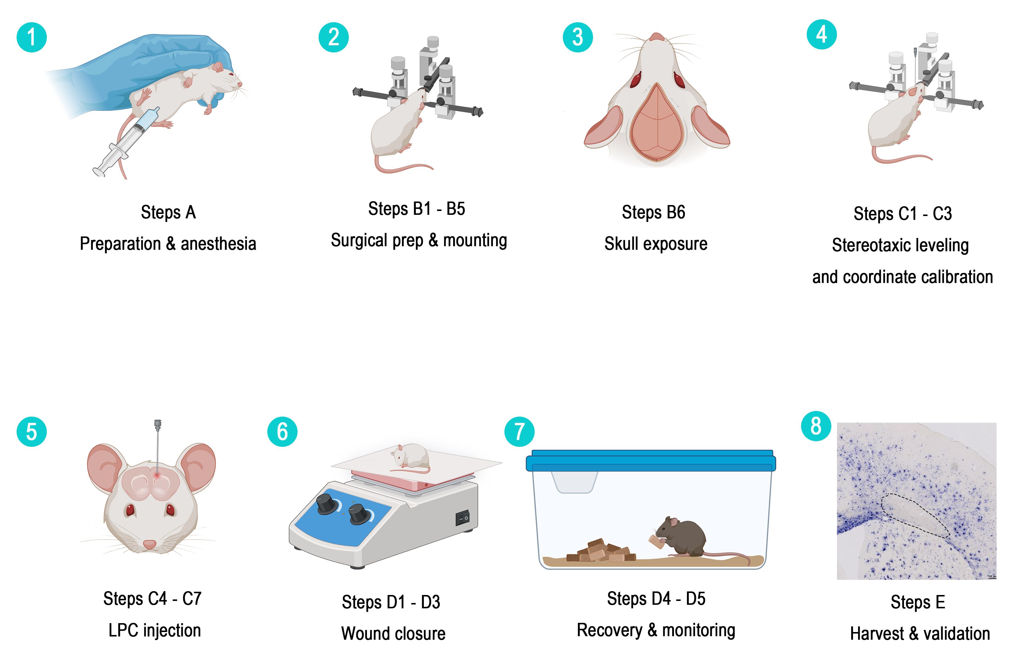
Step-by-step numbered schematic of this protocol. 1) Preparation and anesthesia (section A). 2) Surgical preparation and stereotaxic fixation (steps B1–5). 3) Skull exposure (step B6). 4) Stereotaxic leveling and coordinate calibration (steps C1–3). 5) Controlled LPC injection (steps C4–7). 6) Wound closure (steps D1–3). 7) Recovery and monitoring (steps D4–5). 8) Tissue collection and histological validation (section E).
Background
Demyelinating diseases such as multiple sclerosis (MS) involve damage to the myelin sheath surrounding axons in the central nervous system. Focal demyelination models allow investigators to study the cellular and molecular mechanisms of demyelination and remyelination in a spatiotemporally controlled manner. Current animal models for studying demyelination and remyelination fall mainly into three categories: the experimental autoimmune encephalomyelitis (EAE) model, the cuprizone model, and focal toxin-injection models [such as lysophosphatidylcholine (LPC) and ethidium bromide (EB) injection]. Because no single animal model recapitulates all features of human demyelinating disease, the choice of model should depend on the biological question being addressed. Experimental autoimmune encephalomyelitis (EAE) is suitable for studying autoimmune inflammation, but its lesions are temporally asynchronous, and spatial control is limited. Cuprizone intoxication produces pronounced corpus callosum demyelination after several weeks of systemic feeding, but the lesions are diffuse rather than experimentally targeted. Ethidium bromide can produce focal lesions but is broadly cytotoxic, damaging astrocytes and oligodendrocyte lineage cells in addition to myelin. By contrast, LPC is a membrane-disrupting lysophospholipid that produces spatially confined lesions, relatively spares axons, and exhibits a predictable demyelination–spontaneous remyelination sequence [1–9]. The LPC injection model is widely used because it induces focal demyelination without damaging axons, making it an ideal system for studying remyelination. Stereotaxic injection of LPC into the corpus callosum targets an anatomically well-defined and functionally important white matter tract, which is highly suitable for studying the recruitment and differentiation of oligodendrocyte progenitor cells. Compared with systemic demyelination models (such as the cuprizone model), this protocol allows precise control of lesion location and size, enables paired comparisons with the contralateral hemisphere, and facilitates the correlation of lesions with specific behavioral deficits [10–12]. Based on these advantages, this protocol describes in detail a standardized procedure for establishing a focal demyelination model by stereotaxic injection of LPC into the mouse corpus callosum and provides a complete histological validation method, with the aim of offering researchers in this field—especially beginners—a reproducible and easy-to-perform experimental reference (Table 1).
Table 1. Comparison of commonly used demyelination models
| Model | Primary mechanism | Advantages | Limitations | Representative references |
|---|---|---|---|---|
| EAE | Autoimmune inflammation | Clinically relevant immune component; suitable for immunotherapy studies | Lesions are temporally asynchronous; limited spatial control; predominantly spinal cord involvement in many paradigms | [3,4] |
| Cuprizone [bis(cyclohexanone)oxaldihydrazone] | Systemic oligodendrocyte mitochondrial toxicity | Pronounced corpus callosum demyelination; spontaneous remyelination after withdrawal | Diffuse lesions; requires several weeks of feeding; targeted location cannot be controlled | [4,5] |
| Ethidium bromide | Focal DNA intercalation and cytotoxicity | Large lesions with clear spatial boundaries | Broad glial cytotoxicity; under certain conditions, repair is slower and less complete than with LPC | [5,8] |
| LPC | Focal disruption of myelin lipids | Precise targeting, rapid onset, high reproducibility, relative axonal sparing, synchronized remyelination | Injection-related tissue damage; lack of autoimmune inflammation; local inflammatory responses still occur | [8,9] |
The LPC paradigm is not restricted to the corpus callosum. Focal LPC lesions have been established in spinal cord white matter, the caudal cerebellar peduncle, the internal capsule, the optic nerve, and the optic chiasm [5,13–19]. Therefore, this method is broadly applicable to region-specific questions of myelin injury and repair. However, coordinates, injection volumes, needle gauges, and tissue collection endpoints must be re-optimized for each target structure and species.
In the corpus callosum, demyelination can be detected within the first few days after LPC injection, typically peaking at around 3–7 dpi, followed by recruitment and differentiation of oligodendrocyte progenitor cells and remyelination at approximately 14–21 dpi. The exact time course depends on species, strain, age, injection volume, concentration, and target site [4,12,14,19,20]. Published volumetric analyses using defined mouse corpus callosum protocols report demyelination volumes of approximately 0.2–0.3 mm3 at 4 dpi and 0.05–0.15 mm3 at 14 dpi; these values are protocol-specific and should not be regarded as universal constants [16].
Adult C57BL/6 mice aged 8–10 weeks are commonly used because of their robust baseline remyelination capacity and well-defined time course. Aged mice may also be used when the experimental question concerns age-dependent repair; however, remyelination in aged animals is slower and less efficient, so age-matched controls should be included and tissue collection times adjusted to account for the slower remyelination in aged animals [15,21–23].
The target coordinates used in this protocol—AP +1.10 mm, ML +0.95 mm, DV -2.15 mm from the skull surface—are designed to place the injectate within the corpus callosum at the level of the fornix, dorsal to the dorsal hippocampus, while avoiding the superior sagittal sinus. If bilateral lesions are required, the procedure is repeated at ML -0.95 mm. Coordinates should be verified against the Paxinos and Franklin mouse brain atlas and adjusted according to atlas edition, animal age, body weight, skull geometry, and local instrument calibration [11,12,16,20,32]. The coordinates in this protocol were determined with reference to The Mouse Brain in Stereotaxic Coordinates by Paxinos and Franklin [32] and cross-validated using the Allen Mouse Brain Common Coordinate Framework 3D digital atlas. The AP +1.10 mm plane lies at the level of the frontal/motor cortex, where the corpus callosum has developed into a broad, thick, horizontally oriented, well-demarcated dense white matter tract with a large target cross-sectional area and good tolerance to targeting error; moreover, this plane is rostral to the hippocampus, so vertical needle insertion passes only through the motor/somatosensory cortex without traversing deep nuclei such as the hippocampus or striatum, thereby minimizing needle track damage. ML +0.95 mm corresponds to the lateral segment of the corpus callosum body (approximately dorsal to the lateral ventricle), the region where the white matter tract extends most widely laterally, ensuring adequate diffusion of the injectate within the callosal fibers. DV -2.15 mm (from the skull surface) is set according to the atlas, which shows the corpus callosum at this plane lying approximately 1.3–2.1 mm below the skull surface; placing the needle tip slightly deeper than its ventral border, combined with slow, low-flow-rate injection, allows the injectate bolus to spread within the corpus callosum and reflux mildly along the needle track, covering the full thickness of the corpus callosum while avoiding excessively deep penetration into the lateral ventricle that would cause injectate loss. The reasons for not injecting at the midline are as follows: first, the superior sagittal sinus runs along the sagittal suture directly beneath the midline, and drilling or needle insertion at the midline readily injures this venous sinus, causing difficult-to-control hemorrhage and markedly increasing surgical failure and animal mortality; a lateral offset of approximately 1 mm completely avoids the sinus body and its major bridging veins. Second, the corpus callosum at the midline is a narrow structure connecting the two cerebral hemispheres, and midline needle insertion would simultaneously damage the bilateral cingulate cortex and the corpus callosum on both sides of the midline, resulting in an uncontrollable extent of injury [11,20].
Materials and reagents
Biological materials
1. C57BL/6 mice, 8–10 weeks old (Jackson Laboratory, strain #000664); use age- and sex-matched animals, record body weight, and report sex and source
Reagents
1. L-α-lysophosphatidylcholine (LPC) from egg yolk (Beyotime, catalog number: ST1425-25mg); store at -20 °C, shelf life: 12 months
2. 2,2,2-Tribromoethanol (Aladdin, catalog number: T161626-25g); store at 4 °C, shelf life: 4 weeks
3. 2-Methyl-2-butanol (tert-amyl alcohol) (Aladdin, catalog number: A103417)
4. Sterile 1× phosphate-buffered saline (PBS) (Biosharp, catalog number: BL302A)
5. 75% ethanol (Sinopharm, catalog number: 100092680)
6. Povidone-iodine (Betadine) (Cofoe, catalog number: 100031918984)
7. Erythromycin eye ointment (Cisen, catalog number: H37022025)
8. 4% paraformaldehyde (PFA) solution (Sigma, catalog number: P6148); store at 4 °C and use within one month
9. Sucrose (Sinopharm, catalog number: H-10021463)
10. OCT embedding compound (Epredia, catalog number: 6502: Neg-50)
11. Myelin True Gold Staining kit (Oasis, catalog number: BK-AC001)
12. Sodium thiosulfate (Solarbio, catalog number: S5561-250g)
13. Anti-MBP antibody (Oasis, catalog number: OB-PGP187-01)
14. Fluorescently labeled secondary antibody (Invitrogen, catalog number: A11073)
15. DAPI (Solarbio, catalog number: ID22502)
16. Proteinase K (Roche, catalog number: 03115879001)
17. Anti-Digoxigenin-AP, Fab fragments (Roche, catalog number: 11093274910)
18. NBT/BCIP developing solution (Roche, catalog number: 11681451001)
19. Triton X-100 (Aladdin, catalog number: T109026)
Note: The catalog numbers listed above are representative products and can be replaced with validated equivalent products.
Solutions
1. 1% LPC working solution (see Recipes)
2. 2% tribromoethanol working anesthetic solution (see Recipes)
Recipes
1. 1% LPC working solution
| Reagent | Final concentration | Quantity or volume |
|---|---|---|
| L-α-lysophosphatidylcholine | 1% (w/v) | 10 mg |
| Sterile 1× PBS (Ca2+/Mg2+-free, pH 7.4) | n/a | 1 mL |
| Total | n/a | 1 mL |
Weigh 10 mg of LPC powder using an electronic balance and transfer it into a sterile centrifuge tube. Use a pipette to draw 1 mL of sterile PBS and add it to the centrifuge tube containing LPC. Tighten the cap, then vortex thoroughly for 3–5 min until the powder is completely dissolved and the solution becomes clear. Perform a brief low-speed centrifugation to spin down the residual liquid on the tube wall. The resulting solution is the 1% sterile LPC working solution, which should be stored at −20 °C, protected from repeated freeze/thaw cycles, and used within one week after preparation. Aliquots showing signs of turbidity, precipitation, or contamination should be discarded.
2. 2% Tribromoethanol anesthetic solution
| Reagent | Final concentration | Quantity or volume |
|---|---|---|
| 2,2,2-tribromoethanol | 2% (w/v) | 1 g |
| 2-methyl-2-butanol (tert-amyl alcohol) | 2% (v/v) | 1 mL |
| Sterile PBS | n/a | 49 mL |
| Total | n/a | 50 mL |
Weigh 1 g of tribromoethanol powder with an electronic balance and transfer it into a 50 mL sterile centrifuge tube. Add 1 mL of tert-amyl alcohol using a pipette. Gently shake at room temperature until the powder dissolves completely to form a clear oily stock solution. Gradually add 49 mL of sterile PBS to the stock solution and mix thoroughly. Transfer the entire solution into a sterile light-resistant reagent bottle, store at 4 °C, and use within one month after preparation.
Laboratory supplies
1. Suture, 5-0 (Jinhuan Medical, catalog number: 100040361204)
2. Cotton swabs (Cofoe, catalog number: 100265718600)
Equipment
1. Fine-tip marker (Fisherbrand, catalog number: 13-379-4)
2. Removable needle: 33G, Small Hub Removable Needle (RN) (Hamilton, catalog number: 7803-05 (point style 4, 12° bevel)
3. Hamilton microsyringe, 10 μL (Hamilton, model 1701), fitted with a removable needle
4. Microinjection pump (Harvard, model: PUMP 11 ELITE Nanomite)
5. Small animal stereotaxic instrument (RWD Life Science, catalog number: 68018) (requires a mouse adaptor, 60° mouse ear bars, a digital display manipulator arm, and a cannula holder)
6. Dental drill with a 0.5–0.8 mm drill bit (RWD Life Science, catalog number: 78001)
7. Animal thermostatic heating system (including heating pad) (RWD Life Science, catalog number: 69020)
8. Hair clipper (RWD Life Science, model: CP-5200)
9. Electronic balance (Deli, model: TE910)
10. Dissecting scissors (RWD Life Science, catalog number: S14014-11)
11. Blunt surgical scissors (RWD Life Science, catalog number: S13003-11)
12. Needle holder (RWD Life Science, catalog number: F31047-12)
13. Tissue forceps (RWD Life Science, catalog number: F13030-10)
14. Fluorescence microscope (Leica, model: DM6B)
Note: The catalog numbers listed above are representative products and can be replaced with validated equivalent products.
Software and datasets
1. Fiji/ImageJ 2.x, NIH, RRID: SCR_002285; used for image processing, thresholding, ROI measurement, and lesion quantification [30]
2. GraphPad Prism 9/10 or R 4.x; used for statistical analysis and plotting.
3. Microscope acquisition software specific to the Leica DM6B or other local imaging systems
Procedure
A. Animal preparation and anesthesia
1. Use C57BL/6 mice, 8–10 weeks old, with a body weight of 20–25 g. House animals in a temperature-controlled (20–24 °C) and humidity-controlled (40%–60%) environment with a 12/12 h light/dark cycle. Provide standard rodent chow and water ad libitum. Allow mice to acclimate for at least 7 days prior to any experimental procedure. Randomly assign animals to experimental groups and apply pre-established health inclusion criteria.
2. Administer 2% (g/vol) 2,2,2-tribromoethanol solution by intraperitoneal injection at a dose of 500 mg/kg body weight. For example, 0.5 mL of the 2% solution (25 μL/g) delivers the 500 mg/kg dose to a 20 g mouse. Confirm the institutionally approved dose before use; supplemental dosing should not be given routinely. Tribromoethanol provides anesthesia but does not provide sustained postoperative analgesia.
3. Verify anesthetic depth by performing a toe-pinch reflex test. Firmly compress the toe pad between forceps; absence of withdrawal reflex indicates adequate anesthetic depth. Reassess every 10–15 min during the procedure and monitor respiratory rate and rhythm throughout. If the withdrawal reflex, whisker movement, or spontaneous movement returns, stop the procedure and manage the animal according to the anesthetic protocol approved by the institutional veterinarian [24].
4. Apply ophthalmic lubricant to both eyes immediately after induction of anesthesia to prevent corneal drying and injury during surgery. Anesthesia suppresses the blink reflex and increases the risk of corneal injury [20,24,25].
B. Stereotaxic surgical preparation
1. Scalp preparation: Shave the fur from the dorsal surface of the head using an electric clipper, extending from between the eyes to the occipital region and from ear to ear. Remove all loose hair with gauze.
2. Skin disinfection: Decontaminate the shaved scalp by applying povidone-iodine (Betadine) solution, followed by thorough wiping with 75% ethanol. Repeat this alternating Betadine–ethanol sequence two additional times (total of three cycles).
3. Head fixation: Place the anesthetized mouse in the small-animal stereotaxic apparatus. First, insert the ear bars into the external auditory meatus bilaterally. Ensure the ear bars are inserted to equal depth on both sides by checking that the scale readings on the left and right ear bar holders are identical.
Note: Do not insert the ear bars too deeply. Excessive insertion depth or incorrect angulation can damage the ear canal, compromise surrounding vasculature, or, in severe cases, cause intracranial injury and internal bleeding.
4. Fixation verification: Verify proper head fixation using the following four criteria:
a. The nose is centered in the nose clamp and aligned with the midline.
b. The head is immobile when gentle lateral pressure is applied.
c. The head remains level when the tail is gently lifted (the mouse does not fall or shift position).
d. The skull surface is oriented horizontally, with no visible tilting in the anterior–posterior or medial–lateral axes.
5. Skin incision: Using a sterile scalpel, make a midline incision approximately 1–1.5 cm in length along the sagittal suture, extending from approximately 2 mm anterior to bregma to 2 mm posterior to lambda. Use sterile tissue forceps to gently retract the skin edges and expose the skull surface.
6. Exposure of cranial landmarks: Gently scrape away the periosteal connective tissue and adherent fascia from the skull surface with a cotton swab moistened with sterile PBS. The coronal, sagittal, and lambdoid sutures, as well as the cranial suture intersection points, including bregma and lambda, must be clearly visible.
7. Stereotaxic leveling: Move the stereotaxic manipulator arm to position the needle tip directly over the bregma landmark, then lower the needle until it just touches the bregma point and reset the anterior–posterior (AP) and medial–lateral (ML) coordinates to zero. Verify left–right horizontality by comparing dorsal–ventral (DV) readings at equidistant lateral points from the midline, ensuring the coordinate difference between both sides is no more than 0.03 mm. Next, move the manipulator arm to position the needle tip over the lambda landmark and record the DV reading. Adjust the ear bar height and/or nose clamp angle until the DV coordinate at bregma and lambda differ by no more than 0.03 mm (Figure 1).
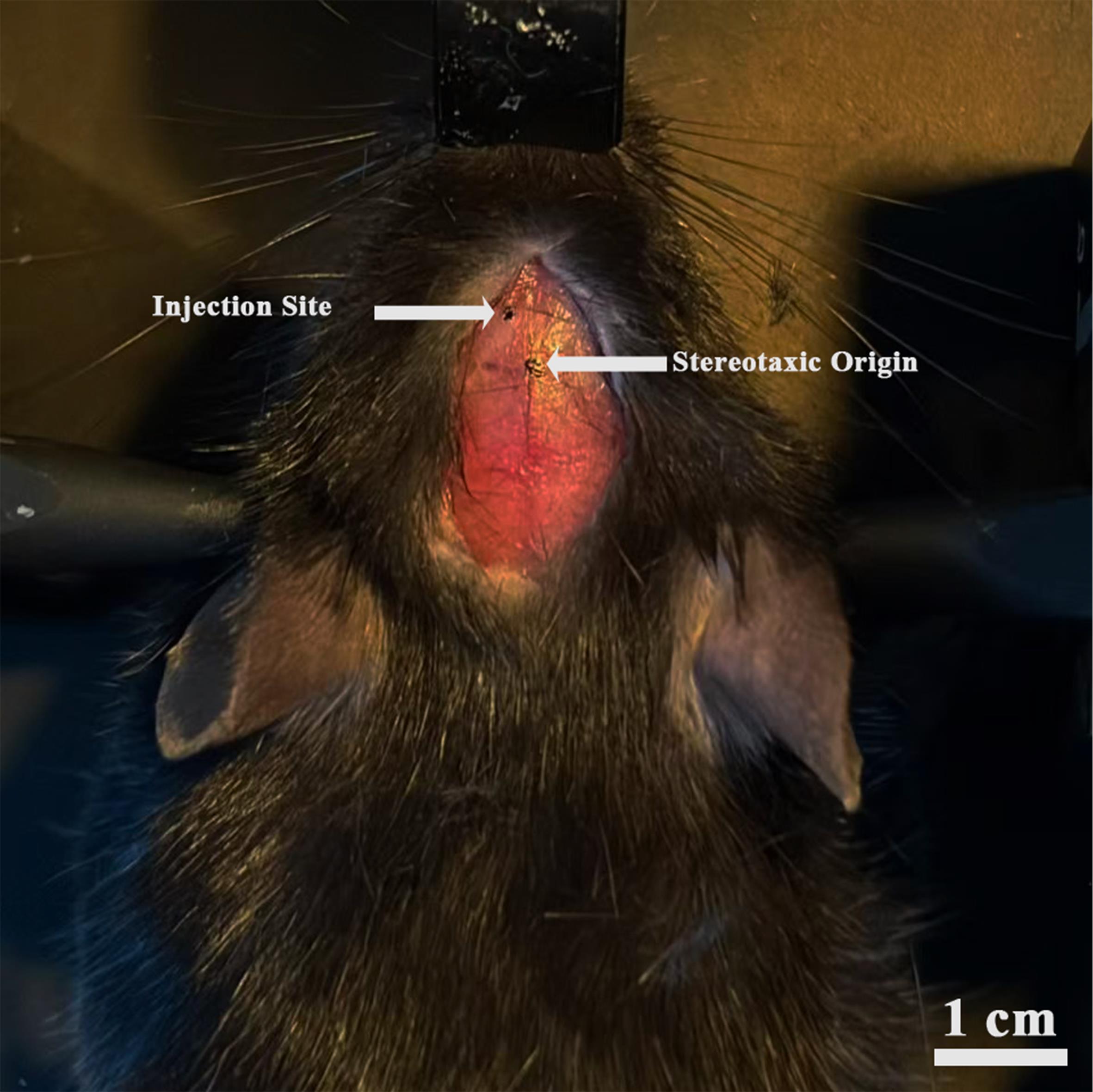
Figure 1. Photograph of head fixation and cranial landmarks. Includes the size of the surgical incision, cranial landmarks, and injection site.
C. LPC stereotaxic injection
Note: The stereotaxic instrument and microinjection pump employed in this procedure are shown in Figure 2.
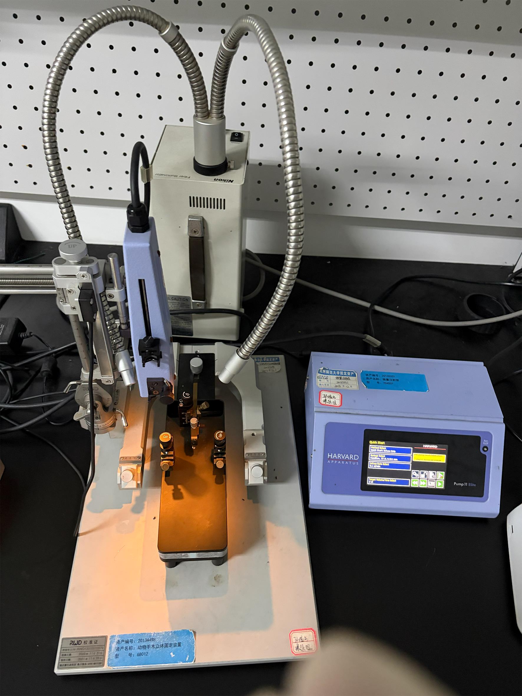
Figure 2. Photographs of the brain stereotaxic instrument and microinjection pump
1. Determine injection coordinates: Position the manipulator arm so that the needle tip is directly above the bregma and set all coordinates to zero. Move the arm to the target coordinates for corpus callosum injection (from bregma):
a. Anterior–posterior (AP): +1.10 mm
b. Medial–lateral (ML): +0.95 mm
c. Dorsal–ventral (DV): -2.15 mm from the skull surface
Note: The above coordinates target the corpus callosum at the level of the fornix, dorsal to the dorsal hippocampus. If bilateral lesions are required, repeat the injection at ML = -0.95 mm. The final coordinates should be verified using a mouse brain stereotaxic atlas [32].
2. Mark the injection site: Use a fine-tip permanent marker to mark the injection point at the determined AP and ML coordinates.
3. Skull drilling: Using a dental drill with a fine burr (approximately 0.8 mm diameter), carefully drill a small circular hole through the skull at the marked injection site. Apply gentle, intermittent pressure to prevent heat buildup and bone damage.
4. Prepare the LPC solution: Immediately before injection, load 1% (w/v) LPC solution (dissolved in sterile PBS) into a 10 μL Hamilton syringe fitted with a 33G beveled metal needle. Ensure the syringe is free of air bubbles.
5. Position the needle: Lower the needle tip until it just contacts the skull surface at the injection coordinates. Reset the DV coordinate to zero at the skull surface. Then, continue lowering the needle slowly to reach the target DV depth of 2.15 mm below the skull surface.
Note: The needle bevel should face caudally in all animals. The corpus callosum extends along the rostrocaudal axis at this level; orienting the bevel (i.e., the direction of preferential spread of the injectate) caudally promotes diffusion of the LPC solution along the longitudinal axis of the corpus callosum while limiting its spread toward the midline and the lateral cortex. Because the injectate tends to diffuse in the direction the bevel faces and to reflux along the needle track on the side of the bevel opening, bevel orientation affects the shape, symmetry, and consistency of the lesion. The bevel orientation should be recorded and kept strictly consistent across all animals and groups.
6. LPC injection: Inject the LPC solution using the following parameters:
a. Injection volume: 1–2 μL per injection site.
b. Injection rate: 0.07–0.15 μL/min. Use a microinjection pump for precise rate control.
Note: Slow injection rate is critical to prevent tissue tearing and excessive mechanical damage to the corpus callosum. Rapid injection causes tissue disruption and inconsistent demyelination volumes.
c. Needle retention: After the full injection volume has been delivered, leave the needle in place for 5–10 min. This step is essential to prevent backflow of the injected LPC solution along the needle track.
d. Needle withdrawal: After the 5–10 min retention period, withdraw the needle slowly (0.02 mm/s).
e. Bilateral injection (if required): If bilateral corpus callosum demyelination is desired, repeat steps on the contralateral hemisphere using a freshly prepared LPC-loaded syringe. The full surgical procedure for stereotaxic LPC injection is shown in Video 1.

D. Wound closure and post-operative care
1. Suture the skin: Close the scalp incision using 5-0 non-absorbable silk sutures with simple interrupted stitches (2–3 sutures).
2. Wound disinfection: Apply a thin layer of povidone-iodine (Betadine) solution to the sutured incision line using a sterile cotton-tipped applicator.
3. Recovery and thermoregulation: Transfer the mouse to a clean, prewarmed recovery cage positioned on a 37 °C heating pad. Monitor continuously until the animal regains the righting reflex and becomes ambulatory (typically 1–3 h post-surgery).
4. Daily monitoring: For the first 7 days post-surgery, monitor all animals at least once daily as follows:
a. Wound integrity: Check for signs of dehiscence, swelling, discharge, or infection.
b. General activity level.
c. Body weight: Record body weight daily. A weight loss exceeding 15% of pre-surgery body weight requires veterinary evaluation.
d. Neurological signs: Monitor for gait abnormalities and limb coordination deficits.
5. Suture removal: Remove skin sutures 7–10 days post-surgery under brief manual restraint.
E. Tissue harvesting and demyelination verification (optional)
1. At the desired post-injection time points, sacrifice mice for histological analysis:
a. 3–7 dpi: Peak demyelination.
b. 7 dpi: Active oligodendrocyte precursor cell recruitment and proliferation.
c. 10–21 dpi: Active remyelination.
d. >28 dpi: Near-complete remyelination.
2. Transcardiac perfusion: Deeply anesthetize the mouse with tribromoethanol (500 mg/kg, intraperitoneal). Confirm deep anesthesia by the absence of toe-pinch and corneal reflexes. Perfuse with ice-cold PBS (approximately 20–30 mL) followed by room-temperature PBS-buffered 4% PFA (approximately 20–30 mL).
3. Brain removal and post-fixation: Carefully remove the brain from the skull. Post-fix the whole brain in 4% PFA at 4 °C for 24 h.
4. Cryoprotection and embedding: Transfer the fixed brain to 20% (w/v) sucrose in PBS at 4 °C for 24 h, then transfer to 30% (w/v) sucrose in PBS at 4 °C for an additional 24–48 h until the brain sinks completely. Embed the brain in optimal cutting temperature (OCT) compound.
5. Cryosectioning: Cut coronal sections at a thickness of 16 μm through the region of the LPC injection site (approximately AP +0.5 to +2.0 mm from bregma). Mount sections onto glass slides.
6. Assessment of demyelination: Optional methods include Myelin Gold staining, anti-MBP immunofluorescence, or Mbp in situ hybridization.
a. Myelin Gold staining: Cover tissue sections with the 1× True Gold Staining kit working solution and incubate at 45 °C for 30 min; wash in water for 3 min, then incubate in sodium thiosulfate solution at 45 °C for 2 min.
Note: Myelin gold staining is a histochemical staining method based on the deposition of gold salts; its principle is that gold ions selectively bind to the basic proteins and lipid components of myelin and, after development, render myelin structures brownish-black to black, whereas demyelinated regions, owing to myelin loss, remain unstained or show markedly lighter staining. This method is simple to perform and low in cost, and it can directly visualize the distribution and integrity of myelin under a conventional brightfield microscope; therefore, it is commonly used to rapidly assess the extent of demyelinating lesions and the degree of remyelination: regions of absent staining correspond to demyelinating lesions, whereas recovery of the staining signal within the lesion area indicates the occurrence of remyelination. For detailed procedures, refer to the published literature [10,26].
b. Anti-MBP immunofluorescence: Brain cryosections are blocked at room temperature for 1 h in PBS containing 5% goat serum and 0.2% Triton X-100; incubate with the anti-MBP antibody overnight at 4 °C. After washing, incubate with a fluorophore-conjugated secondary antibody at room temperature for 1 h. Counterstain nuclei with DAPI for 5 min.
Note: Immunofluorescence staining is a detection method that uses specific antibodies to recognize myelin basic protein (MBP). MBP is one of the major structural proteins of mature myelin; it is synthesized by oligodendrocytes and plays a key role in maintaining the compaction and stability of the myelin lamellar structure. After signal amplification with a fluorophore-conjugated secondary antibody, the distribution of MBP can be visualized at high resolution under a fluorescence or confocal microscope and subjected to precise quantitative analysis (e.g., percentage of MBP-positive area, fluorescence intensity). In this protocol, this staining is used to finely assess the extent of demyelinating lesions and the degree of remyelination [10,16].
c. Mbp in situ hybridization: After permeabilization of brain cryosections with proteinase K and completion of prehybridization blocking, an MBP-specific antisense probe is added for hybridization at constant temperature overnight; stringent graded washes are performed to remove nonspecifically bound probe, followed by incubation with an anti-digoxigenin alkaline phosphatase antibody, and color development with NBT/BCIP under light-protected conditions.
Note: In situ hybridization is a method that uses a labeled probe complementary to the Mbp gene mRNA to detect the distribution and expression level of Mbp transcripts in tissue sections in situ. Unlike immunostaining, which detects protein products, in situ hybridization reflects the transcriptional activity of the gene and can therefore reveal the location and number of oligodendrocytes that are actively synthesizing myelin proteins. In this protocol, this staining is used to track the cellular origin and dynamic process of remyelination: After demyelination, oligodendrocyte progenitor cells are recruited to the lesion and differentiate into mature oligodendrocytes, re-expressing Mbp mRNA; by counting the density of Mbp-positive cells within the lesion, the regeneration of functional myelin-forming cells can be quantitatively assessed. In situ hybridization and MBP immunofluorescence are complementary: the former reflects the number and activation state of myelin-synthesizing cells at the transcriptional level, whereas the latter reflects the actual degree of structural repair of myelin at the protein level. Combining the two enables a more comprehensive evaluation of the remyelination process [10].
7. Image acquisition: Acquire images for all groups using identical magnification, illumination, exposure, gain, laser power, offset, and detector settings. Record the manufacturer/model of the microscope or slide scanner, the objective, numerical aperture, image resolution, acquisition software, and file format. Do not change acquisition settings between groups [27].
8. Confirm that the lesion center is located within the corpus callosum. Pre-establish exclusion criteria before analysis: any animal with an absent lesion, a lesion center located outside the corpus callosum, marked reflux or major hemorrhage, or extension into the cortex or hippocampus in a manner inconsistent with the intended target should be excluded. Apply the same criteria to all groups and report the number of excluded animals [28,29].
Data analysis
1. Experimental design and sample size: At a minimum, the study should include an LPC-injected group and a sham surgery group. The sham surgery group undergoes the same anesthesia, stereotaxic fixation, drilling, and needle insertion procedures and receives an injection of sterile 1× PBS at the same volume and rate. An additional naive group without surgery may be included as a staining baseline control. The contralateral corpus callosum may serve as an internal reference, but cannot replace an independent sham surgery group. For methodological validation, use at least three animals per condition or per time point. When performing quantitative comparisons between groups, a statistical power calculation should be conducted in advance whenever possible; in the absence of pilot data, 5–6 animals per group or per time point is a practical starting range, but it cannot substitute for a power analysis.
2. Image acquisition: Use a calibrated brightfield or fluorescence microscope or a slide scanner. A 10× or 20× objective is generally suitable for lesion mapping; use consistent high magnification only when cellular detail is required. All images obtained with the same staining method must be acquired with identical settings, and the instrument model, objective, numerical aperture, pixel size, exposure, gain, laser power, offset, detector settings, and file format must be recorded.
3. Thresholding and ROI definition: Delineate the corpus callosum based on anatomical landmarks and the atlas. Apply one predefined thresholding method to all images within the same experiment—such as a fixed intensity threshold or a validated automatic threshold. Define the lesion as a contiguous region of low-signal, myelin-negative, or myelin-poor tissue within the corpus callosum. If the needle track is included, record it separately.
4. Quantitative metrics: Report lesion area (mm2), lesion volume (mm3), percentage of corpus callosum involvement, and normalized MBP intensity or integrated density. Lesion volume is the sum of lesion areas across sections multiplied by the intersection spacing. Percentage of corpus callosum involvement is the lesion area divided by the total corpus callosum area within the same anatomical ROI. When analyzing multiple sections, report the intersection spacing and the number of sections per animal. The biological unit is the animal, not the section.
5. Statistics: Data are expressed as mean ± SEM, and exact n values are reported. Test for normality using an appropriate method (e.g., the Shapiro–Wilk test). When assumptions are met, use a two-tailed unpaired Student's t-test for two-group comparisons; otherwise, use nonparametric tests. For comparisons across multiple time points or multiple groups, use one-way or two-way ANOVA with appropriate post hoc corrections. P values less than 0.05 are considered statistically significant. Reporting should follow the ARRIVE 2.0 guidelines, including randomization, blinding, inclusion and exclusion criteria, and attrition of cases [31].
Validation of protocol
Following this protocol, 8-week-old C57BL/6 mice received a unilateral corpus callosum injection of 2 μL of 1% LPC, and brains were collected for histological analysis at 7, 14, and 21 dpi. Representative images are shown in Figures 3 and 4. The two staining methods mutually corroborate each other at the transcriptional level (Mbp mRNA) and the protein/structural level (myelin), revealing a consistent demyelination–remyelination time course. It was observed that lesions from successful modeling were consistently confined within the corpus callosum, without obvious spread to the adjacent cortex, hippocampus, or contralateral hemisphere; needle track injury was minimal, morphologically readily distinguishable from the LPC-induced lesion, and was recorded separately.
A suggested time-course validation scheme for this demyelination model is summarized in Figure 5.
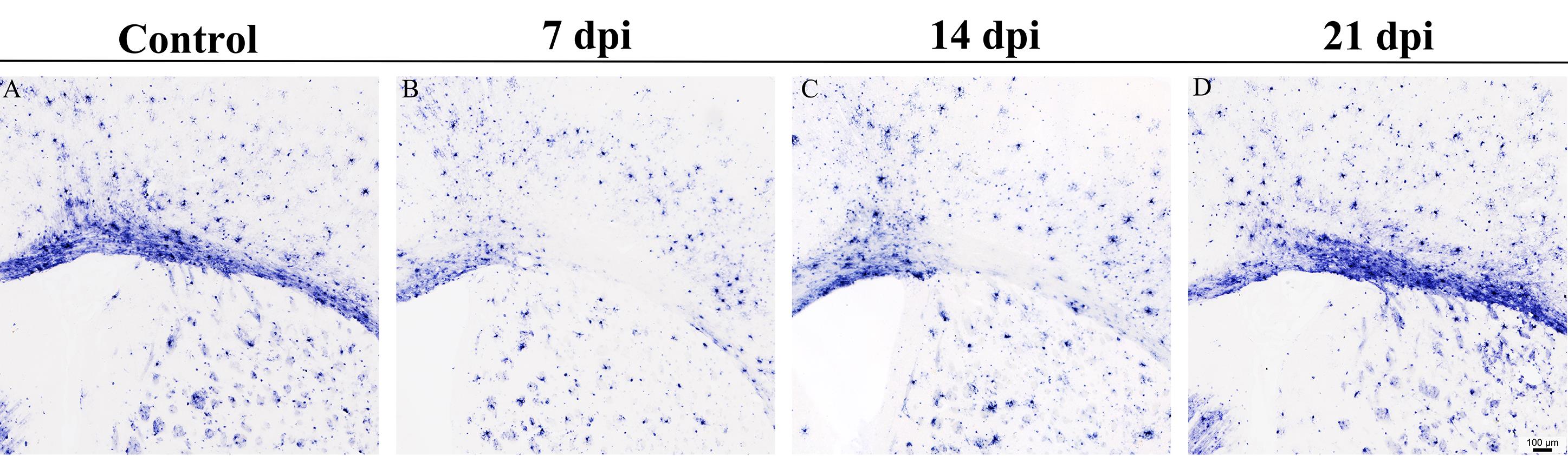
Figure 3. Validation of lysophosphatidylcholine (LPC)-induced demyelination. Representative in situ hybridization (ISH) staining of myelin basic protein (Mbp) mRNA, illustrating the dynamic changes in Mbp expression in (A) control and injured tissue at (B) 7, (C) 14, and (D) 21 days after injury. The corpus callosum of control mice displays a continuous, dense Mbp-positive band. At 7 dpi after LPC injection, the band on the injected side of the corpus callosum is interrupted, and Mbp-positive cells are markedly reduced in the lesion area; at 14 dpi, recovery of the Mbp signal begins at the lesion border zone, while the core region remains low in signal, consistent with the typical pattern of remyelination progressing from the lesion edge toward the core. At 21 dpi, the Mbp signal in the lesion area recovers further, and the band morphology is largely re-established. Scale bar = 100 μm. Images were acquired using a Leica DM6B upright microscope equipped with a 10× objective, and all sections were photographed under constant light source intensity, exposure time (10 ms), and automatic white balance parameters.
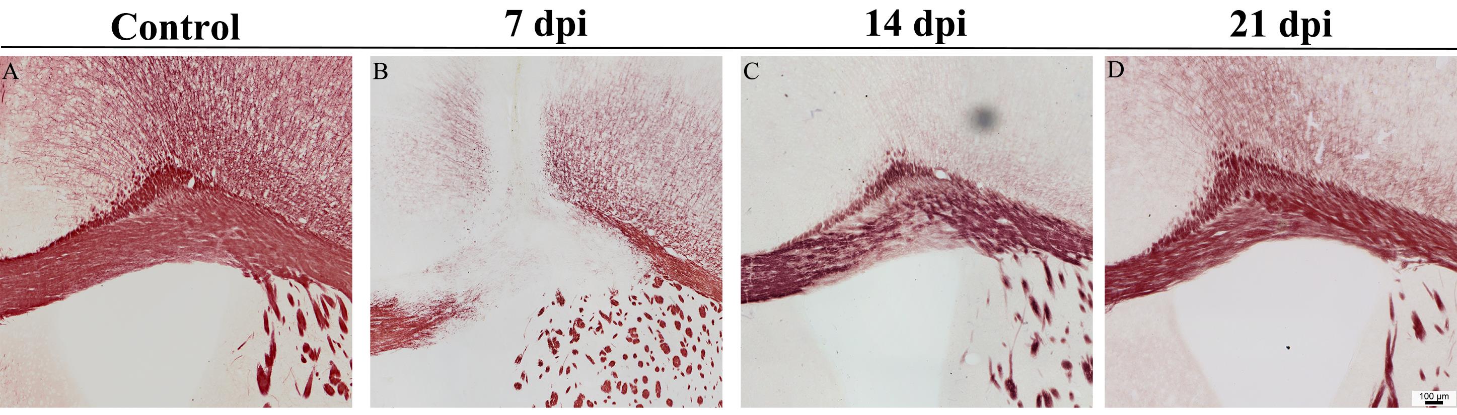
Figure 4. Gold staining reveals the time course of lysophosphatidylcholine (LPC)-induced demyelination in the corpus callosum. A representative schematic timeline of demyelination and remyelination is shown, illustrating the dynamic changes in the pathological state of (A) control and lesioned tissues at (B) 7, (C) 14, and (D) 21 days post-injury. The corpus callosum of control mice exhibits uniform, dense dark-red staining. At 7 dpi, a large, well-demarcated area of light staining appears in the corpus callosum, with staining almost completely absent in the lesion core, indicating marked myelin loss; at 14 dpi, staining in the lesioned area partially recovers but remains lighter and finer than normal; at 21 dpi, staining in the lesioned area further intensifies, approaching control levels, indicating substantial remyelination. Scale bar = 100 μm. Images were acquired using a Leica DM6B upright microscope equipped with a 10× objective; all sections were photographed under constant light source intensity, exposure time (10 ms), and automatic white balance parameters.

Figure 5. Recommended validation timeline. Recommended validation timeline for the lysophosphatidylcholine (LPC) corpus callosum model: the 3 and 7 dpi time points are suitable for observing demyelination, whereas 14, 21, and 28 dpi are suitable for observing remyelination. The exact endpoint should be chosen according to the experimental question and adjusted for aged animals. This schematic summarizes published kinetics data rather than new experimental data [12,14,15,20].
Recommended experimental design and analysis workflow: include an LPC group, a sham surgery group, and an optional naive group; randomly assign animals to groups; perform image analysis in a blinded manner; use constant acquisition settings; quantify lesion area, lesion volume, percentage of corpus callosum involvement, and normalized MBP signal; and apply predefined inclusion and exclusion criteria.
Successful injection produces a focal region of myelin loss centered on the corpus callosum. In published mouse corpus callosum protocols, reported lesion volumes are approximately 0.2–0.3 mm3 at 4 dpi and approximately 0.05–0.15 mm3 at 14 dpi, but these values vary with injection parameters, mouse strain, age, and quantification method. There is no universal threshold that the percentage of corpus callosum involvement must reach; it should be calculated within a defined ROI and reported together with the absolute lesion area or volume. The lesion should remain centered on the corpus callosum, without obvious spread to the adjacent cortex, hippocampus, or contralateral hemisphere. Minor needle track injury should be recorded separately [16].
Monitoring of inter-operator consistency
There is currently no accepted threshold for inter-operator variability in the literature. Therefore, the following exclusion criteria are recommended rather than mandatory in this protocol: The lesion must be centered on the corpus callosum, without obvious spread to the adjacent cortex, hippocampus, or contralateral hemisphere; needle track injury is minor and clearly distinguishable from the lesion; animals in which lesion targeting fails are excluded. We recommend evaluating inter-operator consistency according to the following criteria: (1) targeting accuracy: the deviation of the lesion center coordinates produced by different operators from the target coordinates should be ≤0.2–0.3 mm; (2) consistency of lesion size: the difference in mean lesion area and volume between different operators should be <20%–30%, and the coefficient of variation (CV) within the same operator should be <15%–20%; (3) statistical comparison: differences in lesion location, area, and volume between different operators are not statistically significant (unpaired t-test, p > 0.05); (4) success rate: the injection success rate of each operator (the proportion of animals meeting the inclusion criteria) should be ≥80%. If systematic differences between operators exceed the above criteria, key steps such as coordinate targeting, injection rate, needle gauge, and needle retention time should be retrospectively reviewed, and operators should be retrained and re-evaluated when necessary.
When multiple operators perform this procedure, the same inclusion and exclusion criteria should be applied, and inter-operator variability should be monitored by comparing lesion location, area, and volume across operators.
General notes and troubleshooting
General notes
1. The LPC injection model produces a focal demyelination lesion with minimal axonal damage, making it ideal for studying remyelination. Lesion size can be adjusted by changing the injection volume (1–2 μL) or LPC concentration (0.5%–1%). Any change in concentration, volume, or coordinates should be regarded as a new protocol condition and re-validated.
2. This protocol is applicable to other mouse strains and rat models, but the stereotaxic coordinates must be adjusted accordingly. LPC injection has also been applied to other white matter structures; however, each new target requires independent validation of the coordinates and endpoints [2,13–19].
3. Sources of variability include minor differences in injection coordinates, needle insertion angle, LPC solution preparation, and individual animal anatomical variations. Consistent technique and thorough operator training are essential for reproducible results.
Troubleshooting
Problem 1: Excessive mortality (>20%) post-surgery.
Possible causes: Anesthetic overdose, hypothermia, or surgical trauma.
Solutions: Verify anesthetic dose based on actual body weight; maintain body temperature using a heating pad throughout the procedure; ensure gentle tissue handling and minimize surgical duration. Considering that warming alters the depth of anesthesia in mice and that a single stereotaxic intracerebral injection is brief, it is recommended to provide warming only as needed when the mouse develops hypothermia.
Problem 2: Inconsistent or absent demyelination lesions.
Possible causes: Incorrect stereotaxic coordinates, needle clogging, or LPC degradation.
Solutions: Verify coordinates using a stereotaxic atlas; ensure needle patency before each injection; prepare fresh LPC solution for each experiment; confirm skull is level before injection.
Problem 3: Excessive bleeding during surgery.
Possible cause: Damage to the sagittal sinus or superficial blood vessels.
Solutions: Avoid midline drilling when possible; if bleeding occurs, apply gentle pressure with a cotton swab soaked in cold PBS; ensure proper hemostasis before closing the wound.
Limitations
This focal toxin model does not recapitulate the autoimmune inflammation, relapsing disease course, or diffuse lesion distribution pattern of multiple sclerosis. Therefore, it is best suited for mechanistic studies of focal myelin injury and repair, glial responses, and axon–myelin interactions, as well as for preclinical screening of pro-remyelination interventions.
The procedure requires experience with stereotaxic techniques. Minor differences in skull leveling, needle position, injection rate, bevel orientation, and injection volume can all alter the size and location of the lesion. Therefore, criteria for determining injection deviation, sham surgery controls, blinding, and standardized image acquisition are essential.
Age, sex, strain, anesthetic regimen, injection site, and LPC formulation can all influence lesion dynamics. Remyelination is delayed in aged animals, which should not be analyzed with the same endpoint assumptions as young adult animals.
Acknowledgments
This experimental protocol was formulated by referring to the well-established LPC-induced demyelination modeling methods reported in published literature and making further optimizations and improvements. We sincerely thank the Mengsheng Qiu Laboratory for providing technical assistance and valuable suggestions during the optimization of this protocol.
This work was supported by the research platform of Dr. Yu Tian.
Author contributions
Investigation, R.T.; Conceptualization, Q.C.; Writing—Original Draft, Q.C.; Writing—Review & Editing, Y.T.
Competing interests
The authors declare no conflicts of interest.
Ethical considerations
All animal procedures were approved by the Institutional Animal Care and Use Committee of Hangzhou Normal University (approval number: HSD20211210).
References
- Kipp, M., Clarner, T., Dang, J., Copray, S. and Beyer, C. (2009). The cuprizone animal model: new insights into an old story. Acta Neuropathol. 118(6): 723–736. https://doi.org/10.1007/s00401-009-0591-3
- Matsushima, G. K. and Morell, P. (2006). The Neurotoxicant, Cuprizone, as a Model to Study Demyelination and Remyelination in the Central Nervous System. Brain Pathol. 11(1): 107–116. https://doi.org/10.1111/j.1750-3639.2001.tb00385.x
- Gold, R. (2006). Understanding pathogenesis and therapy of multiple sclerosis via animal models: 70 years of merits and culprits in experimental autoimmune encephalomyelitis research. Brain. 129(8): 1953–1971. https://doi.org/10.1093/brain/awl075
- Lassmann, H. and Bradl, M. (2016). Multiple sclerosis: experimental models and reality. Acta Neuropathol. 133(2): 223–244. https://doi.org/10.1007/s00401-016-1631-4
- Woodruff, R. H. and Franklin, R. J. (1999). Demyelination and remyelination of the caudal cerebellar peduncle of adult rats following stereotaxic injections of lysolecithin, ethidium bromide, and complement/anti-galactocerebroside: A comparative study. Glia. 25(3): 216–228. https://doi.org/10.1002/(sici)1098-1136(19990201)25:3<216::aid-glia2>3.0.co;2-l
- Merrill, J. E. (2008). In Vitro and In Vivo Pharmacological Models to Assess Demyelination and Remyelination. Neuropsychopharmacology. 34(1): 55–73. https://doi.org/10.1038/npp.2008.145
- Torre-Fuentes, L., Moreno-Jiménez, L., Pytel, V., Matías-Guiu, J., Gómez-Pinedo, U. and Matías-Guiu, J. (2020). Experimental models of demyelination and remyelination. Neurología (English Edition). 35(1): 32–39. https://doi.org/10.1016/j.nrleng.2019.03.007
- McMurran, C. E., Zhao, C. and Franklin, R. J. M. (2019). Toxin-Based Models to Investigate Demyelination and Remyelination. In Lyons, D. A. and Kegel, L. Oligodendrocytes: Methods and Protocols. Springer New York, 377–396. https://doi.org/10.1007/978-1-4939-9072-6_21
- Plemel, J. R., Michaels, N. J., Weishaupt, N., Caprariello, A. V., Keough, M. B., Rogers, J. A., Yukseloglu, A., Lim, J., Patel, V. V., Rawji, K. S., et al. (2017). Mechanisms of lysophosphatidylcholine‐induced demyelination: A primary lipid disrupting myelinopathy. Glia. 66(2): 327–347. https://doi.org/10.1002/glia.23245
- Xu, X., Fang, M., Chen, L., Huang, H., Dai, Z. M., Yang, J. and Qiu, M. (2024). Nzf2 promotes oligodendrocyte differentiation and regeneration via repressing HDAC1-mediated histone deacetylation. Sci Adv. 10(50): eadf8405. https://doi.org/10.1126/sciadv.adf8405
- Pang, X. W., Chen, M., Chu, Y. H., Tang, Y., Qin, C. and Tian, D. S. (2022). A Stably Established Two-Point Injection of Lysophosphatidylcholine-Induced Focal Demyelination Model in Mice. J Visualized Exp.: e3791/64059. https://doi.org/10.3791/64059
- Bottes, S. and Jessberger, S. (2021). Live imaging of remyelination in the adult mouse corpus callosum. Proc Natl Acad Sci USA. 118(28): e2025795118. https://doi.org/10.1073/pnas.2025795118
- Hall, S. M. (1972). The effect of injections of lysophosphatidyl choline into white matter of the adult mouse spinal cord. J Cell Sci. 10(2): 535–546. https://doi.org/10.1242/jcs.10.2.535
- Jeffery, N. D. and Blakemore, W. F. (1995). Remyelination of mouse spinal cord axons demyelinated by local injection of lysolecithin. J Neurocytol. 24(10): 775–781. https://doi.org/10.1007/bf01191213
- Keough, M. B., Jensen, S. K. and Yong, V. W. (2015). Experimental Demyelination and Remyelination of Murine Spinal Cord by Focal Injection of Lysolecithin. J Visualized Exp.: e3791/52679. https://doi.org/10.3791/52679
- Bosch-Queralt, M., Tiwari, V., Damkou, A., Vaculčiaková, L., Alexopoulos, I. and Simons, M. (2022). A fluorescence microscopy-based protocol for volumetric measurement of lysolecithin lesion-associated de- and re-myelination in mouse brain. STAR Protoc. 3(1): 101141. https://doi.org/10.1016/j.xpro.2022.101141
- Yamazaki, R., Ohno, N. and Huang, J. K. (2020). Acute motor deficit and subsequent remyelination‐associated recovery following internal capsule demyelination in mice. J Neurochem. 156(6): 917–928. https://doi.org/10.1111/jnc.15142
- Dehghan, S., Aref, E., Raoufy, M. R. and Javan, M. (2021). An optimized animal model of lysolecithin induced demyelination in optic nerve; more feasible, more reproducible, promising for studying the progressive forms of multiple sclerosis. J Neurosci Methods. 352: 109088. https://doi.org/10.1016/j.jneumeth.2021.109088
- Daneshdoust, D., Khalili-Fomeshi, M., Ghasemi-Kasman, M., Ghorbanian, D., Hashemian, M., Gholami, M., Moghadamnia, A. and Shojaei, A. (2017). Pregabalin enhances myelin repair and attenuates glial activation in lysolecithin-induced demyelination model of rat optic chiasm. Neuroscience. 344: 148–156. https://doi.org/10.1016/j.neuroscience.2016.12.037
- Schultz, V., van der Meer, F., Wrzos, C., Scheidt, U., Bahn, E., Stadelmann, C., Brück, W. and Junker, A. (2017). Acutely damaged axons are remyelinated in multiple sclerosis and experimental models of demyelination. Glia. 65(8): 1350–1360. https://doi.org/10.1002/glia.23167
- Sim, F. J., Zhao, C., Penderis, J. and Franklin, R. J. M. (2002). The Age-Related Decrease in CNS Remyelination Efficiency Is Attributable to an Impairment of Both Oligodendrocyte Progenitor Recruitment and Differentiation. J Neurosci. 22(7): 2451–2459. https://doi.org/10.1523/jneurosci.22-07-02451.2002
- Shields, S. A., Gilson, J. M., Blakemore, W. F. and Franklin, R. J. (1999). Remyelination occurs as extensively but more slowly in old rats compared to young rats following gliotoxin-induced CNS demyelination. Glia. 28(1): 77–83. https://doi.org/10.1002/(sici)1098-1136(199910)28:1<77::aid-glia9>3.0.co;2-f
- Cantuti-Castelvetri, L., Fitzner, D., Bosch-Queralt, M., Weil, M. T., Su, M., Sen, P., Ruhwedel, T., Mitkovski, M., Trendelenburg, G., Lütjohann, D., et al. (2018). Defective cholesterol clearance limits remyelination in the aged central nervous system. Science. 359(6376): 684–688. https://doi.org/10.1126/science.aan4183
- Oh, S. S. and Narver, H. L. (2024). Mouse and Rat Anesthesia and Analgesia. Curr Protocol. 4(2): e995. https://doi.org/10.1002/cpz1.995
- Turner, P. V. and Albassam, M. A. (2005). Susceptibility of rats to corneal lesions after injectable anesthesia. Comp Med. 55(2): 175–182. https://pubmed.ncbi.nlm.nih.gov/15884781/
- Jiang, C., Qiu, W., Yang, Y., Huang, H., Dai, Z. m., Yang, A., Tang, T., Zhao, X. and Qiu, M. (2023). ADAMTS4 Enhances Oligodendrocyte Differentiation and Remyelination by Cleaving NG2 Proteoglycan and AttenuatingPDGFRα Signaling. J Neurosci. 43(24): 4405–4417. https://doi.org/10.1523/jneurosci.2146-22.2023
- Mei, R., Fu, J., Jiang, C., Yang, J., Zheng, K., Yang, A., Qiu, M. and Zhao, X. (2020). TAPP1 Represses the Differentiation of Oligodendrocyte and its Deficiency Accelerates Myelin Regeneration after Demyelinating Injuries. Neurosci Bull. 37(3): 385–388. https://doi.org/10.1007/s12264-020-00609-0
- Rangarajan, J. R., Vande Velde, G., van Gent, F., De Vloo, P., Dresselaers, T., Depypere, M., van Kuyck, K., Nuttin, B., Himmelreich, U., Maes, F., et al. (2016). Image-based in vivo assessment of targeting accuracy of stereotactic brain surgery in experimental rodent models. Sci Rep. 6(1): e1038/srep38058. https://doi.org/10.1038/srep38058
- De Vloo, P. and Nuttin, B. (2019). Stereotaxy in rat models: Current state of the art, proposals to improve targeting accuracy and reporting guideline. Behav Brain Res. 364: 457–463. https://doi.org/10.1016/j.bbr.2017.10.035
- Schindelin, J., Arganda-Carreras, I., Frise, E., Kaynig, V., Longair, M., Pietzsch, T., Preibisch, S., Rueden, C., Saalfeld, S., Schmid, B., et al. (2012). Fiji: an open-source platform for biological-image analysis. Nat Methods. 9(7): 676–682. https://doi.org/10.1038/nmeth.2019
- Percie du Sert, N., Hurst, V., Ahluwalia, A., Alam, S., Avey, M. T., Baker, M., Browne, W. J., Clark, A., Cuthill, I. C., Dirnagl, U., et al. (2020). The ARRIVE guidelines 2.0: Updated guidelines for reporting animal research. PLoS Biol. 18(7): e3000410. https://doi.org/10.1371/journal.pbio.3000410
- Paxinos, G., Franklin, K. B. J. (2012). The Mouse Brain in Stereotaxic Coordinates, 4th ed. Academic Press, San Diego.
Article Information
Publication history
Received: Jun 30, 2026
Accepted: Aug 10, 2026
Available online: Aug 25, 2026
Published: Oct 20, 2026
Copyright
© 2026 The Author(s); This is an open access article under the CC BY-NC license (https://creativecommons.org/licenses/by-nc/4.0/).
How to cite
Tan, R., Cheng, Q. and Tian, Y. (2026). Stereotaxic Injection of Lysophosphatidylcholine Into Mouse Corpus Callosum for Establishment of a Focal Demyelination Model. Bio-protocol 16(20): e5818. DOI: 10.21769/BioProtoc.5818.

 Download Citation in RIS Format
Category
Neuroscience > Nervous system disorders > Animal model
Neuroscience > Cellular mechanisms > Myelin
Immunology > Inflammatory disorder > Multiple sclerosis
Do you have any questions about this protocol?
Post your question to gather feedback from the community. We will also invite the authors of this article to respond.
 Post a Question


