


- Protocols

- Articles and Issues
- For Authors
- About
- Become a Reviewer


R-Loop Modification and Quantification by Dot Blot

(*contributed equally to this work) Published: Vol 16, Iss 17, Sep 5, 2026 DOI: 10.21769/BioProtoc.5806 Views: 309
Reviewed by: Anonymous reviewer(s)
 PDF
PDF  Ask a question
Original research article
The authors used this protocol in:

Mar 2025
Advertisement


Protocol Collections
Comprehensive collections of detailed, peer-reviewed protocols focusing on specific topics




Related protocols
Abstract
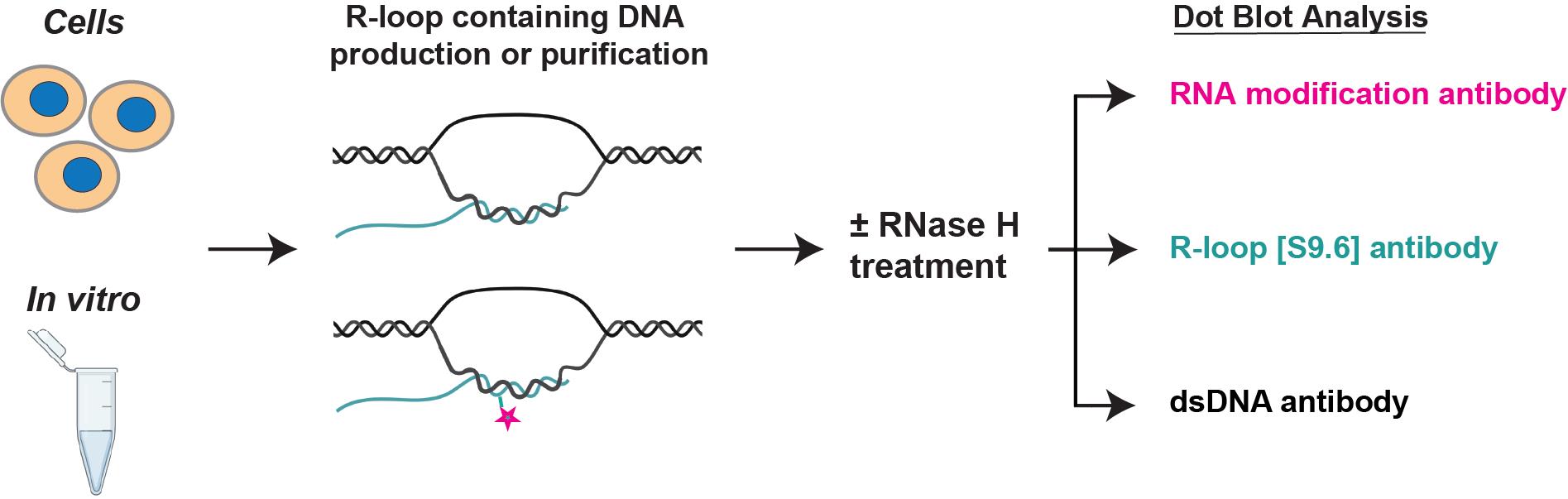
RNA modifications and their “writer,” “eraser,” and “reader” proteins are emerging as key regulators of gene expression and DNA repair through dynamically regulating RNA:DNA hybrids, or R-loops, during transcription. Therefore, it is paramount to develop rigorous techniques for accurate analysis of R-loop modifications. A convenient method for analyzing RNA modifications within total RNA is by dot blot with specific RNA modification antibodies; however, analysis of the modification of the RNA moiety within R-loops presents specific challenges. Here, we provide a detailed protocol for the production or purification of DNA containing R-loops in vitro and from cells, and the analysis of the RNA moiety modifications by dot blot. The DNA containing R-loops is treated with either mock or RNase H, which specifically degrades the RNA within RNA:DNA hybrids, to control for the specificity of the signal as originating from R-loops. Known quantities of the mock or RNase H–treated DNA are then spotted on three membranes, each blotted with antibodies that recognize double-stranded DNA, RNA:DNA hybrids, or the specific RNA modification antibodies of interest, such as m6A or ac4C. Thus, this protocol is useful to both biochemists and cell biologists with scientific interests at the intersection of R-loops and epitranscriptomics.
Key features
• R-loops produced via in vitro transcription of R-loop-forming DNA sequences can serve as substrates for biochemical assays, quantification standards, or antibody specificity controls.
• R-loop-containing DNA can be purified from cells after gene editing or various treatments to analyze how global R-loop levels and modifications are regulated.
• This protocol was initially applied to the study of the role of NAT10 and ac4C modification of the RNA moiety within R-loops in human cells.
• Requires 4–7 days to complete, depending on the origin of R-loops (in vitro transcription vs. from cells).
Keywords: R-loop Epitranscriptomics
Dot blot
ac4C
NAT10
Epitranscriptomics
Dot blot
ac4C
NAT10
Graphical overview
Background
R-loops are non-B nucleic acid structures formed by an RNA:DNA hybrid and the displaced ssDNA of the original DNA duplex [1]. They are formed by reinvasion of the DNA template by the nascent RNA molecule in a co-transcriptional manner. Two major classes of RNAPII-associated R-loops can be distinguished: type I R-loops are smaller (less than 60 nt long) and are associated with paused promoters, while type II R-loops are more variable in size (median size of 300 nt long) and are associated with gene bodies [2]. R-loops regulate key cellular processes, and their dynamic formation, resolution by helicases, or degradation by RNase H and other nucleases fine-tune transcription timing and yield and contribute to genome (in)stability [3]. Several RNA modifications and their “writer,” “eraser,” and “reader” proteins have recently emerged as key regulators of R-loops during transcription and DNA repair [4]. These include m6A [5–9], m5C and its oxidated derivative hm5C [10–12], A-to-I editing [13–16], and, more recently, ac4C [17]. Given the novelty of the R-loop epitranscriptomics field, it is important to develop accurate techniques for the analysis of RNA modifications within R-loops at the global level (e.g., dot blot, mass spectrometry), at the single R-loop loci level (e.g., next generation sequencing or direct sequencing), and at the single-cell level (e.g., immunofluorescence, single cell sequencing).
Here, we provide a detailed protocol for the global analysis of R-loop RNA modification by dot blot. This protocol was developed based on existing protocols for the production or purification of DNA containing R-loops in vitro and from cells [18,19], as well as our own development of the analysis of the RNA moiety modifications (i.e., ac4C) by dot blot [17]. In addition to molecular genetics controls consisting of the knock-out of the RNA-modifying writer (i.e., NAT10-KO), the DNA containing R-loops is treated with either mock or RNase H, which specifically degrades the RNA within RNA:DNA hybrids, to control for the specificity of the signal as originating from R-loops. Known quantities of the mock- or RNase H–treated DNA are then spotted on three membranes, each blotted with antibodies that recognize double-stranded DNA, RNA:DNA hybrids, and the specific RNA modification. In addition to the RNase H treatment control, we add an excess RNase A + T treatment control, which removes any trace of RNA from the samples [17]. This additional control is particularly important for the analysis of m5C and hm5C within R-loops, as these modifications exist at high levels in DNA (5mC and 5hmC). Additionally, as previously mentioned, R-loops from different loci (type I vs. type II) can have different lengths and stabilities, likely making RNA modifications associated with R-loops at paused promoters (type I) more difficult to detect. This is why it is important to use a gentle method of purifying R-loop-containing DNA [18] when probing R-loops for RNA modifications. As mentioned above, our protocol also contains a section for in vitro R-loop production, which can serve as substrates for biochemical assays, quantification standards, or for antibody specificity controls. Thus, this protocol is useful to scientists who aim to decipher how RNA modifications regulate R-loops at both the biochemical and functional levels.
Materials and reagents
Biological materials
1. HeLa control and NAT10-KO cells (gift from Dr. Shalini Oberdoerffer)
Reagents
1. pFC53-mAIRN plasmid (gift from Dr. Frédéric Chédin)
2. T3 RNA polymerase (Promega, catalog number: P2083)
3. 5× transcription optimized buffer (Promega, catalog number: P1181)
4. 1 M dithiothreitol (DTT) (ACROS Organics, catalog number: AC426380100)
5. 100% Tween-20 (Thermo Fisher Scientific, Fisher BioReagents, catalog number: BP337-500)
6. Ribonucleotide tri-phosphates (75 mM rATP, rGTP, rUTP, rCTP) (components of the MEGAscript T7 Transcription kit) (Thermo Fisher Scientific, Invitrogen, catalog number: AM1334)
7. Nuclease-free water (not DEPC-treated) (Thermo Fisher Scientific, Invitrogen, catalog number: AM9937)
8. 1 M Tris-HCl, pH 8.0 (Thermo Fisher Scientific, Invitrogen, catalog number: AM9855G)
9. 0.5 M EDTA, pH 8.0 (Thermo Fisher Scientific, Invitrogen, catalog number: AM9260G)
10. Ribonuclease (RNase) A (10 mg/mL) (Thermo Fisher Scientific, Thermo Scientific, catalog number: EN0531)
11. Ribonuclease (RNase) H (5 U/μL) (New England Biolabs, catalog number: M0297S)
12. Proteinase K (New England Biolabs, catalog number: P8107S)
13. 100% glycerol (MilliporeSigma, catalog number: G5516-500ML)
14. Agarose (low-EEO/multi-purpose/molecular biology grade) (Thermo Fisher Scientific, Fisher BioReagents, catalog number: BP160-500)
15. SYBR Safe DNA gel stain (10,000×) (Thermo Fisher Scientific, Invitrogen, catalog number: S33102)
16. Micro-Bio Spin 6 Columns (Bio-Rad, catalog number: 7326221)
17. 1× Dulbecco's phosphate-buffered saline (DPBS), no calcium, no magnesium (Thermo Fisher Scientific, Gibco, catalog number: 14190250)
18. Trypsin-EDTA (0.05%), phenol red (Thermo Fisher Scientific, Gibco, catalog number: 25300054)
19. Dulbecco’s minimum essential medium (DMEM), high glucose (Thermo Fisher Scientific, Gibco, catalog number: 11965118)
20. FoundationTM fetal bovine serum (FBS) (GeminiBio, catalog number: 900-108)
21. Penicillin-streptomycin-glutamine (PSQ) (100×) (Thermo Fisher Scientific, Gibco, catalog number: 10378016)
22. Trypan blue solution, 0.4% (Thermo Fisher Scientific, Gibco, catalog number: 15250061)
23. Sodium dodecyl sulfate (SDS) (MilliporeSigma, catalog number: L3771-1KG)
24. Phenol-chloroform-isoamyl alcohol mixture (25:24:1) (MilliporeSigma, catalog number: 77617-100ML)
25. Sodium acetate (NaOAc), 3 M aq. soln., pH 5.2, RNAse-free (Thermo Fisher Scientific, catalog number: AAJ61928AK)
26. Ethyl alcohol, pure (MilliporeSigma, catalog number: E7023-500ML)
27. NEBuffer r2.1 (10×) (New England Biolabs, catalog number: B6002S)
28. BsrGI-HF (New England Biolabs, catalog number: R3575S)
29. EcoRI-HF (New England Biolabs, catalog number: R3101S)
30. HindIII (New England Biolabs, catalog number: R0104S)
31. SspI-HF (New England Biolabs, catalog number: R3132S)
32. XbaI (New England Biolabs, catalog number: R0145S)
33. Glycogen (5 mg/mL) (Thermo Fisher Scientific, Invitrogen, catalog number: AM9510)
34. DNA gel loading dye (6×) (Thermo Fisher Scientific, Thermo Scientific, catalog number: R0611)
35. GeneRuler 1 kb DNA ladder, ready-to-use (Thermo Fisher Scientific, Invitrogen, catalog number: SM0313)
36. Ribonuclease (RNase) T1 (1,000 U/μL) (Thermo Fisher Scientific, Thermo Scientific, catalog number: EN0541)
37. PBS (10×), pH 7.4 (Thermo Fisher Scientific, Gibco, catalog number: 70011044)
38. Ethylenediaminetetraacetic acid, Di Na Salt Dihydr. (Na2EDTA·2H2O) (Crystalline powd./electrophor.) (Thermo Fisher Scientific, Fisher BioReagents, catalog number: BP120-500)
39. Anti-DNA-RNA hybrid (S9.6) antibody (Kerafast, catalog number: ENH001)
40. ds DNA marker antibody (HYB331-01) (Santa Cruz Biotechnology, catalog number: sc-58749)
41. Anti-N4-acetylcytidine (ac4C) antibody (EPRNCI-184-128) (Abcam, catalog number: ab252215)
42. Nonfat dry milk (Cell Signaling Technology, catalog number: 9999S)
43. Anti-rabbit IgG, HRP-linked antibody (Cell Signaling Technology, catalog number: 7074V)
44. Anti-mouse IgG, HRP-linked antibody (Cell Signaling Technology, catalog number: 7076V)
45. Pierce ECL western blotting substrate (Thermo Fisher Scientific, catalog number: 32160X4)
46. SuperSignal West Femto maximum sensitivity substrate (Thermo Fisher Scientific, catalog number: 34095)
47. Tris base (Fisher Scientific, catalog number: BP152-500)
48. Boric acid (Fisher Scientific, catalog number: BP168-500)
49. Sodium hydroxide (MilliporeSigma, catalog number: 221465-500G)
Solutions
1. 2.5% Tween-20 (see Recipes)
2. 2.5 mM rNTP (see Recipes)
3. 50% glycerol (see Recipes)
4. 0.5 M EDTA (see Recipes)
5. 10× TBE buffer (see Recipes)
6. TE buffer (see Recipes)
7. 20% SDS (see Recipes)
Recipes
1. 2.5% Tween-20
| Reagent | Final concentration | Volume |
| 100% Tween-20 | 2.5% | 1 mL |
| Nuclease-free water | n/a | 39 mL |
| Total | n/a | 40 mL |
Store at 4 °C.
2. 2.5 mM rNTP
| Reagent | Final concentration | Volume |
| rATP (75 mM) | 2.5 mM | 1 μL |
| rGTP (75 mM) | 2.5 mM | 1 μL |
| rUTP (75 mM) | 2.5 mM | 1 μL |
| rCTP (75 mM) | 2.5 mM | 1 μL |
| Nuclease-free water | n/a | 26 μL |
| Total | n/a | 30 μL |
Store at -20 °C.
3. 50% glycerol
| Reagent | Final concentration | Volume |
| 100% glycerol | 50% | 50 mL |
| ddH2O | n/a | 50 mL |
| Total | n/a | 100 mL |
Autoclave the solution. Store at room temperature.
4. 0.5 M EDTA
| Reagent | Final concentration | Quantity or volume |
| Na2EDTA·2H2O | 0.5 M | 186.12 g |
| ddH2O | n/a | 800 mL |
| Stir the solution vigorously | ||
| NaOH | Adjust to pH 8.0 | ~ 20 g of NaOH pellets |
| ddH2O | to 1 L | |
| Total | n/a | 1,000 mL |
Filter the solution using a 0.22-μm filter. Store at room temperature.
5. 10× TBE buffer
| Reagent | Final concentration | Quantity or volume |
| Tris base | 890 mM | 108 g |
| Boric acid | 890 mM | 55 g |
| ddH2O | n/a | 800 mL |
| 0.5 M EDTA, pH 8.0 (Recipe 4) | 20 mM | 40 mL |
| ddH2O | to 1 L | |
| Total | n/a | 1,000 mL |
Filter the solution using a 0.22-μm filter. Store at room temperature.
6. TE buffer
| Reagent | Final concentration | Volume |
| 0.5 M EDTA, pH 8.0 | 1 mM | 100 μL |
| 1 M Tris-HCl, pH 8.0 | 10 mM | 500 μL |
| Nuclease-free water | n/a | 49.4 mL |
| Total | n/a | 50 mL |
Store at room temperature.
7. 20% SDS
| Reagent | Final concentration | Quantity or volume |
| SDS | 20% | 200 g |
| ddH2O | n/a | 800 mL |
| Stir the solution overnight on a stir plate at 50 °C | ||
| ddH2O | to 1 L | |
| Total | n/a | 1,000 mL |
Filter the solution using a 0.22-μm filter. Store at room temperature.
Laboratory supplies
1. Pipette tips LTS, 1,000 μL (Mettler Toledo, catalog numbers: 30389212 and 30389272)
2. Pipette tips LTS, 200 μL (Mettler Toledo, catalog numbers: 30389240 and 30389276)
3. Pipette tips GP LTS, 20 μL (Mettler Toledo, catalog numbers: 30389226 and 30389274)
4. Serological pipettes, 10 mL (Genesee Scientific, GenClone, catalog number: 12-104C)
5. Serological pipettes, 5 mL (Genesee Scientific, GenClone, catalog number: 12-102C)
6. DNA LoBind tubes, 2 mL (Eppendorf, catalog number: 022431048)
7. DNA LoBind tubes, 1.5 mL (Eppendorf, catalog number: 022431021)
8. 0.2-mL PCR tubes (Eppendorf, catalog number: 951010006)
9. Cell culture/Petri dishes, 150 × 20 mm (Thermo Fisher Scientific, Nunc, catalog number: 168381)
10. Disposable borosilicate glass Pasteur pipettes (Thermo Fisher Scientific, Fisherbrand, catalog number: 13-678-20D)
11. Cell counting slides, 500 slides (Logos Biosystems, Luna, catalog number: L12002)
12. MaXtract high density, 100 × 15 mL (Qiagen, catalog number: 129065)
13. PhaseShield gel tubes (200 × 2 mL) (Biofargo, catalog number: M2302-20-H)
14. 15-mL conical centrifuge tubes (Corning, Falcon, catalog number: 352097)
15. 50-mL conical centrifuge tubes (Corning, Falcon, catalog number: 352070)
16. BrightStar-Plus positively charged nylon membrane, 30 cm × 45 cm (Thermo Fisher Scientific, Invitrogen, catalog number: AM10102)
17. Syringe filters (30 mm, 0.22 µm, PES, PP, non-sterile) (VWR, catalog number: 76479-018)
Equipment
1. Rainin Pipet-Lite XLS LTS single-channel pipettes (100–1,000 μL, 20–200 μL, 2–20 μL, 0.5–10 μL, and 0.1–2 μL)
2. Rainin Pipet-X pipette controller
3. IKA Vortex 4 Digital vortexer
4. Centrifuges (Eppendorf, models: 5810R, 5702, and 5427R)
5. Applied Biosystems ProFlex PCR System
6. Bio-Rad horizontal DNA gel electrophoresis system
7. Syngene G:box
8. DeNovix DS-11 spectrophotometer
9. New Brunswick Galaxy 170S CO2 incubator
10. Inverted laboratory microscope (Leica, model: DM IL LED)
11. LUNA Automated Cell Counter
12. UVP HB-1000 hybridization incubator
13. Eppendorf ThermoMixer C
14. Dual LED blue/white light transilluminator (Thermo Fisher Scientific, Invitrogen, catalog number: LB0100)
15. Stratagene Stratalinker UV 1800 Crosslinker
16. Labnet International Rocker
Software and datasets
1. ImageJ, Version 2.14.0/1.54f
2. Prism 10
Procedure
A. In vitro R-loop synthesis
The validation of the specificity of the S9.6 antibody is recommended before using it to assess the R-loops in different types of cells or under various treatments. This can be achieved by testing its performance with a dot blot using R-loops produced in vitro. The pFC53-mAirn plasmid, which contains the CpG island of the mouse Airn gene, is designed to synthesize stable R-loops in vitro using the T3 RNA polymerase system [19]. Several controls are needed when performing the in vitro R-loop synthesis. Treatment with RNase A removes free RNAs, and additional treatment with RNase H degrades R-loops.
1. Thaw the frozen reagents needed for the in vitro transcription by placing the T3 RNA Polymerase on ice and the other reagents on a nutator at room temperature. Immediately after thawing, quickly spin the rNTP tube for 5 s to collect the solution at the bottom and place it on ice. Keep the 5× Promega Transcription Optimized Buffer at room temperature to avoid precipitation of the DNA template due to the 10 mM spermidine.
2. Prepare a 1.5-mL microcentrifuge tube containing 3 μg of pFC53-mAirn plasmid, Promega 1× Transcription Optimized Buffer, 20 mM DTT, 0.05% Tween-20, and 0.25 mM rNTP in a total volume of 45.5 μL (Table 1) at room temperature.
Table 1. Components for setting up an in vitro transcription reaction
| Components | Final concentration | Volume (μL) |
| Nuclease-free water | - | to 45.5 |
| pFC53-mAirn plasmid | 3 μg/50 μL reaction | |
| 5× Promega Transcription Optimized Buffer | 1× | 10 |
| 1 M DTT | 20 mM | 1 |
| 2.5% Tween-20 | 0.05% | 1 |
| 2.5 mM rNTP | 0.25 mM | 5 |
3. Add 4.5 μL (160 U) of T3 RNA polymerase into the 1.5-mL tube and vortex for 30 s at 500 rpm.
4. Aliquot 25 μL of the reaction into two 0.2-mL microcentrifuge tubes.
5. Initiate the reaction by incubating the 0.2-mL tubes at 37 °C for 30 min in the PCR thermal cycler.
6. Inactivate the reaction by heating the 0.2-mL tubes at 65 °C for 10 min in the same PCR thermal cycler.
7. Take the 0.2-mL tubes out at room temperature. Then, add 5 μL of RNase A at 0.1 μg/μL (0.5 μg total) into one of the 0.2-mL tubes (RNase A sample), followed by 5 μL of RNase A at 0.1 μg/μL (0.5 μg total) and 2 μL of RNase H at 5 U/μL (10 units total) into the other 0.2-mL tube (RNase A+H sample).
8. Incubate both 0.2-mL tubes at 37 °C for 30 min in the PCR thermal cycler.
9. Take the 0.2-mL tubes back out at room temperature. Then, add 2 μL of Proteinase K into each tube.
10. Incubate both tubes at 37 °C for 30 min in the PCR thermal cycler.
11. To check the R-loop quality, take 4 μL from each tube and mix it with 1 μL of 50% glycerol. Load the 5 μL samples on a 0.9% agarose gel in 1× TBE and run at 100 V for 1 h.
12. Stain the agarose gel by submerging it in a box containing 100 mL of 1× TBE with 1× SYBR Safe DNA gel stain for 15 min on a rocker.
13. Image the gel using a Syngene G:box imaging system (Figure 1).
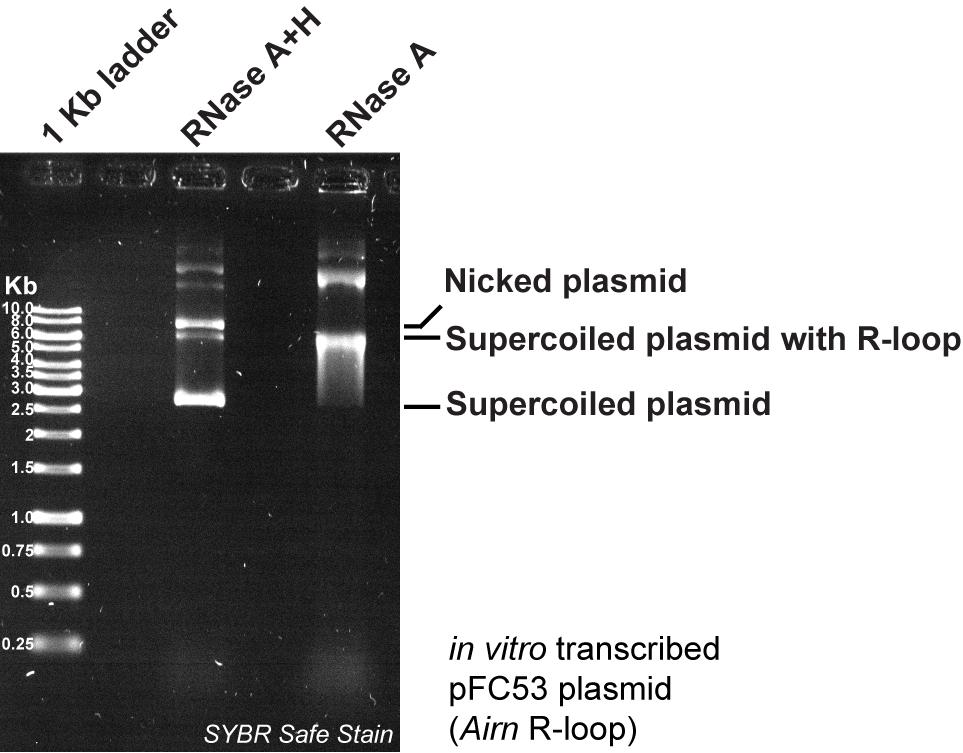
Figure 1. Validation of the in vitro–produced R-loop by agarose gel electrophoresis, followed by SYBR Safe staining
14. Purify the samples using size exclusion chromatography with the Micro-Bio Spin 6 Columns.
15. Quantify the DNA using a DeNovix DS-11 spectrophotometer.
16. Store the samples at -20 °C until further use.
B. Genomic DNA purification from cells
Genomic DNAs from different cell lines or cells at various treatments are purified as described in [18] with slight modifications. More specifically, the example of purifying gDNA from HeLa control and NAT10-KO cells [17] is shown here.
1. Plate 20 mL of 5 × 10⁴ cells/mL for HeLa control and 7.5 × 10⁴ cells/mL for HeLa NAT10-KO cells in a 20-cm diameter plate and culture for 3 days. At this time, the cells should be at 75%–80% cell confluency.
Note: A total of 5–6 million cells is recommended as a starting material for genomic DNA purification, so count the cells and pool multiple dishes if needed.
2. Rinse the cells twice with 20 mL of 1× DPBS prewarmed to 37 °C. Then, dissociate the cells by adding 5 mL of 0.05% Trypsin-EDTA prewarmed to 37 °C.
3. Check the detached status of cells under a microscope. It will take 3–4 min for HeLa cells to be detached after adding 0.05% Trypsin-EDTA.
4. Add 5 mL of DMEM supplemented with 10% FBS and 1× PSQ prewarmed to 37 °C to stop the trypsinization. Then, pipette the detached cells and transfer them into a 15-mL conical tube.
5. Centrifuge the conical tube at 200× g at room temperature for 2 min.
6. Aspirate the supernatant and gently resuspend the cell pellet in 1 mL of 1× DPBS with a P1000 pipette and then another 4 mL of DPBS.
7. Take 10 μL of cell suspension into a 1.5-mL microcentrifuge tube and mix it with 10 μL of 0.4% trypan blue solution by pipetting up and down 10 times gently. Pipette 10 μL of the stained cells into a chamber of the Luna cell counting slide and measure the concentration of cells using the Luna cell counter.
8. Centrifuge the cells in the conical tube at 200× g at room temperature for 2 min.
9. Aspirate all the supernatant and gently resuspend the cell pellet in TE buffer to make the cell density 5 × 10⁶ cells/mL. That is, if a total of 1 × 10⁷ cells is collected, resuspend the cell pellet in 2 mL of TE buffer. Make sure the cells are fully resuspended without any clumps observed.
10. Take 1.6 mL of cell suspension into a 2-mL microcentrifuge tube and add 50 μL of 20% SDS and 5 μL of Proteinase K into the cell suspension. Do not try to pipette the cell suspension after adding SDS. Invert the conical tube gently six times until the cell suspension becomes viscous.
11. Incubate the tube at 300 rpm at 37 °C for 12–14 h in an Eppendorf ThermoMixer with a heated lid.
12. Prepare a 15-mL MaXtract high-density tube and spin it at 1,500× g at room temperature for 1 min to pellet the gel. Then, pour all the cell lysate from step B11 into the 15-mL tube.
13. Add 1.6 mL (1 volume) of phenol-chloroform-isoamyl alcohol mixture (25:24:1) into the 15-mL tube.
Note: Equilibrate the phenol-chloroform-isoamyl alcohol mixture (25:24:1) at room temperature for 1 h before use.
14. Invert the 15-mL tube gently six times and centrifuge the tube at 1,500× g at room temperature for 5 min. If the supernatant is not clear after the centrifugation, centrifuge the tube at 1,500× g at room temperature for an additional 5 min.
15. Check the volume of the aqueous phase by inspecting the scale on the side of the tube.
16. Prepare a 15-mL conical tube containing 1/10 volume of 3 M NaOAc (pH 5.2) and 2.5 volumes of 100% ethanol. That is, since the expected volume of the aqueous phase is 1.6 mL, prepare a 15-mL conical tube containing 160 μL of 3 M NaOAc (pH 5.2) and 4 mL of 100% ethanol.
17. Pour the aqueous phase into the tube prepared in the last step. Invert the tube gently until white (not translucent) precipitate is observed. Do not centrifuge the tube, as it will result in RNA contamination. Transfer the DNA precipitate carefully with a 1,000-mL tip (cut at the 250-μL mark) to a 2-mL microcentrifuge tube.
18. Wash the DNA precipitate by adding 1.5 mL of 80% ethanol, gently inverting the tube three times, and letting it stand for 10 min at room temperature. Do not centrifuge the tube.
19. Carefully remove the supernatant without disturbing the DNA pellet.
20. Repeat steps B18–19 twice. At the last wash, carefully remove as much ethanol as possible.
21. Invert the tube to air-dry the DNA pellet completely, which may take up to 1 h depending on the amount of DNA.
C. Genomic DNA fragmentation
To obtain DNA fragments at 3–5 kb on average but also retain the RNA:DNA hybrids, genomic DNAs are digested, instead of sonicated, using a mix of five restriction enzymes (BsrGI-HF, EcoRI-HF, HindIII, SspI-HF, and XbaI), as described in [17] with slight modifications. Several controls are necessary when interpreting the R-loop dot blot results. The treatment with RNase H will degrade R-loops, and the treatment with excess RNase A/T1 at low-salt concentration will remove any residual RNAs.
1. Add 125 μL of TE buffer directly to the DNA pellet from step B21 and keep the tube on ice for 1 h. Then, gently pipette the DNA up and down three times with a 200-μL tip (cut at the 50-μL mark) and leave the tube on ice for another hour.
Note: Do not try to resuspend the DNA pellet by overpipetting or even vortexing. The genomic DNA should remain viscous at this stage.
2. Prepare the restriction enzyme digestion reactions in 1.5-mL microcentrifuge tubes for the HeLa control and NAT10-KO samples. The components are shown in Table 2.
Table 2. Components for setting up a restriction enzyme digestion
| Components | Final concentration | Volume (μL) |
| Nuclease-free water | - | to 150 |
| Extracted genomic DNA | - | 100 |
| 10× NEBuffer r2.1 | 1× | 15 |
| BsrGI-HF | 30 units | 1.5 |
| EcoRI-HF | 30 units | 1.5 |
| HindIII | 30 units | 1.5 |
| SspI-HF | 30 units | 1.5 |
| XbaI | 30 units | 1.5 |
3. Incubate the digestion reaction tubes at 300 rpm at 37 °C overnight in the Eppendorf ThermoMixer.
4. Prepare two 2-mL PhaseShield gel tubes and spin them at 16,000× g at room temperature for 1 min to pellet the gel. Then, gently pipette all the digested DNA (150 μL) into the PhaseShield gel tubes.
5. Add 100 μL of nuclease-free water to increase the volume to 250 μL and 1 volume (i.e., 250 μL) of phenol-chloroform-isoamyl alcohol mixture (25:24:1) into the PhaseShield gel tubes.
6. Gently invert the PhaseShield gel tubes six times and centrifuge at 16,000× g at room temperature for 10 min.
7. Prepare two 1.5-mL microcentrifuge tubes containing glycogen, 1/10 volume of 3 M NaOAc (pH 5.2), and 2.5 volumes of 100% ethanol. That is, if the volume of the DNA is 250 μL, prepare a microcentrifuge tube containing 1.5 μL of glycogen, 25 μL of 3 M NaOAc (pH 5.2), and 625 μL of 100% ethanol.
8. Transfer the DNA from the PhaseShield gel tubes into the 1.5-mL microcentrifuge tubes prepared in the last step and then invert the tubes six times gently.
9. Incubate the tubes at -20 °C for 1 h to increase precipitation yield.
10. Centrifuge the tube at 16,000× g for 35 min at 4 °C.
11. Remove the supernatant and add 200 μL of 80% ethanol. Then, centrifuge the tube at 16,000× g for 10 min at 4 °C.
12. Carefully remove the supernatant without disturbing the DNA pellet.
13. Repeat steps C11–12.
14. Air-dry the pellet at room temperature for 15–25 min.
15. Add 50 μL of TE buffer directly to the DNA pellet and keep the tube on ice for 30 min. Then, gently resuspend the DNA and measure the DNA concentration using the DeNovix DS-11 spectrophotometer.
Note: Do not try to resuspend the DNA pellet by overpipetting or even vortexing. Leave the tube on ice longer to help the resuspension.
16. To check the digestion quality, take 1 μg of digested DNA into a 1.5-mL microcentrifuge tube and then add 1 μL of 6× DNA loading buffer and TE buffer to make a final volume of 6 μL. Run those samples with 5 μL of GeneRuler 1 kb DNA ladder in a 0.8% agarose gel in 1× TBE with 1× SYBR Safe DNA gel stain at 100 V for 45 min. Image it using a Syngene G:box (see Figure 2).
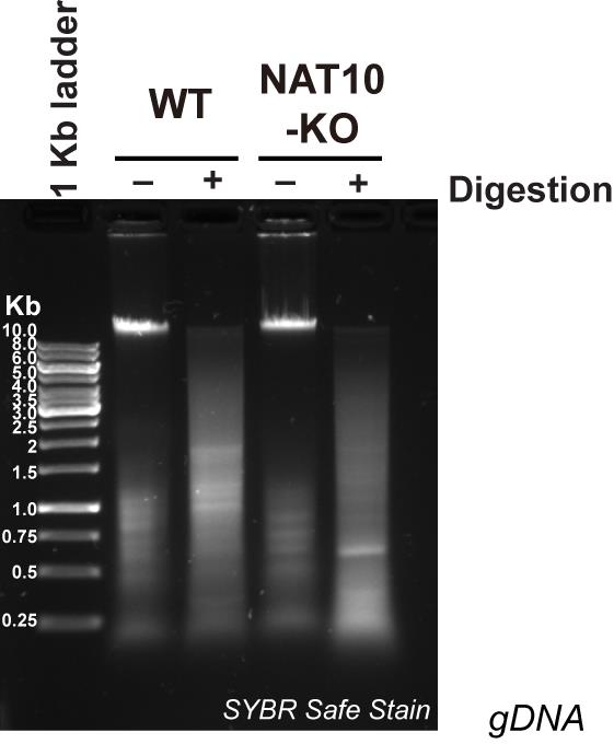
Figure 2. Example of a 0.8% agarose gel showing 1 μg of gDNA after mock treatment (-) or treatment with restriction enzymes (+)
17. For each sample, prepare six 1.5-mL microcentrifuge tubes, labeling two as mock, two as RNase H, and two as RNase A/T1 on the lids. Add 5 μg of DNA into each tube.
18. For the tubes labeled as mock, add 10 μL of 10× RNase H Reaction Buffer and nuclease-free water to make a final volume of 100 μL. For the tubes labeled as RNase H, add 5 μL of RNase H (5U/μL), 10 μL of 10× RNase H reaction buffer, and nuclease-free water to make a final volume of 100 μL. For the tubes labeled RNase A/T1, add 2 μL of RNase A (10 μg/μL), 2 μL of RNase T1 (1,000 U/μL), and 1× PBS to make a final volume of 100 μL.
19. Incubate the tubes at 300 rpm at 37 °C for 30 min in the Eppendorf ThermoMixer.
20. Add 1 μL of 0.5 M EDTA into each tube to stop the reactions.
21. Prepare three 2-mL PhaseShield gel tubes for each sample and centrifuge at 16,000× g at room temperature for 1 min to pellet the gel. Then, combine the duplicate tubes for each of the three groups (mock, RNase H, and RNase A/T1) by transferring all treated DNA (totaling 202 μL) into each of the PhaseShield gel tubes. Add 48 μL of nuclease-free water to increase the volume to 250 μL and 1 volume (i.e., 250 μL) of phenol-chloroform-isoamyl alcohol mixture (25:24:1) into the PhaseShield gel tubes.
22. Gently invert the PhaseShield gel tubes six times and centrifuge at 16,000× g at room temperature for 10 min.
23. Prepare six 1.5-mL microcentrifuge tubes containing 1.5 μL of glycogen, 25 μL of 3 M NaOAc (pH 5.2), and 625 μL of 100% ethanol.
24. Transfer the DNA from the PhaseShield gel tubes into the 1.5-mL tubes prepared in the last step and then invert the tubes six times gently.
25. Incubate the tubes at -20 °C for at least 2 h to increase precipitation yield.
26. Centrifuge the tube at 16,000× g for 35 min at 4 °C.
27. Remove the supernatant and add 200 μL of 80% ethanol. Then, centrifuge the tube at 16,000× g for 10 min at 4 °C.
28. Carefully remove the supernatant without disturbing the DNA pellet.
29. Repeat steps C27–28.
30. Air-dry the pellet at room temperature for 15–25 min.
31. Add 25 μL of TE buffer directly to the DNA pellet and keep the tubes on ice for 30 min. Then, gently resuspend the DNA and measure DNA concentration using the DeNovix DS-11 spectrophotometer.
32. To check treatment quality, take 2.5 μL of DNA from step C31 into a 1.5-mL microcentrifuge tube and then add 2.5 μL of TE buffer and 1 μL of 6× DNA loading buffer to make a final volume of 6 μL. Run those samples with 5 μL of GeneRuler 1 kb DNA ladder in a 0.8% agarose gel in 1× TBE with 1× SYBR Safe at 100 V for 45 min. Image it using a Syngene G:box (see Figure 3).
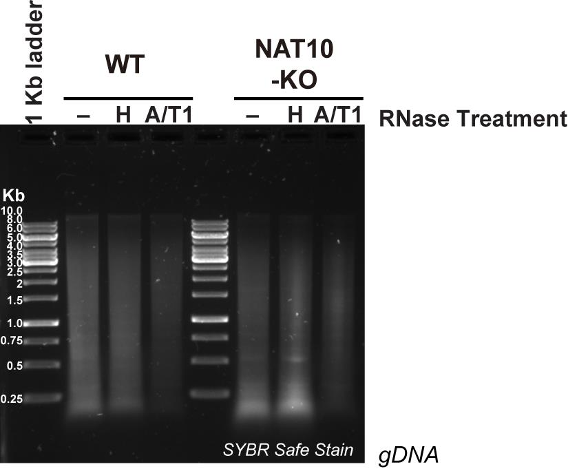
Figure 3. Example of a 0.8% agarose gel showing 1 μg of gDNA solubilized with the cocktail of restriction enzymes after mock treatment (-), treatment with RNase H (H), or with excess RNase A/T1 (A/T1)
D. R-loop dot blot (ac4C/S9.6/dsDNA)
1. For each mock-, RNase H-, or RNase A/T1-treated sample, prepare three dilutions of genomic DNA in a final volume of 10 μL (e.g., 200 ng/μL, 100 ng/μL, and 50 ng/μL).
2. Prepare three pieces of BrightStar-Plus positively charged nylon membrane of sufficient size to accommodate all samples. For orientation, mark an X in the upper left corner of each membrane using a pencil (Figures 4 and 5).
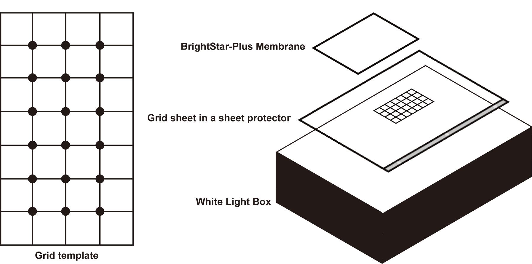
Figure 4. Grid template and setup for loading the samples for subsequent dot blotting
3. Place a membrane on the top of a grid sheet as shown in Figure 4. Then, slowly and gently blot 2.5 μL of each dilution on the membrane at the grid intersection. Repeat this step until all three membranes have the samples in the same order (Figure 5).
Note: Ensure that the pipette tip does not contact the membrane during DNA application; a distance of approximately 2 mm should be maintained. Although a multichannel pipette may be used, manual dispensing with a single-channel pipette yields more consistent and well-defined dots.
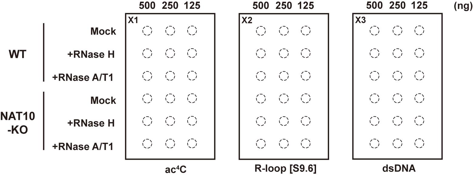
Figure 5. Dot blot loading example
4. Let the membranes dry at room temperature for 30 min or until no spots are visible.
5. Crosslink DNA on the membranes with 150,000 μJ (150 mJ/cm2) of UV254 nm using a Stratalinker UV crosslinker.
6. Block the membranes with 10 mL of 1× PBS containing 0.1% Tween-20 and 5% nonfat dry milk for 30 min on a rocker.
7. Dilute the primary antibodies (ac4C, S9.6, or dsDNA) at a 1:1,000 ratio in 5 mL of 1× PBS containing 0.1% Tween-20 and 1% nonfat dry milk. After aspirating the blocking solution, incubate the membranes overnight at 4 °C on a rocker with the diluted antibody solution.
8. Discard the primary antibody solutions and wash the membranes three times with 10 mL of 1× PBS containing 0.1% Tween-20 for 10 min on a rocker for each wash.
9. Dilute the HRP-linked secondary antibodies at a 1:10,000 ratio in 10 mL of 1× PBS containing 0.1% Tween-20 and 1% nonfat dry milk. Incubate the membranes with secondary antibody solutions overnight at 4 °C on a rocker.
10. Discard the secondary antibody solutions and wash the membranes three times with 10 mL of 1× PBS containing 0.1% Tween-20 for 10 min on a rocker for each wash.
11. Aspirate the solutions and add 2 mL of a 1:1 mixture of the ECL reagent directly on top of the membrane. Incubate the membrane for 2 min and place it in a reaction folder. Rub the folder with a Kimwipe to eliminate air bubbles.
Note: Apply the Pierce ECL western blotting substrate to the membranes treated with dsDNA and S9.6 antibodies, and the SuperSignal West Femto maximum sensitivity substrate to the membrane treated with the ac4C antibody.
12. Image the membranes using a chemiluminescent imaging system, acquiring exposure times at 1 s, 10 s, 30 s, 1 min, 2 min, 5 min, and 20 min, along with a final membrane image.
Data analysis
Data analysis is done through quantification of the dot blot images (step D12) using ImageJ.
1. Select the image from the stack in step D12 where the signal is not saturated and remains linear between the three dilutions. Create a circle with a diameter 1 mm larger than the dots, place it over the first dot, and click Analyze → Measure. Continue doing the same for each dot as well as an empty area of the dot blot (i.e., the blank sample). Save the results as a .csv file first and then as .xlsx after opening it in Excel. Subtract the “Mean” value of the blank sample from the “Mean” value of each dot.
2. To quantify the ac4C signal originating from R-loops in HeLa control and HeLa NAT10-KO cells, normalize the ac4C signal of each mock sample over the corresponding +RNase H sample and over HeLa control. The dot blots with the S9.6 and dsDNA antibodies from the same samples are used to make sure that equal amounts of R-loops and dsDNA are loaded (see step 3 below). ac4C dot blots from three biological replicates are analyzed in this way, and a paired two-tailed ratio t-test is used to calculate the statistical significance. See Figure 4A from [17] for an example.
3. To quantify the S9.6 signal in HeLa control and HeLa NAT10-KO cells, normalize the S9.6 signal of each mock and RNase H sample over the dsDNA signal of the corresponding +RNase A/T1 sample and further normalize the S9.6 signal over HeLa control. S9.6 and dsDNA dot blots from three biological replicates are analyzed in this way, and a paired two-tailed t-test is used to calculate the statistical significance. See Figure 3D from [17] for an example.
Note: The value of +RNase A/T1 sample is used for normalization, as this sample does not have any trace of R-loop or RNA left. R-loops could interfere with the dsDNA antibody owing to the melting of the DNA duplex at R-loop regions (see Background section).
Validation of protocol
This protocol has been used and validated in the following research article:
• Debnath et al. [17]. NAT10 and N4-acetylcytidine restrain R-loop levels and related inflammatory responses. Science Advances (Figures 3C–D and 4A).
A dot blot example performed independently is shown in Figure 6.
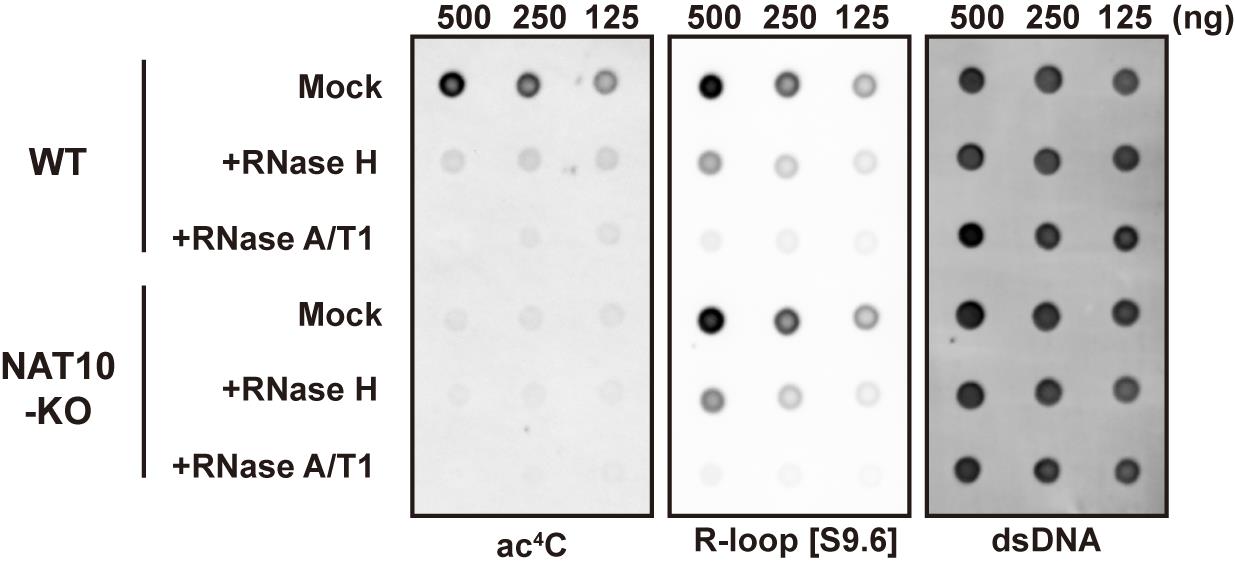
Figure 6. Dot blot example
General notes and troubleshooting
General notes
1. When working with R-loops, make sure to only use DNase/RNase-free reagents and filtered tips.
2. As indicated in the protocol, there are several potential stopping points. However, we have noticed that we obtain the best results when we do not freeze the R-loop-containing dsDNA and use it directly for the subsequent assays. However, we have observed that flash-freezing in liquid nitrogen at step C15 does not affect the quality of R-loops or their modification with ac4C.
3. In addition to checking the specificity of the S9.6 antibody, as in [17], the in vitro–produced R-loops (see section A) could be used in in vitro RNA modification assays. Additionally, one of the rNTPs could be substituted with a modified nucleotide (such as ac4CTP) either entirely (100% ac4CTP) or at defined proportions (i.e., 12.5% ac4CTP and 87.5% CTP, etc.) to serve as positive controls or standards for the RNA modification R-loop dot blots, or to be used in other assays.
Troubleshooting
Problem 1: The in vitro R-loop production protocol may not provide good yields.
Possible cause: The plasmid used for the in vitro transcription is of lower quality, i.e., not supercoiled. This will be visible on the gel in the RNase A+H sample, with the ratio of nicked vs. supercoiled plasmid being over 25%.
Solution: Produce fresh plasmid and check its quality on agarose gel. Using a plasmid that is at least 90% supercoiled is highly recommended.
Problem 2: The S9.6 or the RNA modification antibodies may not work well in dot blot with samples purified from cells.
Possible cause 1: The R-loops can be degraded during cell lysis and subsequent DNA purification due to harsh handling or RNase contamination.
Possible cause 2: The antibodies are no longer working.
Solution: R-loops produced in vitro with or without modified rNTPs can be used as a spike-in during the procedure or as a positive control for the dot blots.
Acknowledgments
The original research in the Xhemalçe lab that served as the basis for this Bio-protocol paper [17] was supported by NIH Grant R01 GM127802, and an APX grant from the office of the Vice President for Research at the University of Texas at Austin, USA. Y.R.L. and B.X. were supported by NSF Grant 2418919.
The original research paper in which the protocol was described and validated is: Debnath et al. Science Advances (2025) [17]. We would like to thank Dr. Turja Debnath and Enrique Navedo for initially setting up the assays published in [17] and described here.
Previous works from which the protocol was developed are [17–19]. Additionally, personal communications from the Chedin (UC Davis) and Finkelstein (UT Austin) labs assisted our development of the in vitro R-loop production protocol.
Author contributions
Specific contributions of each author: Conceptualization, Y.R.L. and B.X.; Investigation, T.Y., Y.R.L., and B.X.; Writing—Original Draft, Y.R.L. and B.X.; Writing—Review & Editing, T.Y. and B.X.; Funding acquisition, B.X.; Supervision, B.X.
Competing interests
The authors declare no conflicts of interest.
References
- Gaillard, H. and Aguilera, A. (2016). Transcription as a Threat to Genome Integrity. Annu Rev Biochem. 85(1): 291–317. https://doi.org/10.1146/annurev-biochem-060815-014908
- Castillo-Guzman, D. and Chédin, F. (2021). Defining R-loop classes and their contributions to genome instability. DNA Repair. 106: 103182. https://doi.org/10.1016/j.dnarep.2021.103182
- Aguilera, A. and García-Muse, T. (2012). R Loops: From Transcription Byproducts to Threats to Genome Stability. Mol Cell. 46(2): 115–124. https://doi.org/10.1016/j.molcel.2012.04.009
- Xhemalçe, B., Miller, K. M. and Gromak, N. (2024). Epitranscriptome in action: RNA modifications in the DNA damage response. Mol Cell. 84(19): 3610–3626. https://doi.org/10.1016/j.molcel.2024.09.003
- Abakir, A., Giles, T. C., Cristini, A., Foster, J. M., Dai, N., Starczak, M., Rubio-Roldan, A., Li, M., Eleftheriou, M., Crutchley, J., et al. (2019). N6-methyladenosine regulates the stability of RNA:DNA hybrids in human cells. Nat Genet. 52(1): 48–55. https://doi.org/10.1038/s41588-019-0549-x
- Yang, X., Liu, Q. L., Xu, W., Zhang, Y. C., Yang, Y., Ju, L. F., Chen, J., Chen, Y. S., Li, K., Ren, J., et al. (2019). m6A promotes R-loop formation to facilitate transcription termination. Cell Res. 29(12): 1035–1038. https://doi.org/10.1038/s41422-019-0235-7
- Hao, J. D., Liu, Q. L., Liu, M. X., Yang, X., Wang, L. M., Su, S. Y., Xiao, W., Zhang, M. Q., Zhang, Y. C., Zhang, L., et al. (2024). DDX21 mediates co-transcriptional RNA m6A modification to promote transcription termination and genome stability. Mol Cell. 84(9): 1711–1726.e11. https://doi.org/10.1016/j.molcel.2024.03.006
- Zhang, C., Chen, L., Peng, D., Jiang, A., He, Y., Zeng, Y., Xie, C., Zhou, H., Luo, X., Liu, H., et al. (2020). METTL3 and N6-Methyladenosine Promote Homologous Recombination-Mediated Repair of DSBs by Modulating DNA-RNA Hybrid Accumulation. Mol Cell. 79(3): 425–442.e7. https://doi.org/10.1016/j.molcel.2020.06.017
- Chen, L., Zhang, C., Ma, W., Huang, J., Zhao, Y. and Liu, H. (2022). METTL3-mediated m6A modification stabilizes TERRA and maintains telomere stability. Nucleic Acids Res. 50(20): 11619–11634. https://doi.org/10.1093/nar/gkac1027
- Chen, H., Yang, H., Zhu, X., Yadav, T., Ouyang, J., Truesdell, S. S., Tan, J., Wang, Y., Duan, M., Wei, L., et al. (2020). m5C modification of mRNA serves a DNA damage code to promote homologous recombination. Nat Commun. 11(1): 2834. https://doi.org/10.1038/s41467-020-16722-7
- Yang, H., Lachtara, E. M., Ran, X., Hopkins, J., Patel, P. S., Zhu, X., Xiao, Y., Phoon, L., Gao, B., Zou, L., et al. (2023). The RNA m5C modification in R-loops as an off switch of Alt-NHEJ. Nat Commun. 14(1): e1038/s41467–023–41790–w. https://doi.org/10.1038/s41467-023-41790-w
- Yang, H., Wang, Y., Xiang, Y., Yadav, T., Ouyang, J., Phoon, L., Zhu, X., Shi, Y., Zou, L., Lan, L., et al. (2022). FMRP promotes transcription-coupled homologous recombination via facilitating TET1-mediated m5C RNA modification demethylation. Proc Natl Acad Sci USA. 119(12): e2116251119. https://doi.org/10.1073/pnas.2116251119
- Zheng, Y., Lorenzo, C. and Beal, P. A. (2017). DNA editing in DNA/RNA hybrids by adenosine deaminases that act on RNA. Nucleic Acids Res. 45(6): 3369–3377. https://doi.org/10.1093/nar/gkx050
- Shiromoto, Y., Sakurai, M., Minakuchi, M., Ariyoshi, K. and Nishikura, K. (2021). ADAR1 RNA editing enzyme regulates R-loop formation and genome stability at telomeres in cancer cells. Nat Commun. 12(1): e1038/s41467–021–21921–x. https://doi.org/10.1038/s41467-021-21921-x
- Jimeno, S., Prados-Carvajal, R., Fernández-Ávila, M. J., Silva, S., Silvestris, D. A., Endara-Coll, M., Rodríguez-Real, G., Domingo-Prim, J., Mejías-Navarro, F., Romero-Franco, A., et al. (2021). ADAR-mediated RNA editing of DNA:RNA hybrids is required for DNA double strand break repair. Nat Commun. 12(1): 5512. https://doi.org/10.1038/s41467-021-25790-2
- Zhang, B., Li, Y., Zhang, J., Wang, Y., Liang, C., Lu, T., Zhang, C., Liu, L., Qin, Y., He, J., et al. (2023). ADAR1 links R-loop homeostasis to ATR activation in replication stress response. Nucleic Acids Res. 51(21): 11668–11687. https://doi.org/10.1093/nar/gkad839
- Debnath, T. K., Abell, N. S., Li, Y. R., Devanathan, S. K., Navedo, E. and Xhemalçe, B. (2025). NAT10 and N4-acetylcytidine restrain R-loop levels and related inflammatory responses. Sci Adv. 11(13): eads6144. https://doi.org/10.1126/sciadv.ads6144
- Sanz, L. A. and Chédin, F. (2019). High-resolution, strand-specific R-loop mapping via S9.6-based DNA–RNA immunoprecipitation and high-throughput sequencing. Nat Protoc. 14(6): 1734–1755. https://doi.org/10.1038/s41596-019-0159-1
- Stolz, R., Sulthana, S., Hartono, S. R., Malig, M., Benham, C. J. and Chedin, F. (2019). Interplay between DNA sequence and negative superhelicity drives R-loop structures. Proc Natl Acad Sci USA. 116(13): 6260–6269. https://doi.org/10.1073/pnas.1819476116
Article Information
Publication history
Received: Jan 19, 2026
Accepted: Jul 27, 2026
Available online: Aug 14, 2026
Published: Sep 5, 2026
Copyright
© 2026 The Author(s); This is an open access article under the CC BY-NC license (https://creativecommons.org/licenses/by-nc/4.0/).
How to cite
Readers should cite both the Bio-protocol article and the original research article where this protocol was used:
- Yang, T., Li, Y. and Xhemalçe, B. (2026). R-Loop Modification and Quantification by Dot Blot. Bio-protocol 16(17): e5806. DOI: 10.21769/BioProtoc.5806.
- Debnath, T. K., Abell, N. S., Li, Y. R., Devanathan, S. K., Navedo, E. and Xhemalçe, B. (2025). NAT10 and N4-acetylcytidine restrain R-loop levels and related inflammatory responses. Sci Adv. 11(13): eads6144. https://doi.org/10.1126/sciadv.ads6144

 Download Citation in RIS Format
Category
Molecular Biology > RNA > Epitranscriptome
Molecular Biology > RNA > RNA Modification
Molecular Biology > RNA > RNA detection
Do you have any questions about this protocol?
Post your question to gather feedback from the community. We will also invite the authors of this article to respond.
 Post a Question


