


- Protocols

- Articles and Issues
- For Authors
- About
- Become a Reviewer


Analysis of Bacterial-Mediated c-di-AMP Degradation by Thin-Layer Chromatography

Published: Vol 16, Iss 16, Aug 20, 2026 DOI: 10.21769/BioProtoc.5796 Views: 384
Reviewed by: Qing TangAnonymous reviewer(s)
 PDF
PDF  Ask a question
Original research article
The authors used this protocol in:
May 2026
Advertisement


Protocol Collections
Comprehensive collections of detailed, peer-reviewed protocols focusing on specific topics




Related protocols
Abstract
Cyclic di-AMP is a bacterial second messenger nucleotide required for the regulation of numerous cellular functions, including potassium and osmolyte homeostasis, DNA repair, cell wall integrity, central metabolism, and stress adaptation. This second messenger is synthesized from two ATP molecules by diadenylate cyclases (DAC) and degraded by cytoplasmic and surface-associated phosphodiesterases (PDE) to phosphoadenylyl adenosine (5′ pApA), adenosine monophosphate (AMP), and, in some instances, adenosine and inorganic phosphate (Pi). Levels of c-di-AMP in bacteria can be determined using different methods, including liquid chromatography–mass spectrometry (LC-MS/MS), enzyme-linked immunosorbent assay (ELISA), and luminescent and fluorescent biosensors. Thin-layer chromatography (TLC) is another method routinely used to monitor c-di-AMP synthesis and degradation by purified DAC and PDE enzymes and is particularly useful for monitoring c-di-AMP degradation products. Here, we devised a TLC-based method to monitor extracellular c-di-AMP stability and degradation by intact bacterial cells using radiolabeled c-di-AMP. We show that bacterial strains of Enterococcus faecalis and Streptococcus agalactiae that possess surface-associated PDEs can rapidly degrade extracellular c-di-AMP. In addition, we demonstrate that this method can be used to indirectly identify alternative enzyme substrates through competition assays. We propose that this TLC-based assay is an efficient method to analyze bacterial-mediated degradation of c-di-AMP and is amenable to testing other radiolabeled nucleotides.
Key features
• This method makes use of TLC, a well-established technique, to visualize c-di-AMP degradation products.
• Bypasses the need to obtain purified protein and instead uses live bacterial cells to monitor extracellular substrate degradation at the cell surface interface.
• This protocol can be adapted to other organisms and cell types and to detect other radiolabeled substrates.
Keywords: Thin-layer chromatography Bacteria
Radiolabeled nucleotides
c-di-AMP
Nucleotide degradation
Bacteria
Radiolabeled nucleotides
c-di-AMP
Nucleotide degradation
Graphical overview
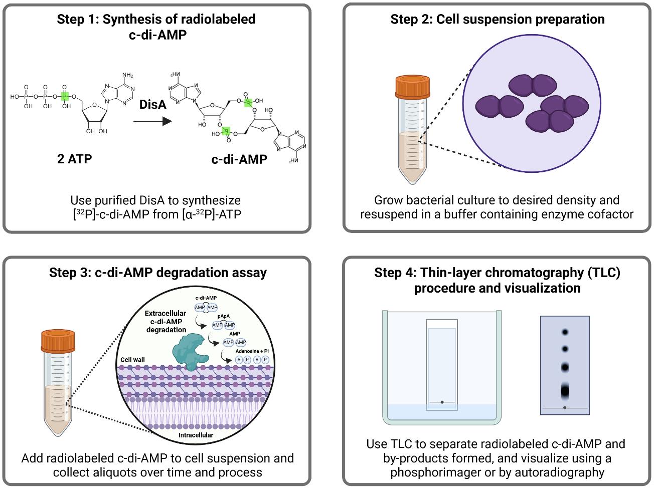
Background
Second messengers are small regulatory molecules, many of them nucleotides, that are produced to modulate cell adaptation to specific stimuli or stressors. While second messengers are conserved across the different domains of life, they are particularly important to bacteria, with some of them deemed essential for cell viability and virulence expression. Some examples of second messenger nucleotides shown to modulate bacterial adaptation include (p)ppGpp, cyclic AMP (cAMP), cyclic di-GMP (c-di-GMP), 3′3′-cyclic GMP-AMP (3′3′-cGAMP), and cyclic di-AMP (c-di-AMP). Cyclic di-AMP was serendipitously discovered in 2008, during a crystallization study of the DNA integrity scanning protein (DisA) of Thermotoga maritima [1]. Considered an essential poison [2] due to its essentiality and toxicity at elevated levels, c-di-AMP regulates a variety of cellular processes, including osmoregulation, cell wall synthesis and homeostasis, central metabolism, DNA integrity, and stress responses [3–9]. Not surprisingly, c-di-AMP regulation has also been linked to bacterial pathogenesis through its roles in mediating antibiotic tolerance and regulating virulence factor expression [5,8,9]. As a second messenger, c-di-AMP binds to specific proteins, such as transcriptional regulators, various transporters, and riboswitches, exerting allosteric control over their activities [3–9].
Intracellular levels of c-di-AMP are maintained by diadenylate cyclases (DAC) that synthesize c-di-AMP from two molecules of ATP and by phosphodiesterases (PDE) that degrade c-di-AMP to 5′ phosphoadenylyl adenosine (pApA), adenosine monophosphate (AMP), and, in some instances, adenosine (Ado) and inorganic phosphate (Pi) [3–9]. In addition, multidrug efflux (MDF)-type transporters have been implicated in c-di-AMP export [10] and, once outside the cell, c-di-AMP is rapidly degraded by extracellular phosphodiesterases (ePDE) identified in a few bacterial species [11–14]. The ability of bacteria to modulate extracellular c-di-AMP levels is particularly relevant during infection, given that c-di-AMP is a potent pathogen-associated molecular pattern (PAMP) molecule. Despite major progress in c-di-AMP research since its discovery, our current understanding of the extent of its influence on bacterial physiology and its role as a PAMP remains limited. Thus, developing new approaches to detect and quantify c-di-AMP and its degradation products may potentially accelerate future discoveries.
Currently, c-di-AMP can be detected and quantified using several methods, including liquid chromatography–mass spectrometry (LC-MS/MS), enzyme-linked immunosorbent assay (ELISA), luminescent and fluorescent biosensors, and thin-layer chromatography (TLC). Benefits of LC-MS/MS include high sensitivity, reproducibility, and the ability to determine the presence of different types of nucleotides within one sample. However, performing this analysis requires highly specialized equipment and knowledge of analytical chemistry techniques. The ELISA-based method is also sensitive and does not require sophisticated instrumentation, yet it can only quantify c-di-AMP and does not provide information on c-di-AMP degradation products. More recently, c-di-AMP-responsive riboswitches have been used to develop sensitive biosensors that allow real-time monitoring of c-di-AMP fluctuations. Still, c-di-AMP biosensors are not easily adaptable from species to species and often require additional genetic manipulations.
Here, we describe an adaptation of the traditional TLC-based method, which normally requires purified protein, to assess extracellular degradation of c-di-AMP using live bacteria. Our goal was to investigate EecP, a unique cell wall–anchored nucleotidase in E. faecalis that degrades c-di-AMP extracellularly, which was challenging to purify despite several attempts with different expression systems [11]. Therefore, this method was developed to analyze the degradation of c-di-AMP by live bacterial cells of Enterococcus faecalis and Streptococcus agalactiae strains. Even so, this accessible and low-cost method can be easily adapted to test a variety of other organisms, including Gram-negative bacteria, archaea, yeasts, and eukaryotic cells. Furthermore, the method can also be used to monitor degradation of other nucleotides and to identify factors that interfere with extracellular nucleotide levels.
Materials and reagents
Biological materials
Bacterial strains
| Strain | Relevant characteristics | Source |
|---|---|---|
| E. faecalis | ||
| OG1RF | Laboratory/reference strain Rifr, Fusr | Lab stock |
| V583 | Laboratory/reference strain, Vanr | Lab stock |
| E. coli | ||
| Rosetta (DE3) pLysS pSpeedET:: Akr1c13 | Host for AKR1C13, protein herein referred to as RECON, protein expression, Kanr | Woodward Lab at the University of Washington [15] |
| Rosetta (DE3) pLysS pET20b:: disA | Host for DisA protein expression (disA from Bacillus subtilis), Ampr | Woodward Lab at the University of Washington [1,16] |
| S. agalactiae | ||
| COH1 | Laboratory/reference strain, serotype III | Brady Lab at the University of Florida |
| NGBS93 | WGS clinical blood isolate, serotype V, BioSample: SAMN03329865 | Fittipaldi Lab at the University of Montreal |
Reagents
1. Luria-Bertani (LB) broth, Lennox (Fisher Bioreagents, catalog number: BP1427)
2. Todd Hewitt broth (THB) (Becton Dickinson, catalog number: 249240)
3. Chemically defined medium (CDM) (see recipe in [11])
4. Kanamycin monosulfate (Fisher Scientific, CAS number: 25389-94-0)
5. Ampicillin sodium salt (Sigma-Aldrich, CAS number: 69-52-3)
6. D-(+)-Glucose (Sigma-Aldrich, CAS number: 50-99-7)
7. Isopropyl β-D-thiogalactoside (IPTG) (Fisher Scientific, CAS number: 367-93-1)
Note: Prepare 1 M stock in sterile ddH2O and freeze aliquots at -20 °C. Do not re-use thawed aliquots.
8. Halt protease inhibitor cocktail (100×) (Thermo Scientific, catalog number: 78429)
9. Ni-NTA agarose (Marvelgent Biosciences, catalog number: 11-0224-100)
10. Sodium phosphate monobasic (NaH2PO4) (ICN Biomedicals, CAS number: 10049-21-5)
11. Sodium chloride (NaCl) (Fisher Scientific, CAS number: 7647-14-5)
12. Imidazole (Acros Organics, CAS number: 288-32-4)
13. Triton X-100 (Fisher Bioreagents, CAS number: 9002-93-1)
14. Tris base (Fisher Bioreagents, CAS number: 77-86-1)
15. Manganese (II) chloride (MnCl2·4H2O) (Sigma-Aldrich, CAS number: 13446-34-9)
16. Magnesium chloride (MgCl2·6H2O) (Fisher Bioreagents, CAS number: 7791-18-6)
17. Glycerol (Fisher Bioreagents, CAS number: 56-81-5)
18. [α-32P]-ATP, 3,000 Ci/mmol, 10 mCi/mL, 250 μCi (Revvity Health Sciences, catalog number: BLU003H250UC)
19. c-di-AMP (InvivoGen, catalog number: tlrl-nacda)
20. c-di-GMP (InvivoGen, catalog number: tlrl-nacdg)
21. 2′3′-cGAMP (InvivoGen, catalog number: tlrl-nacga23-02)
22. Methanol (Fisher Chemical, CAS number: 67-56-1)
23. Ammonium sulfate [(NH4)2SO4] (Sigma-Aldrich, CAS number: 7783-20-2)
24. Monopotassium phosphate (KH2PO4) (Fisher Bioreagents, CAS number: 7778-770)
25. Phosphoric acid (H3PO4) (Sigma-Aldrich, CAS number: 7664-38-2)
26. Hydrochloric acid (HCl) (Fisher Chemical, CAS number: 7664-01-0)
27. Sodium hydroxide (NaOH) (Fisher Chemical, CAS number: 1310-73-2)
28. Pierce BCA Protein Assay kit (Thermo Scientific, catalog number: 23225)
29. Econo-Safe Biodegradable counting cocktail (Research Products International, catalog number: 111175)
30. Double-distilled water (ddH2O)
Solutions
1. Protein purification and storage buffers (see Recipes)
a. Equilibration/lysis buffer
b. Wash buffer 1
c. Wash buffer 2
d. Wash buffer 3
e. Elution buffer
f. Protein storage/binding buffer
2. Reaction buffer (see Recipes)
3. TLC mobile phase buffer (see Recipes)
Recipes
Note: All buffers can be prepared at room temperature with constant stirring unless specified. Adjust the pH of components or buffers as needed using either HCl or NaOH before adding water up to the final volume. Filter-sterilize components as needed and store at room temperature unless otherwise specified.
1. Protein purification and storage buffers
To prepare buffers for protein purification, first prepare each stock component separately as follows:
| Stock components | Molecular weight (g/mol) | Preparation |
|---|---|---|
| 1 M NaH2PO4 | 137.99 | • Dissolve 68.99 g of NaH2PO4 in 300 mL of ddH2O. • Add ddH2O to obtain a final volume of 500 mL. |
| 5 M NaCl | 58.44 | • Dissolve 146.1 g of NaCl in 300 mL of ddH2O. • Add ddH2O to obtain a final volume of 500 mL. |
| 1 M imidazole | 68.08 | • Dissolve 34.04 g of imidazole in 300 mL of ddH2O. • Add ddH2O to obtain a final volume of 500 mL. |
| 1 M MgCl2 | 203.30 | • Dissolve 101.65 g of MgCl2 in 300 mL of ddH2O. • Add ddH2O to obtain a final volume of 500 mL. |
| 1 M Tris-Cl pH 7.5 | 121.136 (Tris base) | • Dissolve 60.568 g of Tris base in 300 mL of ddH2O. • Add ddH2O to obtain a final volume of 500 mL after pH adjustment with HCl. |
| 1 M MnCl2 | 197.9 | • Dissolve 98.95 g of MnCl2 in 300 mL of ddH2O. • Add ddH2O to obtain a final volume of 500 mL. |
a. Equilibration/lysis buffer (pH 7.5)
| Stock components | Volume | Final concentration |
|---|---|---|
| 1 M NaH2PO4 | 20 mL | 20 mM |
| 5 M NaCl | 60 mL | 300 mM |
| 1 M imidazole | 10 mL | 10 mM |
| ddH2O | up to 1,000 mL after pH adjustment |
b. Wash buffer 1 (pH 8.0)
| Stock components | Volume | Final concentration |
|---|---|---|
| 1 M NaH2PO4 | 20 mL | 20 mM |
| 5 M NaCl | 60 mL | 300 mM |
| 1 M imidazole | 25 mL | 25 mM |
| Concentrated Triton X-100 | 10 mL | 1% |
| ddH2O | up to 1,000 mL after pH adjustment |
c. Wash buffer 2 (pH 8.0)
| Stock components | Volume | Final concentration |
|---|---|---|
| 1 M NaH2PO4 | 20 mL | 20 mM |
| 5 M NaCl | 100 mL | 500 mM |
| 1 M imidazole | 25 mL | 25 mM |
| ddH2O | up to 1,000 mL after pH adjustment |
d. Wash buffer 3 (pH 8.0)
| Stock components | Volume | Final concentration |
|---|---|---|
| 1 M NaH2PO4 | 20 mL | 20 mM |
| 5 M NaCl | 100 mL | 500 mM |
| 1 M imidazole | 50 mL | 50 mM |
| ddH2O | up to 1,000 mL after pH adjustment |
e. Elution buffer (pH 8.0)
| Stock components | Volume | Final concentration |
|---|---|---|
| 1 M NaH2PO4 | 20 mL | 20 mM |
| 5 M NaCl | 100 mL | 500 mM |
| 1M imidazole | 250 mL | 250 mM |
| ddH2O | up to 1,000 mL after pH adjustment |
f. Protein storage/binding buffer (pH 7.5)
| Stock components | Volume | Final concentration |
|---|---|---|
| 1 M Tris-Cl pH 7.5 | 40 mL | 40 mM |
| 5 M NaCl | 20 mL | 100 mM |
| 1 M MgCl2 | 20 mL | 20 mM |
| ddH2O | up to 1,000 mL after pH adjustment |
2. Reaction buffer
| Stock components | Volume | Final concentration |
|---|---|---|
| 1 M Tris-Cl pH 7.5 | 50 mL | 50 mM |
| 1 M MnCl2 | 5 mL | 5 mM |
| ddH2O | up to 1,000 mL after pH adjustment |
3. TLC mobile phase buffer
Prepare each component separately as follows:
| Stock components | Molecular weight (g/mol) | Preparation |
|---|---|---|
| 1.5 M KH2PO4, pH 3.6 | 136.086 | • Dissolve 102.06 g of KH2PO4 in 400 mL of ddH2O with stirring. • Adjust pH to 3.6 using H3PO4 (phosphoric acid). • Add ddH2O to obtain a final volume of 500 mL. |
| 4.1 M saturated (NH4)2SO4 | 132.14 | • Dissolve 270.887 g of ammonium sulfate in 200 mL of ddH2O with stirring and gentle heating. • Add ddH2O to obtain a final volume of 500 mL. • Cool to room temperature before filter sterilizing and store at 4 °C. |
Note: A saturated ammonium sulfate solution is a highly concentrated mixture where no more of the salt [(NH4)2SO4] can be dissolved in water.
To prepare 250 mL of TLC mobile phase buffer, mix 100 mL of saturated (NH4)2SO4 and 150 mL of KH2PO4 in a 1:1.5 ratio (v/v) and use immediately.
Laboratory supplies
1. Microcentrifuge tubes (1.5 and 2 mL)
2. Individual PCR tubes (0.5 mL)
3. Micropipette tips (10, 200, and 1,000 μL)
4. Micropipettes (P2, P20, P200, and P1000)
5. Serological pipettes (10 and 25 mL)
6. Micropipette filtered tips (10, 200, and 1,000 μL)
7. 1.5 mL polystyrene semi-micro cuvette (Fisher, catalog number: 14-955-127)
8. Falcon tubes (15 and 50 mL)
9. Econo-column glass chromatography columns, 1.0 × 30 cm (Bio-Rad, catalog number: 7371032)
10. Pierce protein concentrator PES, 10K MWCO (5–20 mL) (Thermo Scientific, catalog number: 88528)
11. Pierce concentrator, PES, 10K MWCO (0.5 mL) (Thermo Scientific, catalog number: 88513)
12. Zeba spin desalting columns, 7K MWCO (Thermo Scientific, catalog number: 89891)
13. Pierce spin columns (Thermo Scientific, catalog number: 69705)
14. TLC polyethylenimine (PEI) cellulose F plates (EMD Millipore, catalog number: 1.05579.0001)
15. Nitrocellulose membrane, 0.45 μm (Cytiva, catalog number: GE10600048)
16. Acrylic benchtop beta radiation shield (Thermo Scientific Nalgene, catalog number: 6700-1812)
17. TLC glass chambers (Supelco, Sigma-Aldrich, catalog number: Z204161)
18. Fume hood
19. X-ray exposure cassette (Wolf X-Ray Corporation, part number: 11319)
20. Clear blue X-ray film (CL-X Posure Film, Thermo Scientific, catalog number: 34091)
21. Clear plastic cling film (Boardwalk, catalog number: BWK 7202)
Equipment
1. Shaking incubator (Benchmark, model: Incu-Shaker 10L)
2. Spectrophotometer (Thermo Scientific, model: Genesys 30)
3. Sonicator (Fisher Scientific, Sonic Dismembrator model: 500)
4. Tube rotator (VWR, catalog number: 1016-084)
5. Benchtop large centrifuge (Eppendorf, model: 5910 Ri)
6. Benchtop microcentrifuge (Eppendorf, model:5415 C)
7. High-speed centrifuge (Beckman, model: J2-21)
8. Liquid scintillation counter (Beckman, model: LS 6000IC)
Procedure
Note: The procedure described in sections A and B is intended for the expression and purification of recombinant 6×-His tagged proteins DisA and RECON from E. coli host strains. These proteins are then used for the synthesis and affinity purification of radiolabeled [32P]-c-di-AMP as described in section C.
A. Recombinant protein induction and harvesting of bacterial cell culture
Note: We recommend using a suitable host strain for optimal expression and induction of recombinant proteins. For both recombinant proteins used in this protocol, the same procedure can be used.
1. Starter culture: Inoculate 5 mL of LB media containing appropriate antibiotics (100 μg/mL ampicillin for pET20b:: disA; 300 μg/mL kanamycin for pSpeedET:: Akr1c13) with a single colony of bacteria and incubate in a 37 °C shaking incubator (19 mm orbit) set to 200 rpm for 15–18 h.
2. Main culture: The following day, dilute the starter culture (1:100) into fresh LB supplemented with 0.4% glucose and appropriate antibiotic(s) (~OD600 0.05) and incubate at 37 °C in a shaking incubator (19 mm orbit) set to 200 rpm to allow bacteria to reach the exponential growth phase. Measure the density of the cultures at OD600 using a spectrophotometer periodically and, once cultures reach an ~OD600 of 0.5, add IPTG to a final concentration of 500 μM, allowing bacteria to further grow at 30 °C under shaking for 3 h.
3. Following induction, save 1 mL of the induced sample to confirm protein expression by SDS-PAGE, followed by Coomassie blue staining. Collect the remaining bacterial cell pellets by centrifugation at 18,000× g for 15 min at 4 °C. Store the bacterial cell pellet at -20 °C until use.
Note: Bacterial cell pellets are stable for at least 2–3 months at -20 °C and for up to 6 months at -80 °C. We generally prepare 1 L batches of bacteria, collect, freeze 250–500 mL culture pellets in aliquots, and take single aliquots for protein purification as needed.
B. Bacterial cell lysis and protein purification
1. Remove a single frozen bacterial cell pellet made from a 500 mL culture and thaw it over ice.
2. Resuspend the pellet thoroughly in 20 mL of equilibration/lysis buffer until it becomes homogenous. Add 200 μL of Halt protease inhibitor cocktail (1× final concentration) to the suspension. Then, transfer the cell suspension to a 50 mL Falcon tube and keep it on ice until the next step.
Note: Equilibration/lysis buffer should be freshly prepared.
3. Sonicate bacterial cell lysate using a 10-min cycle of alternating pulses of 15-s on/30-s off at 15% amplitude.
Note: Suspension tubes should remain submerged in ice throughout the sonication cycle to protect the cell lysate from the heat generated during sonication cycles.
4. Centrifuge the lysate at 24,000× g for 30–40 min at 4 °C to pellet insoluble material and debris. After centrifugation, transfer the supernatant containing soluble proteins (cleared lysate) to a new Falcon tube and keep it on ice until use.
5. Preparation of nickel-nitrilotriacetic acid (Ni-NTA) agarose matrix: Take 1–2 mL of uniformly mixed Ni-NTA agarose beads and centrifuge at 3,400× g for 5 min at room temperature. Discard supernatant. Wash Ni-NTA agarose beads by centrifugation with 1–2 mL of the equilibration buffer twice.
Note: Ni-NTA agarose beads should be stored at 4 °C and uniformly mixed before use.
6. Mix supernatant (cleared lysate) from step B4 with washed Ni-NTA agarose beads from step B5 in a Falcon tube and mix on a tube rotator with fixed speed (18 rpm) at 4 °C for 2 h.
7. Transfer the Ni-NTA agarose–lysate mix onto an empty glass column and allow it to settle by gravity. In this step, the His-tagged protein binds to Ni2+ ions and is retained in the column while untagged proteins pass through. Collect the flowthrough lysate into a new tube.
8. Wash the column containing protein-bound beads with 40 mL each of three different buffer preparations: wash buffer 1, 2, and 3 (see Recipes) consecutively. Collect the washthrough from each wash step in separate Falcon tubes.
9. Elute the bound protein with 5 mL of elution buffer two to four times. Collect all elution fractions.
Note: All purification steps (steps B6–9) can be carried out either at 4 °C or at room temperature. The temperature used for purification depends on the stability of the protein of interest. If your protein is prone to aggregation at 4 °C, these steps should be performed at room temperature.
10. Pool all elution fractions and concentrate using a 10 kDa MWCO Pierce protein concentrator to 1–2 mL at 4 °C following the manufacturer’s instructions.
11. Exchange the buffer of the concentrated protein using Zeba Spin columns 7K MWCO with the protein storage/binding buffer following the manufacturer’s instructions.
12. If required, concentrate buffer-exchanged proteins further to 400–500 μL using Pierce protein concentrator PES 10K MWCO following the manufacturer’s instructions.
13. Add glycerol to the purified protein to a final concentration of 20%. Aliquot 50–100 μL of purified protein in 0.5 mL PCR tubes and store protein samples at -20 °C for short-term and at -80 °C for long-term use.
14. Save a small aliquot (100–200 μL) from each step and run on 12% SDS-PAGE, followed by Coomassie staining to evaluate the purity and stability of the purified protein (Figure 1). Determine protein concentration using the BCA Assay kit following the manufacturer’s instructions.
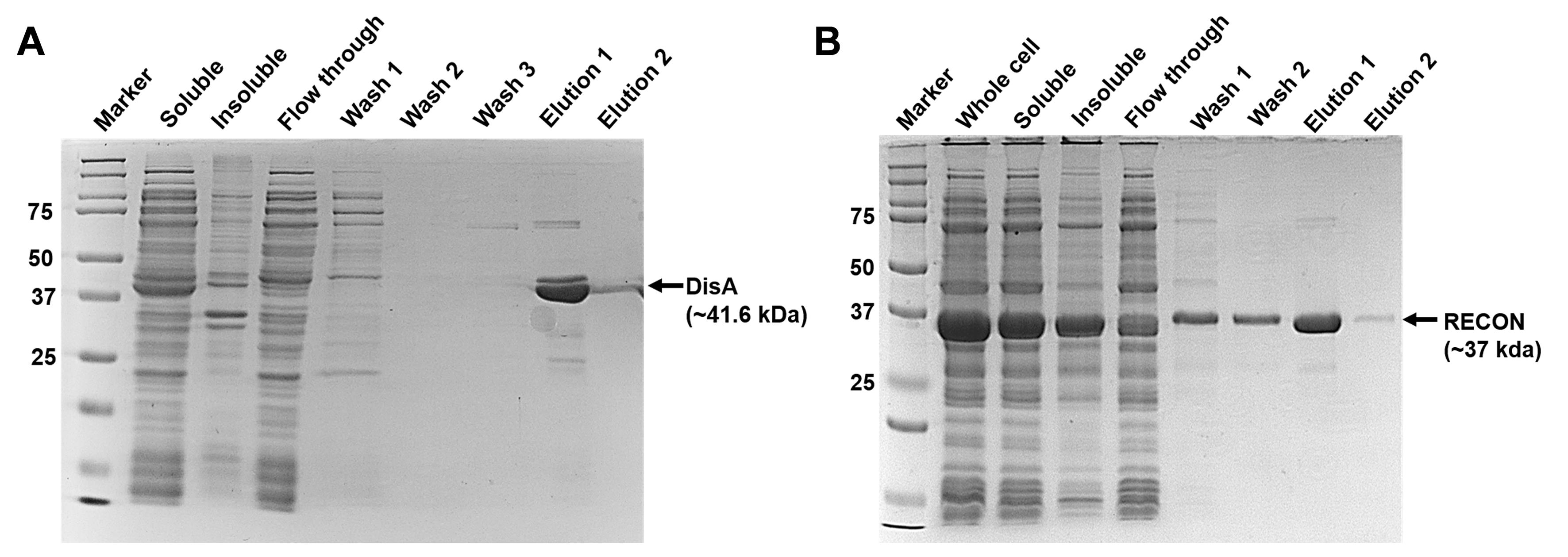
Figure 1. Representative 12% Coomassie-stained SDS-PAGE gel showing all fractions from purification steps of recombinant (A) DisA and (B) RECON proteins. The arrows indicate the band corresponding to the specific protein.
C. Synthesis and purification of [α-32P]-labeled c-di-AMP
Note: The following procedure is adapted from a previously developed method [17] with modifications as needed.
Caution: All steps involving radioisotopes should be carried out in a designated isotope-use restricted area only by trained personnel using an acrylic benchtop beta radiation shield to prevent radiation exposure.
1. Synthesis of [32P]-c-di-AMP: On day 1, mix [α-32P]-ATP with DisA to a final concentration of 1 μM for both components in storage/binding buffer in a 50 μL final reaction volume in an Eppendorf tube. Incubate the reaction mix in a water bath set to 30 °C overnight.
2. The next day, spike the reaction mix with another 1 μM DisA and incubate for an additional 4 h at 30 °C. After incubation, boil the reaction mix for 5 min. Use a beaker with boiling water for this step.
3. Then, spin down the reaction mix at 16,000× g for 5 min to remove DisA. Transfer the supernatant containing synthesized crude [32P]-c-di-AMP into a new Eppendorf tube and keep at room temperature until use.
4. Affinity purification of [32P]-c-di-AMP: Recombinant His-tagged RECON, a c-di-AMP binding protein, is used for the affinity purification of the synthesized radiolabeled c-di-AMP.
5. Prepare the Ni-NTA resin by adding 100 μL of Ni-NTA agarose into an Eppendorf tube, then spin down the beads at 3,400× g to remove ethanol. Wash beads by centrifugation twice with 1 mL of water and centrifuge at 3,400× g. Thaw one aliquot of purified RECON protein over ice.
6. To bind RECON with Ni-NTA beads, add 1 mL of RECON (final concentration 30 μM) to Ni-NTA resin and mix on a tube rotator at fixed speed (18 rpm) for 30 min at 4 °C. After incubation, spin down beads at 16,000× g for 1 min and wash resin by centrifugation twice with 1 mL of binding buffer. Aspirate supernatant to remove any unbound protein and keep RECON-bound beads on an ice bucket until use.
7. Add the supernatant containing crude [32P]-c-di-AMP (from step C3) to the RECON-bound beads and incubate at room temperature for 30 min on a tube rotator with fixed speed (18 rpm), followed by a 10-min incubation on ice with vortexing at 5-min intervals for 10 s each.
8. Spin down beads at 16,000× g for 1 min and discard supernatant. Wash Ni-NTA beads by centrifugation twice with 1 mL of ice-cold binding buffer and discard supernatant again. Add 100 μL of binding buffer and heat beads for 10 min in a boiling beaker.
9. Transfer the bead slurry to a mini spin column and centrifuge at 16,000× g for 1 min.
10. Add another 50 μL of binding buffer to the mini spin column and spin again at 16,000× g for 1 min. Reserve the flowthrough that contains the purified c-di-AMP and store at -20 °C.
Note: [32P] has a physical half-life of 14.29 days. Thus, [32P]-c-di-AMP should be used within 14 days or less.
11. Determination of [32P]-c-di-AMP quantity and purity: Determine the final yield of the synthesis by measuring the radioactivity counts of a series of diluted preparations of [32P]-c-di-AMP and [α-32P]-ATP in a liquid scintillation counter.
12. Spot 1 μL of each dilution onto a nitrocellulose membrane and transfer the membrane to a vial containing scintillation counting cocktail. Count the vials in the scintillator counter using a program for [32P]. Plot dilutions versus disintegrations per minute (DPM) and fit each count data to a linear equation to calculate the concentration. The yield is ~3.3 nM [32P]-c-di-AMP on average.
13. Evaluate the purity of the final preparation by thin-layer chromatography (TLC) as described in section F. Spot 1 μL each of diluted [32P]-c-di-AMP and [α-32P]-ATP on the prepared TLC plate to check the purity, as shown in Figure 2.
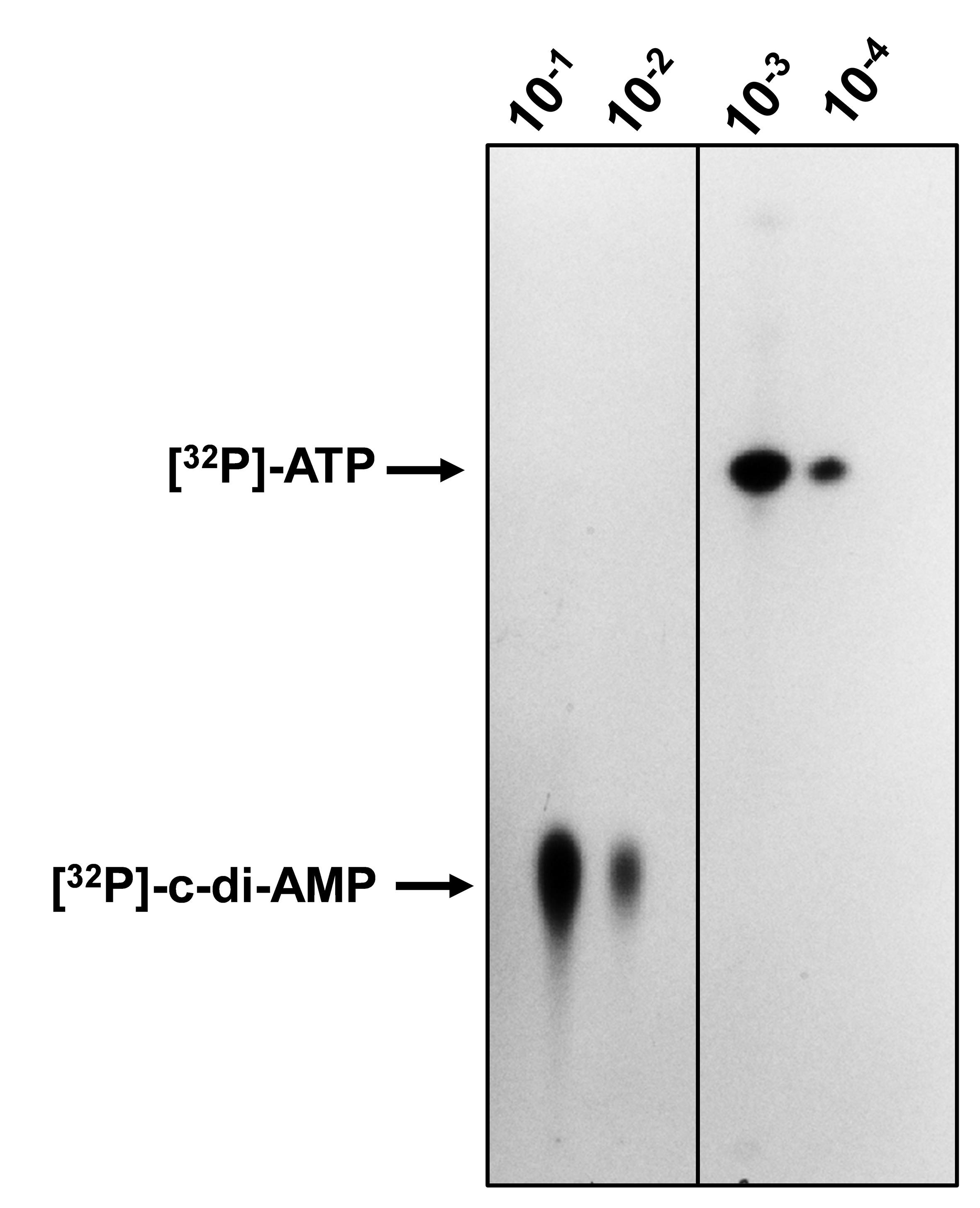
Figure 2. Thin-layer chromatography (TLC) of freshly synthesized [32P]-c-di-AMP from [32P]-ATP. Dilutions of [32P]-c-di-AMP (10-1 and 10-2) and [32P]-ATP (10-3 and 10-4) were prepared in binding buffer, and 1 μL from each sample was spotted on a polyethylenimine (PEI)-cellulose TLC plate. Upon completion of migration, the TLC plate was dried and exposed to X-ray film overnight, followed by development and visualization of radioactive spots using a film processor.
D. Degradation of radiolabeled c-di-AMP using live bacteria
1. Inoculate single bacterial colonies of E. faecalis OG1RF and V583 strains in 5 mL of CDM and S. agalactiae COH1 and S. agalactiae NGBS93 strains in 5 mL of THB. Incubate at 37 °C under static conditions overnight.
2. Next day, dilute overnight cultures (1:20 to 1:40) in 50 mL of fresh CDM (or THB) and incubate at 37 °C under static conditions, allowing cultures to reach mid-log exponential growth phase (~OD600 0.4–0.5).
3. Centrifuge cultures at 3,500× g for 10 min at 4 °C. Discard supernatant and save cell pellets.
4. Resuspend cell pellets in 1 mL of reaction buffer, transfer to an Eppendorf tube, and centrifuge at 16,000× g for 1 min. Repeat the wash step twice.
5. Finally, resuspend cell pellets in 0.5 mL of reaction buffer to a final cell density of 108–109 CFU/mL. Determine the cell density (CFU/mL) of bacterial suspensions by enumerating colonies on appropriate agar plates.
Note: The remaining steps should be performed in an area specifically designated for working with isotopes.
6. Aliquot 16 μL of [32P]-c-di-AMP in separate Eppendorf tubes and to each, add 144 μL of cell suspension in a 1:10 v/v ratio (~0.333 nM final concentration). Dilute [32P]-c-di-AMP with reaction buffer in a 1:10 v/v ratio to use as a control.
7. Incubate cell suspension and [32P]-c-di-AMP mix in a 37 °C water bath.
8. At the selected time points, transfer 40 μL of the suspension to a new tube, immediately placing the original tube back in the water bath.
Note: The ratio of [32P]-c-di-AMP to live cell suspension can be adjusted depending on the final concentration of [32P]-c-di-AMP synthesized, duration of the experiment, and bacterial cell culture density.
9. Process collected aliquots by immediately centrifuging at 16,000× g for 3 min and transferring the supernatant to a new tube. Stop the reaction by boiling for 5 min.
10. Let samples cool to room temperature and store at -20 °C until use.
E. Competition assay using cold nucleotides
1. The c-di-AMP degradation assay described in Section D can be modified to perform a competition assay using an excess of cold nucleotides to measure the substrate specificity of the reaction. Prepare the cell suspension in reaction buffer as described in steps D1–5.
2. Aliquot 180 μL of each cell suspension in an Eppendorf tube and spike with 20 μL of diluted [32P]-c-di-AMP to a final concentration of ~0.06 nM in the reaction mix.
3. Add an excess of cold (unlabeled) c-di-AMP or other nucleotides (c-di-GMP, 2′3′-cGAMP, etc.) to the reaction mix. As a control, add the same volume of water instead of the cold nucleotide to the reaction mix and process similarly.
Note: The concentration of competing nucleotides to be added should be empirically decided. Based on our experience, we recommend starting with 10,000× (~600 nM) and 100,000× (~6,000 nM) excess of the competitor nucleotide.
4. Incubate the reaction mix at 37 °C in a water bath. Collect 40 μL aliquots at pre-selected time points and process as described in steps D8–10. Store samples at -20 °C until use.
F. Analytical thin-layer chromatography (TLC)
1. Label the PEI plate gently with a pencil ~2 cm from the bottom, drawing a straight line. Mark equidistant points on the line to serve as a guide for spotting samples.
Note: PEI cellulose TLC plates are usually stored at 4 °C until use, and a 20 × 20 cm plate can fit up to 15 samples. Do not use a pen for marking, as ink will diffuse and develop spots on the plate.
2. After labeling, soak the PEI plate in 90% methanol for 15–20 min and then air-dry completely. Once dried, transfer the PEI plates to the fume hood for the next step.
3. At the same time, thaw samples collected from both assays described in sections D and E at room temperature.
4. Aliquot 3 μL of the reaction products along the bottom line on the TLC plate using separate tips for each sample. Care should be taken to avoid merging samples across the marked line. Alternatively, spot 1.5 μL of the sample; then, allow the sample to dry before spotting again in the same location. The sample will remain concentrated, and this will prevent merging. Spot equal volumes of samples from control reactions on the same TLC plate.
5. Allow the plates to air dry for 15–20 min within the fume hood.
6. Concurrently, prepare the TLC mobile phase buffer and add 80–100 mL of this buffer to the TLC glass chamber; keep covered for saturation.
7. Place the PEI plate inside the chromatography chamber using a stainless-steel TLC plate rack so that only the bottom of the plate (~1 cm) is submerged in the buffer. Keep the tank covered and allow the buffer front to reach almost the top of the plate.
Caution: Buffer solvent should always be below the level of the spots; otherwise, the spotted samples will dissolve in the buffer.
8. Remove the PEI plate from the chamber using forceps and allow it to air-dry for 15–20 min.
9. Once completely dried, wrap the PEI plate in plastic cling film and expose it to clear X-ray film in an exposure cassette at room temperature overnight (or several days, depending on signal strength).
10. To visualize radioactive spots, develop the film in an X-ray film processor or use standard film-developing techniques. Once the image is captured, discard the PEI plates in the radioactive waste bin.
Note: Alternatively, the PEI TLC plate can also be exposed to a phosphor storage screen, which records a latent image produced by ionizing radiation. Subsequently, the image can be read by laser scanning and converted into digital format using an Amersham Typhoon phosphorimager. The response of a phosphor storage screen to radiation is linear, unlike that of autoradiography film, facilitating the accurate densitometric quantification of spots on the TLC plate.
Validation of protocol
This adapted TLC-based method was developed and validated to monitor the extracellular degradation of radiolabeled c-di-AMP using live bacterial cell suspensions of E. faecalis and S. agalactiae. The representative TLC image shown in Figure 3 demonstrates that both E. faecalis strains OG1RF and V583, which encode the ectonucleotidase EecP, rapidly degrade c-di-AMP, generating byproducts corresponding to pApA, AMP, and inorganic phosphate (Pi) [11]. Comparable results were obtained using two S. agalactiae strains that encode the c-di-AMP ectonucleotidase CdnP [11] (Figure 3).
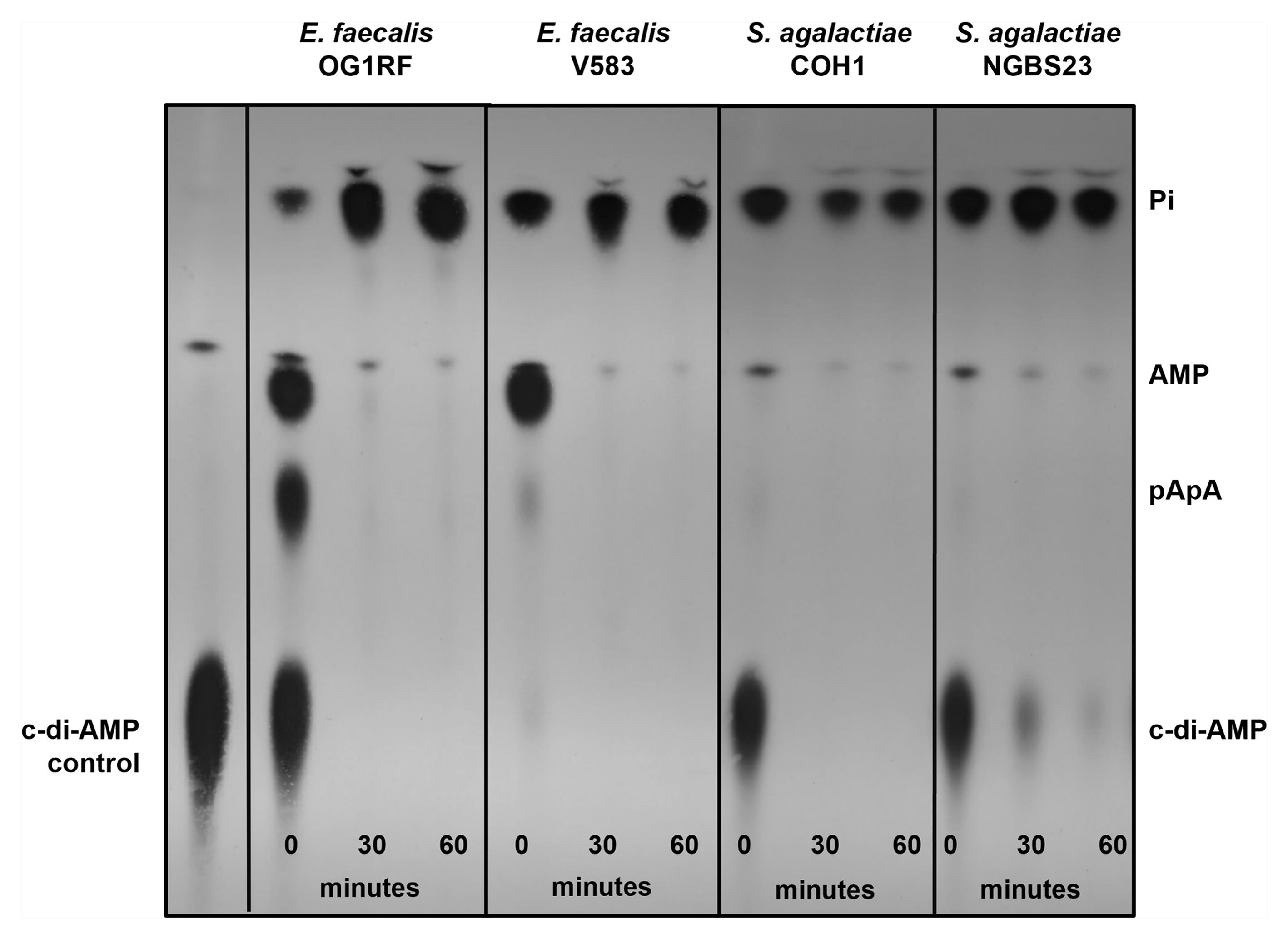
Figure 3. Extracellular [32P]-c-di-AMP is degraded by cell suspensions of E. faecalis and S. agalactiae strains that express cell-surface anchored ectonucleotidases EecP and CdnP, respectively. Mid-log cultures of respective strains grown in THB (~OD600 0.4) and suspended in 50 mM Tris-Cl containing 5 mM MnCl2 were spiked with [32P]-c-di-AMP in a 1:10 v/v ratio and sampled over time for c-di-AMP degradation. Reaction aliquots were collected at the indicated time points and inactivated by boiling for 5 min. The TLC image is representative of experiments conducted at least two times with independent biological replicates. This figure is also featured in Morales Rivera et al. [11].
Next, we assessed the capacity of unlabeled (cold) nucleotides to competitively inhibit the degradation of radiolabeled c-di-AMP by E. faecalis OG1RF. In a control experiment, we determined that 100,000× excess of unlabeled c-di-AMP stabilized the [32P]-c-di-AMP, indicating that cold c-di-AMP can readily compete with [32P]-c-di-AMP (Figure 4). Furthermore, excess unlabeled c-di-GMP also stabilized the [32P]-c-di-AMP signal, almost to the same extent as c-di-AMP; however, excess unlabeled cGAMP was a poor competitor for [32P]-c-di-AMP (Figure 4).
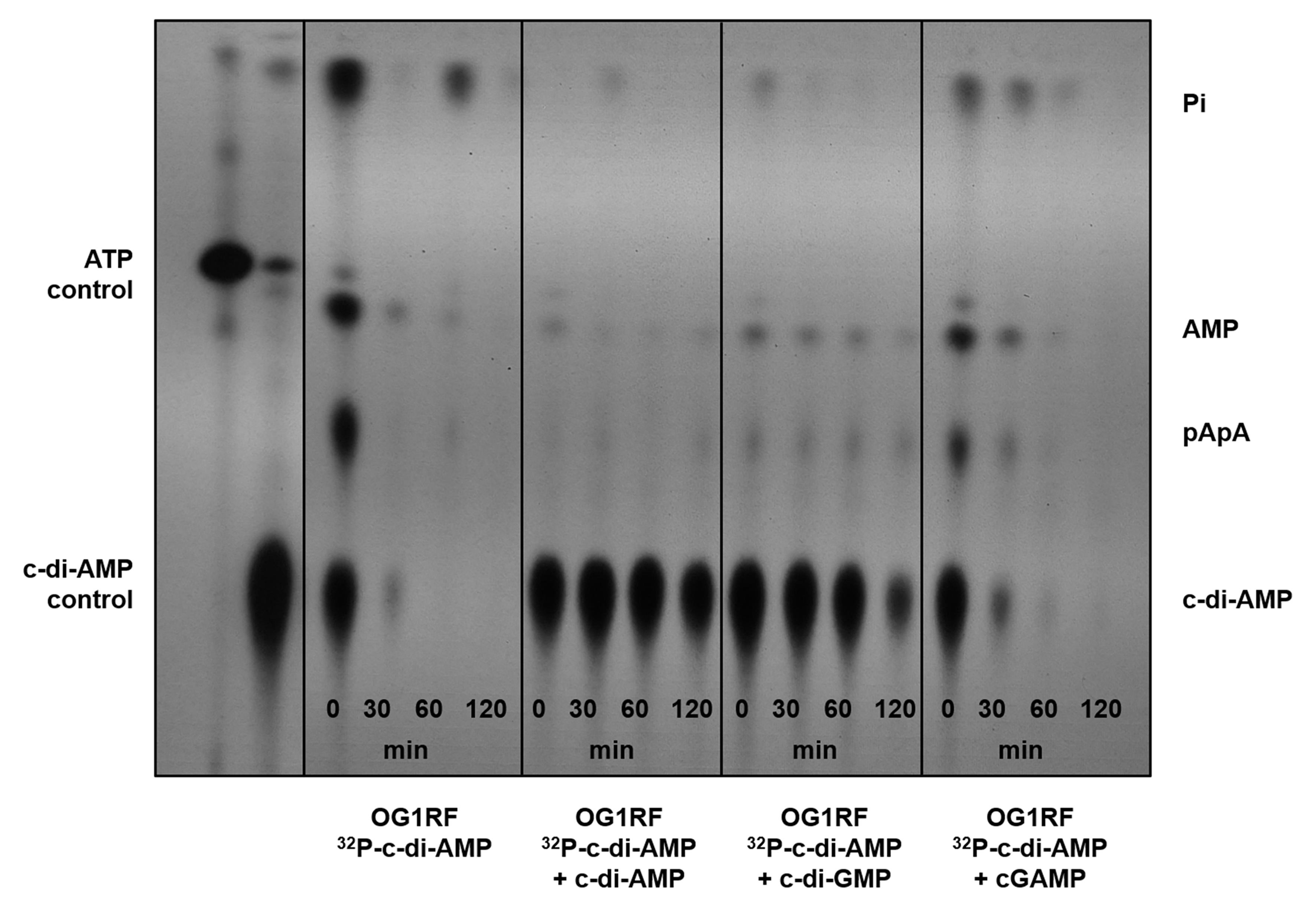
Figure 4. Extracellular [32P]-c-di-AMP degradation can be inhibited by the cold nucleotide competitor c-di-GMP. Thin-layer chromatography (TLC) of cell-free supernatants of OG1RF cultures grown in CDM to OD600 0.4, suspended in 50 mM Tris-Cl containing 5 mM MnCl2, and spiked with [32P]- c-di-AMP and 100,000× excess of cold competitor nucleotides c-di-AMP, c-di-GMP, or cGAMP. Supernatants were sampled over time for c-di-AMP degradation. Reaction aliquots were collected at the indicated time points and inactivated by boiling before spotting on a PEI-cellulose plate for TLC separation. TLC image is a representative of experiments conducted at least two times with independent biological replicates. This figure is also featured in Morales Rivera et al. [11].
General notes and troubleshooting
Troubleshooting
• Since some proteins may lose their activity upon storage for prolonged periods, we recommend that both DisA and RECON be freshly purified to achieve the highest enzymatic activity for the c-di-AMP synthesis reaction.
• Concentration of radiolabeled c-di-AMP synthesized depends on factors such as the starting concentration of [α-32P]-ATP used in the reaction, the activity of the DisA enzyme, and the elution volume. Thus, depending on the radiolabeled c-di-AMP concentration required, these conditions can be varied to achieve the maximum yield.
Limitations
While we have described the advantages and versatile applications of this method, we acknowledge that there are limitations to this technique, as listed below:
• Our assay relies on the use of radiolabeled substrates, which require specialized facilities to handle radioisotopes, training, and oversight, which may not be accessible at all institutions. Although radiolabeled materials can be used safely if handled in accordance with safety guidelines, their use requires additional precautions not necessary for other methods, such as LC-MS/MS.
• Given the use of intact cells for this assay, we cannot exclude the possibility of more than one protein contributing to the observed phenotypes.
• Lastly, this method is mostly performed manually; therefore, it could be considered labor-intensive. Nonetheless, the protocol incorporates natural stopping points, allowing users to scale up certain portions, such as the protein isolation steps, to make the workflow more accessible.
Acknowledgments
Conceptualization, A.B., A.G.M.R., J.A.L.; Investigation, A.B., A.G.M.R.; Writing—Original Draft, A.B., A.G.M.R., J.A.L.; Writing—Review & Editing, A.B., A.G.M.R., J.A.L.; Funding acquisition, J.A.L.; Supervision, J.A.L.
We thank Dr. Joshua Woodward at the University of Washington for the E. coli strains expressing DisA and RECON, Dr. Jeannine Brady at the University of Florida for the COH1 S. agalactiae strain, and Dr. Nahuel Fittipaldi at the University of Montreal for the NGBS93 S. agalactiae strain. This study was supported by NIH-NIAID R01 AI172179 to J.A.L. A.G.M.R. was supported by the Florida Fund of Education McKnight Doctoral Fellowship. The funders had no role in study design, data collection and analysis, decision to publish, or preparation of the manuscript.
This protocol was used in [11].
Competing interests
The authors declare no conflicts of interest.
References
- Witte, G., Hartung, S., Büttner, K. and Hopfner, K. P. (2008). Structural Biochemistry of a Bacterial Checkpoint Protein Reveals Diadenylate Cyclase Activity Regulated by DNA Recombination Intermediates. Mol Cell. 30(2): 167–178. https://doi.org/10.1016/j.molcel.2008.02.020
- Gundlach, J., Mehne, F. M. P., Herzberg, C., Kampf, J., Valerius, O., Kaever, V. and Stülke, J. (2015). An Essential Poison: Synthesis and Degradation of Cyclic Di-AMP in Bacillus subtilis. J Bacteriol. 197(20): 3265–3274. https://doi.org/10.1128/jb.00564-15
- Wright, M. J. and Bai, G. (2023). Bacterial second messenger cyclic di‐AMP in streptococci. Mol Microbiol. 120(6): 791–804. https://doi.org/10.1111/mmi.15187
- Stülke, J. and Krüger, L. (2020). Cyclic di-AMP Signaling in Bacteria. Annu Rev Microbiol. 74(1): 159–179. https://doi.org/10.1146/annurev-micro-020518-115943
- Commichau, F. M., Heidemann, J. L., Ficner, R. and Stülke, J. (2019). Making and Breaking of an Essential Poison: the Cyclases and Phosphodiesterases That Produce and Degrade the Essential Second Messenger Cyclic di-AMP in Bacteria. J Bacteriol. 201(1): e00462–18. https://doi.org/10.1128/jb.00462-18
- Yin, W., Cai, X., Ma, H., Zhu, L., Zhang, Y., Chou, S. H., Galperin, M. Y. and He, J. (2020). A decade of research on the second messenger c-di-AMP. FEMS Microbiol Rev. 44(6): 701–724. https://doi.org/10.1093/femsre/fuaa019
- Zarrella, T. M. and Bai, G. (2020). The Many Roles of the Bacterial Second Messenger Cyclic di-AMP in Adapting to Stress Cues. J Bacteriol. 203(1): e00348–20. https://doi.org/10.1128/jb.00348-20
- Turner, M. S., Xiang, Y., Liang, Z. X., Marcellin, E. and Pham, H. T. (2023). Cyclic-di-AMP signalling in lactic acid bacteria. FEMS Microbiol Rev. 47(3): e1093/femsre/fuad025. https://doi.org/10.1093/femsre/fuad025
- Fahmi, T., Port, G. and Cho, K. (2017). c-di-AMP: An Essential Molecule in the Signaling Pathways that Regulate the Viability and Virulence of Gram-Positive Bacteria. Genes (Basel). 8(8): 197. https://doi.org/10.3390/genes8080197
- Huynh, T. N. and Woodward, J. J. (2016). Too much of a good thing: regulated depletion of c-di-AMP in the bacterial cytoplasm. Curr Opin Microbiol. 30: 22–29. https://doi.org/10.1016/j.mib.2015.12.007
- Morales Rivera, A. G., Bala, A., Casella, L. G., Brunson, D. N., Wongso, E., Patel, A., Cuvilly, I. E., Walker, A. R., Wallet, S. M., Flores-Mireles, A. L., et al. (2026). Characterization of a novel cell wall-associated nucleotidase of Enterococcus faecalis that degrades extracellular c-di-AMP. PLoS Pathog. 22(5): e1014206. https://doi.org/10.1371/journal.ppat.1014206
- Yao, J., Li, S., Wu, X., Qin, S., Lei, C., Sun, X. and Hu, J. (2025). Pleiotropic functions of CpdB in Bacillus anthracis. World J Microbiol Biotechnol. 41(5): 143. https://doi.org/10.1007/s11274-025-04353-3
- Cabezas, A., Costas, M. J., Canales, J., Pinto, R. M., Rodrigues, J. R., Ribeiro, J. M. and Cameselle, J. C. (2022). Enzyme Characterization of Pro-virulent SntA, a Cell Wall-Anchored Protein of Streptococcus suis, With Phosphodiesterase Activity on cyclic-di-AMP at a Level Suited to Limit the Innate Immune System. Front Microbiol. 13: e843068. https://doi.org/10.3389/fmicb.2022.843068
- Andrade, W. A., Firon, A., Schmidt, T., Hornung, V., Fitzgerald, K. A., Kurt-Jones, E. A., Trieu-Cuot, P., Golenbock, D. T. and Kaminski, P. A. (2016). Group B Streptococcus Degrades Cyclic-di-AMP to Modulate STING-Dependent Type I Interferon Production. Cell Host Microbe. 20(1): 49–59. https://doi.org/10.1016/j.chom.2016.06.003
- McFarland, A. P., Luo, S., Ahmed-Qadri, F., Zuck, M., Thayer, E. F., Goo, Y. A., Hybiske, K., Tong, L. and Woodward, J. J. (2017). Sensing of Bacterial Cyclic Dinucleotides by the Oxidoreductase RECON Promotes NF-κB Activation and Shapes a Proinflammatory Antibacterial State. Immunity. 46(3): 433–445. https://doi.org/10.1016/j.immuni.2017.02.014
- Sureka, K., Choi, P. H., Precit, M., Delince, M., Pensinger, D. A., Huynh, T. N., Jurado, A. R., Goo, Y. A., Sadilek, M., Iavarone, A. T., et al. (2014). The Cyclic Dinucleotide c-di-AMP Is an Allosteric Regulator of Metabolic Enzyme Function. Cell. 158(6): 1389–1401. https://doi.org/10.1016/j.cell.2014.07.046
- Corrigan, R. M., Campeotto, I., Jeganathan, T., Roelofs, K. G., Lee, V. T. and Gründling, A. (2013). Systematic identification of conserved bacterial c-di-AMP receptor proteins. Proc Natl Acad Sci USA. 110(22): 9084–9089. https://doi.org/10.1073/pnas.1300595110
Article Information
Publication history
Received: Jun 11, 2026
Accepted: Jul 14, 2026
Available online: Aug 3, 2026
Published: Aug 20, 2026
Copyright
© 2026 The Author(s); This is an open access article under the CC BY-NC license (https://creativecommons.org/licenses/by-nc/4.0/).
How to cite
Bala, A., Morales Rivera, A. G. and Lemos, J. A. (2026). Analysis of Bacterial-Mediated c-di-AMP Degradation by Thin-Layer Chromatography. Bio-protocol 16(16): e5796. DOI: 10.21769/BioProtoc.5796.

 Download Citation in RIS Format
Category
Microbiology > Microbial signaling > Secondary messenger
Biochemistry > Other compound > Nucleoside triphosphate
Microbiology > Microbial metabolism > Other compound
Do you have any questions about this protocol?
Post your question to gather feedback from the community. We will also invite the authors of this article to respond.
 Post a Question


