


- Protocols

- Articles and Issues
- For Authors
- About
- Become a Reviewer


Clonal Analysis in Drosophila Tissues With an Enhanced MAGIC Transgenesis Method

Published: Vol 16, Iss 16, Aug 20, 2026 DOI: 10.21769/BioProtoc.5787 Views: 202
Reviewed by: Olga ZimmermannovaSucheta ChopraAnonymous reviewer(s)
 PDF
PDF  Ask a question
Original research article
The authors used this protocol in:

Mar 2026
Advertisement


Protocol Collections
Comprehensive collections of detailed, peer-reviewed protocols focusing on specific topics




Abstract
Mosaic animals are highly valuable for investigating complex biological processes and cell lineages in vivo. Traditional mosaic techniques in Drosophila, such as the FRT/Flp system, rely on exogenous site-specific recombination sequences, preventing their application to unmodified mutant chromosomes or wild-derived strains. Mosaic analysis by gRNA-induced crossing-over (MAGIC) overcomes this limitation by utilizing the CRISPR/Cas9 system to generate targeted double-strand breaks (DSBs) that induce somatic homologous recombination in precursor cells. Here, we describe a comprehensive protocol for applying MAGIC with a newly developed, genome-wide MAGIC kit. This protocol utilizes optimized gRNA-markers with the Qtg2.1 scaffold for high-efficiency clone induction, alongside improved fluorescent labeling strategies for both positive MAGIC (pMAGIC) and negative MAGIC (nMAGIC). The procedure details the genetic crossing schemes, temporal induction of clones, and tissue processing for diverse Drosophila cell types. This method enables convenient mosaic analysis across all chromosomes and allows for the study of pericentromeric genes, deficiency chromosomes, and species-specific alleles in interspecific hybrids.
Key features
• Recombinase-independent: Generates somatic mosaic clones using CRISPR/Cas9 without requiring pre-inserted FRT sequences on the test chromosome.
• Genome-wide application: Includes a complete toolkit of pMAGIC and nMAGIC gRNA-markers optimized for all Drosophila chromosomal arms (X, 2L, 2R, 3L, 3R, and 4).
• Optimized labeling: Employs destabilized Gal80 for brighter pMAGIC clones and tub-3xHA-BFP/IFP for unambiguous visualization of nMAGIC clones.
• Broad compatibility: Applicable to a wide variety of tissues (e.g., neurons, glia, imaginal discs, polyploid tissues) and complex genetic backgrounds, including pericentromeric mutations and deficiencies.
Keywords: Drosophila melanogaster CRISPR/Cas9
Mosaic analysis
MAGIC
Mitotic recombination
Somatic clones
Genetic screens
CRISPR/Cas9
Mosaic analysis
MAGIC
Mitotic recombination
Somatic clones
Genetic screens
Graphical overview
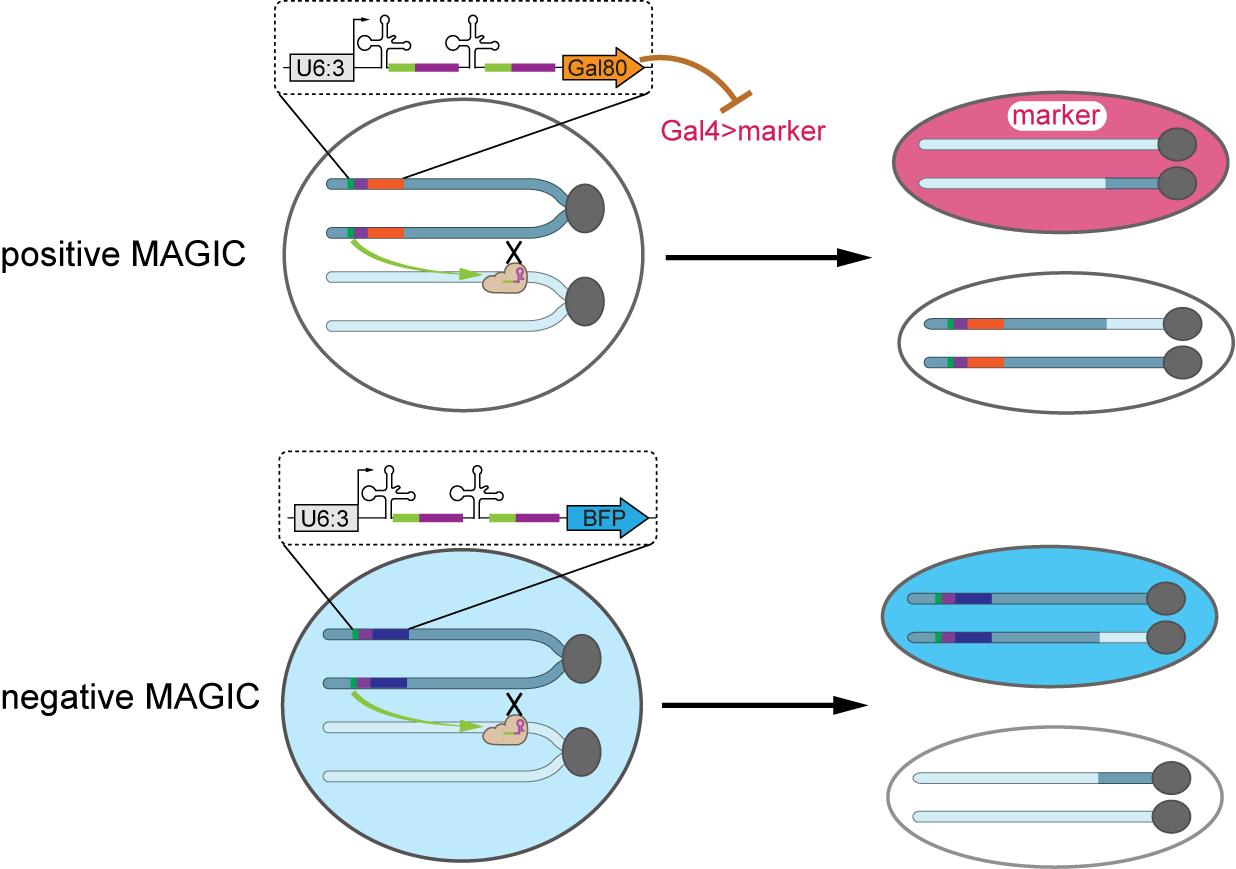
Diagrams of positive MAGIC (pMAGIC) and negative MAGIC (nMAGIC). The terms “positive” and “negative” define the visual labeling strategy used to identify the target mutant clones. pMAGIC (positive labeling) involves ubiquitously expressed Gal80 that suppresses Gal4-driven expression of a fluorescent marker in Gal80-containing cells. Only homozygous cells lacking the gRNA-marker will be labeled. nMAGIC (negative labeling) utilizes ubiquitously expressed blue fluorescent protein (BFP) to distinguish two populations of twin-spot cells, one with two copies of BFP and the other lacking BFP entirely. MAGIC, mosaic analysis by gRNA-induced crossing-over.
Background
Mosaic analysis is a cornerstone of Drosophila genetics, classically driven by FRT/Flp site-specific recombination systems. However, these systems require the laborious introduction of recombination sites onto the specific chromosomal arms of interest. Consequently, existing mutant libraries, uncharacterized wild-derived strains, and genes located proximal to centromeres relative to established FRT sites remain largely inaccessible to standard clonal analysis.
To bypass these constraints, we recently established MAGIC (mosaic analysis by gRNA-induced crossing-over), which relies on CRISPR/Cas9 to generate double-strand breaks (DSBs) at predefined genomic locations, inducing homologous recombination during the S/G2 phase of dividing precursor cells. Segregation of these recombinant chromatids generates "twin spots," which can be visualized using carefully designed genetic markers.
While our initial proof-of-concept focused on chromosome 2L, this expanded protocol utilizes a comprehensive, genome-wide MAGIC kit. We have significantly optimized the gRNA-marker designs to overcome previous limitations of low clone frequency and weak fluorescence. The incorporation of the Qtg2.1 gRNA scaffold enhances DSB induction, while the use of a tubulin enhancer coupled with protein and mRNA destabilization sequences (DE) and an SV40 polyA tail ensures complete suppression of Gal4 in heterozygous cells and bright labeling of pMAGIC clones. For nMAGIC, replacing nuclear blue fluorescent protein (BFP) with cytosolic 3xHA-BFP or infrared fluorescent protein (IFP) allows for clearer visualization of cell morphology. This protocol describes the genetic setups and subsequent imaging techniques to reliably generate and analyze mosaic clones across diverse Drosophila tissues.
Materials and reagents
Biological materials
Fly strains
1. Cas9 driver lines: Tissue-specific, ubiquitous, or inducible Cas9 transgenic lines (Table 1)
Table 1. Cas9 driver lines
| Cas9 | Reference and source | Expression pattern in progenitor cells | Purpose |
|---|---|---|---|
| vas-Cas9 | RRID: BDSC_55821 | Germline and ubiquitous | To induce clones in any tissue |
| zk-Cas9 | [1]; RRIDs: BDSC_607569 and BDSC_607570 | Embryonic dorsal ectoderm | To induce clones in larval epidermis, imaginal discs, larval sensory neurons, and motor neurons |
| ey-Cas9 | [2]; RRID: BDSC_606039 | Eye and antennal discs | To induce clones in cells derived from the eye and antennal discs |
| hs-Cas9 | [3] | Ubiquitous (inducible) | To induce clones in any tissue |
| gcm-Cas9 | [4]; RRID: BDSC_606774 | Glial progenitor | To induce clones in glia |
| Act5C-Cas9 | [5]; RRID: BDSC_54590 | Ubiquitous | To induce clones in any tissue |
| tub-Cas9 | [6]; RRID: BDSC_81930 | Ubiquitous | To induce clones in any tissue |
| hh-Cas9 | [7]; RRIDs: BDSC_81927, BDSC_81928, and BDSC_81929 | Embryonic epidermal cells; imaginal tissues | To induce clones in imaginal discs |
2. MAGIC gRNA-marker lines: Positive MAGIC (pMAGIC) or negative MAGIC (nMAGIC) lines from a genome-wide kit covering all chromosomal arms (X, 2L, 2R, 3L, 3R, and 4)
Note: The complete list of Bloomington Drosophila Stock Center (BDSC) catalog numbers and corresponding target sequences for all lines are provided in Table S1.
3. Fluorescent reporter lines (required for pMAGIC, optional for nMAGIC): Standard Gal4/UAS reporter lines (e.g., tub-Gal4, UAS-mCD8-GFP, UAS-tdTom)
4. User-defined mutant lines: Any Drosophila line carrying a mutation of interest or a chromosomal deletion
Critical: The mutation or deleted segment must be located on the same chromosomal arm and distal to the chosen MAGIC gRNA target site.
Materials and reagents
1. Flies can be cultured on any commonly used Drosophila medium, such as the Bloomington formulation yeast–cornmeal–molasses medium (e.g., Nutri-Fly, Genesee Scientific, catalog number: 66-112) and commercial ready-made medium (such as LabExpress fly food B)
2. Genetic crosses can be set up in standard fly culture vials (e.g., Genesee Scientific, catalog number: 32-113)
3. 60 mm Petri dishes (VWR, catalog number: 25373-085)
4. PYREX® 9-depression glass spot plate (Corning, catalog number: 7220-85)
Equipment
1. Standard stereomicroscope for sorting genetic markers and balancing flies
2. Fluorescence-equipped stereomicroscope (e.g., Nikon, model: SMZ18) for in vivo prescreening and direct visualization of fluorescent clones in live animals
3. Incubators (capable of maintaining 18 and 25 °C) with a 12/12 light/dark cycle; 18 °C is used for preventing activation of hs-Cas9, while 25 °C is for the standard culture of Drosophila
4. Water bath (e.g., VWR, catalog number: 10128-126) set to 37 °C for activating hs-Cas9
5. Fly pushing pads (e.g., Genesee Scientific, catalog number: 59-172) connected to CO2 delivery systems and fine-tipped paintbrushes (e.g., Thermo Fisher Scientific, catalog number: 03-665A) for fly sorting
Procedure
A. Experimental design and component selection
Note: The success of MAGIC relies on correctly pairing four genetic components: Cas9, gRNA-marker, fluorescent reporters, and the test mutation. This system does NOT require FRT sites on the test chromosome.
1. Define the crossover site: Determine the chromosomal location of your mutant gene. Select a MAGIC gRNA-marker line targeting a pericentromeric region proximal to your gene.
Note: The genome-wide kit provides three target sites per arm with varying efficiencies. Select a high-efficiency gRNA for dense clone generation or a low-efficiency gRNA for sparse clones.
2. Select the Cas9 driver: Choose a Cas9 line expressed in the precursor cells of your target tissue (Table 1).
a. Use ubiquitous Cas9 (vas-Cas9, Act5C-Cas9) for broad multi-tissue clones.
b. Use enhancer-driven Cas9 (e.g., ey-Cas9, zk-Cas9) for tissue-specific induction.
c. Use hs-Cas9 for precise temporal control of mitotic recombination.
3. Choose the MAGIC modality (pMAGIC vs. nMAGIC):
a. pMAGIC (positive labeling) uses a Gal80 marker [e.g., tub-Gal80(DE)] to repress Gal4 in heterozygous cells. Clones homozygous for the mutant arm lose Gal80 and become positively labeled by the Gal4-driven fluorescent reporter.
b. nMAGIC (negative labeling) uses a ubiquitously expressed BFP or IFP (e.g., tub-3xHA-BFP). Clones homozygous for the mutant arm are identified by the absence of the fluorescent marker (loss of BFP/IFP).
B. Genetic crossing schemes
Caution: Do not maintain Cas9 and gRNA transgenes together in the same stock long-term, as leaky germline Cas9 activity can prematurely mutate the gRNA target site.
Scheme 1: Generating pMAGIC clones
1. P0 cross (building the components): Place 10–15 virgin females carrying the gRNA-marker and reporter components into a vial alongside 5–8 males carrying the Cas9 and the user-defined mutant allele. Allow the flies to mate and reproduce at 25 °C. Transfer the flies to a new vial after three days initially and then every day to prevent crowding of progeny in the vials.
a. Female genotype example: gRNA-marker(Gal80)/Balancer 1; Gal4, UAS reporter/Balancer 2
b. Male genotype example: mutant_allele/Balancer 1; Cas9/Balancer 2
2. F1 generation (clone induction): Maintain the P0 cross at 25 °C. The somatic crossing-over events will occur during the development of the F1 progeny.
a. Target F1 genotype: gRNA-marker(Gal80)/mutant_allele; Gal4, UAS reporter/Cas9
b. Mechanism: In the precursor cells of the F1 animal, Cas9 induces a DSB at the gRNA target site. Following G2-X segregation, the resulting homozygous mutant cell (mutant_allele/mutant_allele) will lack the gRNA-marker (Gal80) transgene. Without Gal80, the Gal4 component activates the UAS reporter, permanently illuminating the mutant clone.
Scheme 2: Generating nMAGIC clones
1. P0 cross: Following the same physical setup parameters as in Scheme 1, place 10–15 virgin females carrying the gRNA-marker into a vial with 5–8 males carrying the Cas9 and the mutant allele. Transfer the flies to new vials as needed.
a. Female genotype example: gRNA-marker(BFP)/gRNA-marker(BFP)
b. Male genotype example: mutant_allele/Balancer 1; Cas9/Balancer 2
2. F1 generation (clone induction):
a. Target F1 genotype: gRNA-marker(BFP)/mutant_allele; Cas9/+
b. Mechanism: Homozygous mutant clones generated via somatic recombination will lack the gRNA-marker (BFP) transgene and will be visualized as distinct "dark" (BFP-negative) patches amidst the intermediate (heterozygous) and bright (homozygous wild-type twin spot) background.
Genetic crossing schemes and expected visual readouts for MAGIC clone induction are shown in Figure 1.
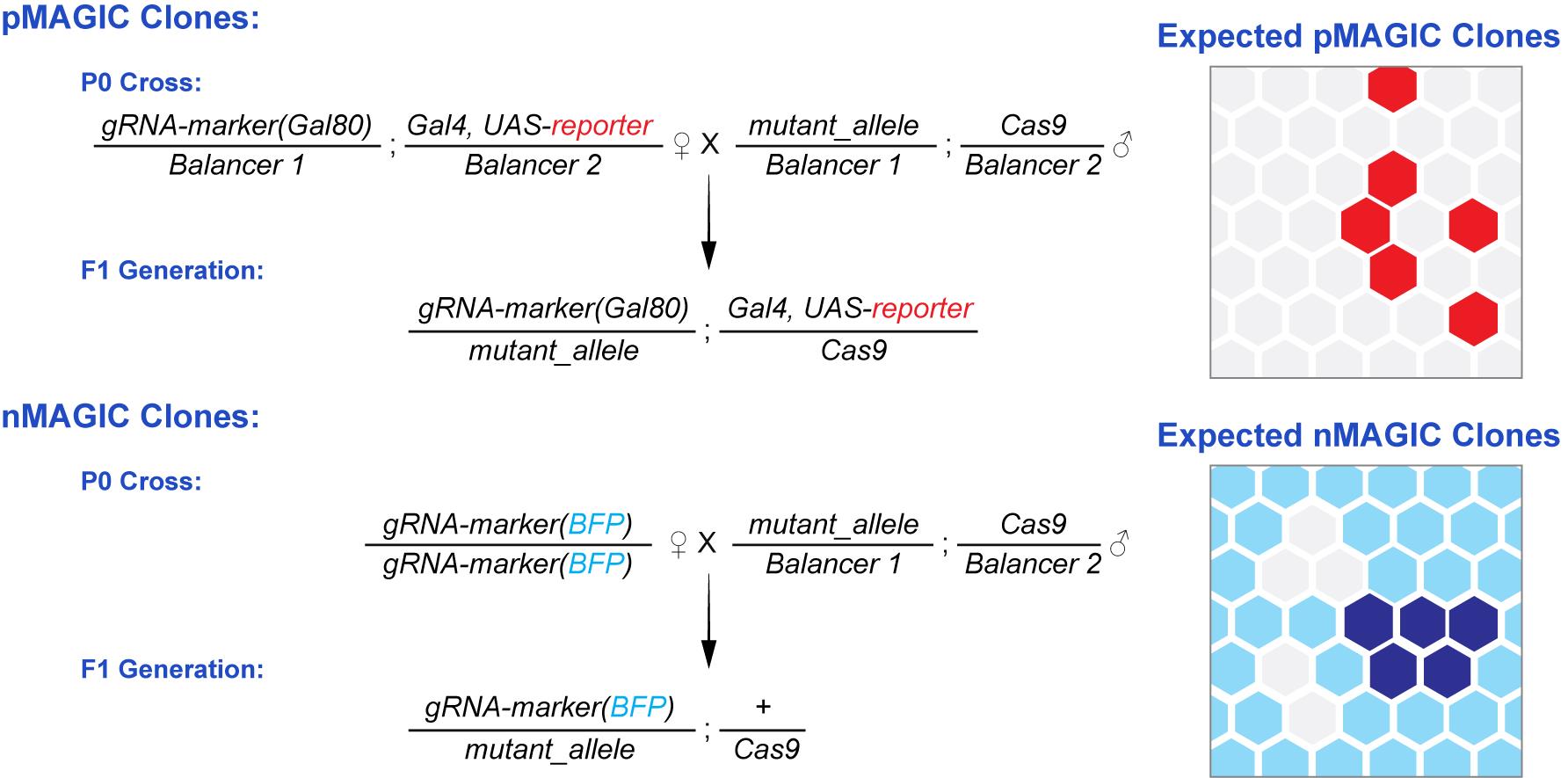
Figure 1. Genetic crossing schemes and expected visual readouts for pMAGIC and nMAGIC. (Top) Positive MAGIC (pMAGIC). The schematic details the P0 cross and the resulting F1 genotype. In F1 somatic cells, Cas9-induced homologous recombination generates homozygous mutant cells that lose the gRNA-marker (Gal80) transgene. As illustrated in the right panel, the de-repression of Gal4 activates the UAS reporter, allowing the mutant clones to be "positively" labeled (e.g., brightly fluorescent red cells) against a non-fluorescent heterozygous background. (Bottom) Negative MAGIC (nMAGIC). The lower panel outlines the crossing strategy to generate nMAGIC clones. Following somatic recombination in the F1 progeny, the resulting homozygous mutant clones lack the gRNA-marker (BFP) entirely. The schematic on the right demonstrates this "negative" labeling readout: mutant clones appear as distinctly dark (BFP-negative) cell patches, which are easily distinguishable from the intermediate (heterozygous, light blue) background and the bright (homozygous wild-type twin spot, white) cells. MAGIC, mosaic analysis by gRNA-induced crossing-over.
C. Temporal clone induction via heat shock (optional)
Note: Temporal control of clone induction is achieved by activating hs-Cas9 via heat shock. Consult Table 2 to determine the timing of the heat shock based on the specific developmental stage at which you wish to activate Cas9. Since hs-Cas9 has leaky activity at room temperature, for tight temporal control, the target genotype should be kept at 18 °C until the desired developmental stage has been reached.
Table 2. Heat shock timing at different developmental stages
| Developmental stage | Approximate time at 18 °C (hours after egg laying, AEL) |
|---|---|
| Embryogenesis ends/first instar begins | 48 h |
| Second instar larva | 96 h |
| Third instar larva | 144 h |
| Pupariation | 240 h |
1. Allow the F1 animals from the P0 cross to develop at 18 °C until they reach the desired developmental stage (e.g., first instar larvae).
2. Submerge the vials in a 37 °C water bath for 1 h. Extend this duration if too few clones are induced; shorten this duration if too many animals die after the heat shock.
3. Immediately return the vials to an 18 °C incubator and allow the F1 animals to continue development.
D. Animal screening and downstream assay
1. To prepare F1 larvae for screening, gently pick them up from the culture medium using a pair of forceps. Rinse the larvae with water in a 60 mm Petri dish to remove food debris. Transfer the cleaned larvae into a water-filled well on a 9-depression glass spot plate. Screen for larvae that do not contain any balancer chromosome under a standard or fluorescence stereomicroscope. Transfer the larvae of the correct genotype to another well for downstream assays.
2. To screen F1 pupae, examine them under a standard or fluorescence stereomicroscope while they are attached to the wall of the culture vial. Mark those that do not contain any balancer chromosome. Gently pick them up using a pair of forceps or a moistened brush for downstream assays.
3. To screen F1 adults, knock them out on a CO2 flypad. Examine them under a standard or fluorescence stereomicroscope. Pick those that do not contain any balancer chromosome for downstream assays.
4. Proceed with your desired downstream phenotypic assay on these mosaic animals (e.g., confocal imaging of intact larvae or dissected tissues).
Data analysis
1. Clone frequency assessment: To evaluate the efficiency of a specific Cas9/gRNA-marker combination, count the number of independent fluorescent (pMAGIC) or non-fluorescent (nMAGIC) clones within a defined anatomical region (e.g., segments A1–A7 in larvae) using a fluorescence stereomicroscope.
2. Phenotypic comparison: Compare the phenotype (e.g., viability, developmental timing, structural morphology) of mosaic animals carrying homozygous mutant clones against control mosaic animals generated using wild-type chromosomes.
3. Statistical analysis: Analyze clone counts or phenotypic penetrance using standard statistical software (e.g., RStudio). Apply Student's t-tests for pairwise comparisons or one-way ANOVA for multiple gRNA efficiency comparisons.
Validation of protocol
This protocol (or parts of it) has been used and validated in the following research articles:
• Shen et al. [8]. A genome-wide MAGIC kit for recombinase-independent mosaic analysis in Drosophila. eLife (2026). DOI: 10.7554/eLife.108453. To view the functional validation strategy—including quantitative comparisons of clone induction frequencies across different chromosomal arms and tissue types (such as wing discs and larval brain)—refer specifically to Figures 2, 3, and Supplementary Figure S1 in this publication.
• Allen et al. [1]. Versatile CRISPR/Cas9-mediated mosaic analysis by gRNA-induced crossing-over for unmodified genomes. PLOS Biology 19(1): e3001061 (2021). DOI: 10.1371/journal.pbio.3001061. Validation of the core MAGIC principle, mapping its efficiency in sensory dendrite arborization (da) neurons, was documented in Figures 1–4 of this study.
General notes and troubleshooting
General notes
1. Transgene variegation on chromosome 4: Transgenes inserted on the fourth chromosome (such as the nMAGIC tub-3xHA-BFP marker) may exhibit uneven or variegated expression in certain tissues due to heterochromatin silencing. pMAGIC is generally recommended for neuronal analysis on the fourth chromosome as the Gal80 repressor remains highly efficient.
2. Off-target considerations: Because perfect DSB repair recreates the gRNA target site, Cas9 will repeatedly cut until an indel forms. Even cells that do not undergo mitotic recombination will likely carry an indel at the gRNA locus. Therefore, the gRNA target sites in this kit were specifically selected within non-critical intergenic sequences to prevent confounding phenotypic artifacts.
Troubleshooting
Problem 1: The desired genotype is difficult to obtain in the F1 generation.
Possible cause: Too many homozygous clones of a lethal mutation in critical tissues could kill animals at an early developmental stage.
Solution: Switch to a tissue-specific Cas9 driver to restrict DSB induction solely to the somatic tissue of interest.
Problem 2: No clones observed in the F1 generation.
Possible cause 1: The chosen gRNA target sequence has inherently low cleavage efficiency.
Solution: Switch to a different MAGIC gRNA-marker line targeting a different locus on the same chromosomal arm (e.g., switch from gRNA-40E1 to gRNA-40D2).
Possible cause 2: The Cas9 driver is not expressed in the appropriate precursor cells during the G2 phase.
Solution: Validate the temporal and spatial expression pattern of your chosen Cas9 driver, or switch to an inducible hs-Cas9 and optimize the timing of the heat shock.
Problem 3: Difficulty in identifying larvae of the correct genotype-carrying clones.
Possible cause: Identifying clone-carrying larvae can be difficult if the clones are not brightly labeled. Additionally, unlike traditional FRT-based mosaic methods, mitotic recombination can be induced between a gRNA-marker chromosome and a balancer chromosome in the MAGIC system. Therefore, simply observing a clone in a larva does not guarantee it has the correct experimental genotype.
Solution: When screening at the larval stage, it is critical to use balancer chromosomes equipped with markers that can be easily distinguished in larvae (such as Tubby or specific fluorescent reporters). This allows for accurate negative selection; without these larval-specific balancers, there is no reliable way to distinguish the desired experimental larvae from those carrying the balancer chromosome.
Supplementary information
The following supporting information can be downloaded here:
1. Table S1. Comprehensive list of genome-wide MAGIC gRNA-marker lines, target sites, and BDSC catalog numbers.
Acknowledgments
Conceptualization: Y.S., C.H.; Methodology: Y.S., C.H.; Visualization: Y.S.; Supervision: C.H.; Funding acquisition: C.H.; Writing—original draft: Y.S., C.H.; Writing—review & editing: Y.S., C.H.
This work and the generation of the MAGIC toolkit were supported by the NIH Office of the Director (R24OD031953) to C.H. Previous development of the MAGIC methodology was supported by NIH grants (R01NS099125, R21OD023824, R01/R37-HD038921, and R03-HD101732) and Cornell University start-up funds.
This protocol is a detailed methodological extension of the original research article in which the genome-wide MAGIC kit was described and validated: Shen et al. [8] eLife (2026) (DOI: 10.7554/eLife.108453). The core MAGIC principles and initial methodology were originally developed and modified from Allen et al. [1] PLOS Biology (2021) (DOI: 10.1371/journal.pbio.3001061). We acknowledge the Bloomington Drosophila Stock Center (NIH P400D018537) and the Drosophila Genetic Reference Panel for providing essential fly stocks.
Competing interests
The authors declare no competing interests.
Ethical considerations
The research described in this protocol utilizes the fruit fly, Drosophila melanogaster, which is an invertebrate model organism. According to institutional and national guidelines in the United States, specific approval from an Institutional Animal Care and Use Committee (IACUC) or equivalent ethics committee is not required for research involving Drosophila. All laboratory practices and animal husbandry were conducted following standard ethical guidelines for the humane care and handling of invertebrate model organisms.
References
- Allen, S. E., Koreman, G. T., Sarkar, A., Wang, B., Wolfner, M. F. and Han, C. (2021). Versatile CRISPR/Cas9-mediated mosaic analysis by gRNA-induced crossing-over for unmodified genomes. PLoS Biol. 19(1): e3001061. https://doi.org/10.1371/journal.pbio.3001061
- Ji, H., Sapar, M. L., Sarkar, A., Wang, B. and Han, C. (2022). Phagocytosis and self-destruction break down dendrites of Drosophila sensory neurons at distinct steps of Wallerian degeneration. Proc Natl Acad Sci USA. 119(4): e2111818119. https://doi.org/10.1073/pnas.2111818119
- Garcia-Marques, J., Espinosa-Medina, I., Ku, K. Y., Yang, C. P., Koyama, M., Yu, H. H. and Lee, T. (2020). A programmable sequence of reporters for lineage analysis. Nat Neurosci. 23(12): 1618–1628. https://doi.org/10.1038/s41593-020-0676-9
- Chen, X., Perry, S., Fan, Z., Wang, B., Loxterkamp, E., Wang, S., Hu, J., Dickman, D. and Han, C. (2024). Tissue-specific knockout in the Drosophila neuromuscular system reveals ESCRT’s role in formation of synapse-derived extracellular vesicles. PLoS Genet. 20(10): e1011438. https://doi.org/10.1371/journal.pgen.1011438
- Port, F., Chen, H. M., Lee, T. and Bullock, S. L. (2014). Optimized CRISPR/Cas tools for efficient germline and somatic genome engineering in Drosophila. Proc Natl Acad Sci USA. 111(29): E2967–76. https://doi.org/10.1073/pnas.1405500111
- Chen, X., Wang, B., Sarkar, A., Huang, Z., Ruiz, N. V., Yeung, A. T., Chen, R. and Han, C. (2025). Phagocytosis-driven neurodegeneration through opposing roles of an ABC transporter in neurons and phagocytes. Sci Adv. 11(11): eadr5448. https://doi.org/10.1126/sciadv.adr5448
- Poe, A. R., Wang, B., Sapar, M. L., Ji, H., Li, K., Onabajo, T., Fazliyeva, R., Gibbs, M., Qiu, Y., Hu, Y., et al. (2018). Robust CRISPR/Cas9-Mediated Tissue-Specific Mutagenesis Reveals Gene Redundancy and Perdurance in Drosophila. Genetics. 211(2): 459–472. https://doi.org/10.1534/genetics.118.301736
- Shen, Y., Yeung, A. T., Ditchfield, P., Korn, E., Clements, R., Chen, X., Wang, B., Huang, Z., Sheen, M., Jarman, P. A., et al. (2026). A genome-wide MAGIC kit for recombinase-independent mosaic analysis in Drosophila. eLife. RP108453. https://doi.org/10.7554/elife.108453.3
Article Information
Publication history
Received: May 22, 2026
Accepted: Jul 8, 2026
Available online: Jul 24, 2026
Published: Aug 20, 2026
Copyright
© 2026 The Author(s); This is an open access article under the CC BY license (https://creativecommons.org/licenses/by/4.0/).
How to cite
Readers should cite both the Bio-protocol article and the original research article where this protocol was used:
- Shen, Y. and Han, C. (2026). Clonal Analysis in Drosophila Tissues With an Enhanced MAGIC Transgenesis Method. Bio-protocol 16(16): e5787. DOI: 10.21769/BioProtoc.5787.
- Shen, Y., Yeung, A. T., Ditchfield, P., Korn, E., Clements, R., Chen, X., Wang, B., Huang, Z., Sheen, M., Jarman, P. A., et al. (2026). A genome-wide MAGIC kit for recombinase-independent mosaic analysis in Drosophila. eLife. RP108453. https://doi.org/10.7554/elife.108453.3

 Download Citation in RIS Format
Category
Molecular Biology > DNA > DNA recombination
Biological Sciences > Biological techniques > CRISPR/Cas9
Cell Biology > Cell engineering > CRISPR-cas9
Do you have any questions about this protocol?
Post your question to gather feedback from the community. We will also invite the authors of this article to respond.
 Post a Question


