


- Protocols

- Articles and Issues
- For Authors
- About
- Become a Reviewer


A Streamlined and Time-Saving Approach to Generate HLA-DR15 MHC Class II Tetramers via In Vivo Biotinylation

Published: Vol 16, Iss 14, Jul 20, 2026 DOI: 10.21769/BioProtoc.5755 Views: 252
Reviewed by: Joel PraneethAnonymous reviewer(s)
 PDF
PDF  Ask a question

Protocol Collections
Comprehensive collections of detailed, peer-reviewed protocols focusing on specific topics




Related protocols
Abstract
This protocol describes an optimized strategy for the efficient generation of peptide-loaded major histocompatibility complex (MHC) class II (pMHC) tetramers, which are essential tools for detecting and characterizing antigen-specific T cells in immunological research. Traditional methods require separate expression of MHC proteins followed by in vitro biotinylation—a multi-step process that is time-consuming and prone to protein loss. Here, we present an integrated approach based on co-expression of MHC monomers and BirA biotin ligase in Expi293F T cells, enabling site-specific biotinylation in vivo during protein synthesis. At the same time, the incorporation of a thrombin-cleavable class II–associated invariant chain peptide (CLIP) peptide into the MHC construct allows flexible loading of any antigenic peptide of interest without the need for re-cloning or re-expression of the MHC molecule. Pre-biotinylated MHC molecules are subsequently purified, loaded with antigenic peptides, and assembled into fluorescent tetramers via streptavidin conjugation. This streamlined workflow significantly reduces handling steps, improves protein yield, and enhances reproducibility. The resulting tetramers are suitable for sensitive detection and isolation of antigen-specific T cells by flow cytometry, supporting applications in T-cell immunogenicity studies, vaccine development, and autoimmune disease research.
Key features
• One-step in vivo biotinylation.
• Eliminates purchased BirA enzyme, saves ~16 h, and reduces protein loss.
• CLIP-based peptide exchange without re-cloning.
• Produces antigen-specific tetramers validated by flow cytometry.
Keywords: MHC tetramer HLA-DRB1*15:01
Biotinylation
T cells
CLIP peptide
HLA-DRB1*15:01
Biotinylation
T cells
CLIP peptide
Graphical overview
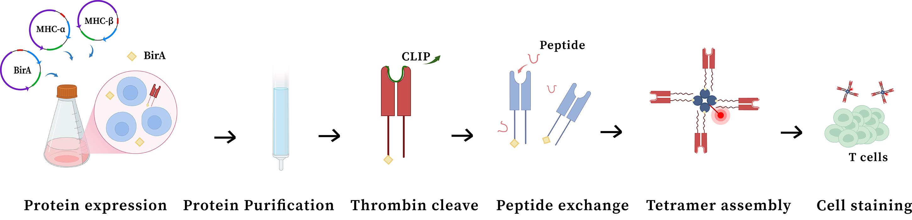
The whole process of preparation and application of HLA-DR15 major histocompatibility complex (MHC) class II tetramer. The sequential process of the experiment is represented, starting from protein expression to preparation of the tetramer and staining for flow cytometry analysis.
Background
Major histocompatibility complex (MHC) class II tetramers are indispensable tools for tracking and characterizing antigen-specific T-cell responses, with broad applications in vaccine development, autoimmune disease research, allergy studies, and cancer immunotherapy [1–4]. However, the widespread adoption of these reagents has been hampered by technical challenges associated with their production. Unlike MHC class I molecules, MHC class II molecules are heterodimeric and inherently unstable in the absence of bound peptide, making them difficult to produce as recombinant proteins [5]. Traditional preparation methods involve multiple sequential steps—including separate MHC protein expression, in vitro biotinylation, peptide loading, and multimer assembly—each requiring purification or buffer exchange [6]. These cumulative handling steps result in substantial protein loss, batch-to-batch variability, and overall low yields, limiting access to high-quality MHC class II tetramers for many research applications.
To overcome these limitations, we developed an integrated expression system that combines MHC class II monomer production with in vivo biotinylation. By co-expressing the engineered MHC α/β heterodimer and BirA biotin ligase in Expi293F cells, site-specific biotinylation occurs concurrently with protein synthesis, eliminating the need for a separate enzymatic reaction and associated purification steps. Compared with conventional in vitro biotinylation, this approach eliminates the need for purchased BirA enzyme, saves approximately 16 h of hands-on time, and avoids approximately half of protein loss caused by buffer exchange and concentration steps, while maintaining comparable tetramer quality. Furthermore, the incorporation of a thrombin-cleavable CLIP peptide allows for flexible loading of any antigenic peptide of interest without requiring re-cloning or re-expression of the MHC construct. This streamlined workflow significantly reduces processing time, minimizes protein loss through fewer handling steps, and ensures consistent, efficient biotinylation. Nevertheless, this method is primarily suitable for small-to-medium scale protein preparation; for large-scale production, the reliance on Ni-NTA gravity column purification and repeated ultrafiltration concentration steps makes the process labor-intensive and time-consuming.
The resulting human leukocyte antigen (HLA) tetramers exhibit excellent staining performance for antigen-specific T cells by flow cytometry, enabling sensitive detection and isolation of rare T-cell populations. Beyond conventional immunophenotyping, this method supports a range of applications, including longitudinal immune monitoring in clinical studies, epitope mapping in infectious and autoimmune diseases, and evaluation of T-cell responses in immunotherapy settings.
Materials and reagents
Biological materials
1. Expi293F (Gibco, catalog number: A14527CN)
2. pcDNA3.4 vector (TSINGKE Biological Technology, catalog number: ZT000101)
Reagents
1. RPMI 1640 (VivaCell, catalog number: C3010-0500); store at 4 °C
2. Fetal bovine serum (FBS) (Gibco, catalog number: 10091148); store at -20 °C
3. PBS pH 7.4 basic (1×) (Gibco, catalog number: C10010500BT); store at 4 °C
4. ExpiFectamineTM 293 Transfection kit, 1 L (Gibco, catalog number: A14524); store at 4 °C
5. Trypan Blue stain (0.4%) (Gibco, catalog number: 15250-061); store at room temperature
6. GibcoTM basic DMEM (Gibco, catalog number: C11995500BT); store at 4 °C
7. 20× tris-buffered saline (TBS) concentrate buffer (Thermo Scientific, catalog number: 28358); store at room temperature
8. Ampicillin (Beyotime, catalog number: ST008); store at -20 °C
9. Thrombin (Sigma, catalog number: T9326-150UN); store at -20 °C
10. PE Streptavidin (high concentration) (BioLegend, catalog number: 405245); store at 4 °C
11. Horseradish peroxidase (HRP)-labeled streptavidin (Beyotime, catalog number: A0305); store at -20 °C
12. Ni-NTA elution buffer (Sangon, catalog number: C600304); store at 4 °C
13. Ni-NTA binding/wash buffer (Sangon, catalog number: C600303); store at 4 °C
14. His Tag (C-terminal specific) mouse monoclonal antibody (HRP conjugated) (Beyotime, catalog number: AF2873); store at -20 °C
15. StrepII Tag mouse monoclonal antibody (HRP conjugate) (Beyotime, catalog number: AF2927); store at -20 °C
16. Skim milk (YEASEN, catalog number: 36120ES60); store at room temperature
17. Tris-MES-SDS running buffer power (Beyotime, catalog number: M00677); store at room temperature
18. Broad Multi Color Pre-Stained Protein Standard (Beyotime, catalog number: M00624); store at -20 °C
19. SDS-PAGE protein loading buffer (5×, odorless) (Beyotime, catalog number: P0286-2 mL); store at 4 °C
20. EDTA (0.5 M), pH 8.0 (Thermo Scientific, catalog number: AM9260G); store at room temperature
21. Octyl-β-D-glucopyranoside (≥98%, reagent grade) (Beyotime, catalog number: ST2546-1 g); store at -20 °C
22. Pepstatin A (YEASEN, catalog number: 20113ES08); store at -20 °C
23. Leupeptin (YEASEN, catalog number: 20112ES08); store at -20 °C
24. PMSF solution (100 mM) (Beyotime, catalog number: ST507-10 mL); store at -20 °C
25. DMSO (Sigma-Aldrich, catalog number: D5879); store at room temperature
26. Sodium citrate (absin, catalog number: abs42015609); store at room temperature
27. NaCl (Sigma-Aldrich, catalog number: S5886); store at room temperature
28. Opti-MEMTM I reduced serum medium (Gibco, catalog number: 31985070); store at 4 °C
29. UltraPureTM DNase/RNase-free distilled water (Thermo Scientific, catalog number: 10977015); store at room temperature
30. XbaI (NEB, catalog number: R0145V); store at -20 °C
31. AgeI-HF (NEB, catalog number: R3552S); store at -20 °C
Solutions
1. FACS buffer (see Recipes)
2. 10% octyl glucoside (see Recipes)
3. 1 mg/mL leupeptin (see Recipes)
4. 1 mg/mL pepstatin (see Recipes)
5. Sodium citrate buffer (see Recipes)
6. Peptide solution (see Recipes)
7. TBS buffer (see Recipes)
8. Peptide exchange buffer (see Recipes)
Recipes
1. FACS buffer (500 mL)
| Reagent | Final concentration | Quantity or volume |
|---|---|---|
| PBS | N/A | 490 mL |
| FBS | 2% | 10 mL |
Store at 4 °C.
2. 10% octyl glucoside (1 mL)
| Reagent | Final concentration | Quantity or volume |
|---|---|---|
| Octyl-β-D-glucopyranoside | N/A | 0.1 g |
| Ultrapure water | 10% | 1 mL |
Store at -20 °C unless using the day of.
3. 1 mg/mL leupeptin (5 mL)
| Reagent | Final concentration | Quantity or volume |
|---|---|---|
| Leupeptin | 1 mg/mL | 5 mg |
| Ultrapure water | N/A | 5 mL |
Store at -20 °C unless using the day of.
4. 1 mg/mL pepstatin (5 mL)
| Reagent | Final concentration | Quantity or volume |
|---|---|---|
| Pepstatin | 1 mg/mL | 5 mg |
| DMSO | N/A | 5 mL |
Store at -20 °C unless using the day of.
5. Sodium citrate buffer (50 mL)
| Reagent | Final concentration | Quantity or volume |
|---|---|---|
| NaCl | 150 mM | 0.4383 g |
| Sodium citrate | 50 mM | 0.645 g |
| Ultrapure water | N/A | 50 mL |
Store at room temperature.
6. Peptide solution
| Reagent | Final concentration | Quantity or volume |
|---|---|---|
| Peptide (1.64 kDa) | 20 mM | 2 mg |
| DMSO | N/A | 61 μL |
Store at -20 °C unless using the day of.
7. TBS buffer (500 mL)
| Reagent | Final concentration | Quantity or Volume |
|---|---|---|
| 20× TBS concentrate buffer | 1× | 25 mL |
| Ultrapure water | N/A | 475 mL |
Store at room temperature.
8. Peptide exchange buffer (1 mL)
| Reagent | Final concentration | Quantity or volume |
|---|---|---|
| 10% octyl glucoside | 1% | 100 μL |
| 1 mg/mL leupeptin | 2 μg/mL | 2 μL |
| 1 mg/mL pepstatin | 1 μg/mL | 1 μL |
| 0.5 M EDTA | 2 mM | 4 μL |
| 100 mM PMSF | 0.5 mM | 5 μL |
| 20 mM peptide | 100 μM | 5 μL |
| Cleaved pMHC | 1 μM | - |
| Sodium citrate buffer | N/A | Add to 1 mL |
Make fresh and keep on ice for use on the same day.
Laboratory supplies
1. Axygen® 1.5 mL MaxyClear Snaplock (Axygen, catalog number: MCT-150-C)
2. Corning® 125 mL polycarbonate Erlenmeyer flask with vent cap (Corning, catalog number: 431143)
3. Corning® flask, canted neck, 25 cm, vented (Corning, catalog number: CLS431463)
4. Amicon® Ultra 15 mL, 30 kDa cutoff (Merck, catalog number: UFC903096)
5. Amicon® Ultra centrifugal filters, 10 kDa molecular weight cutoff (MWCO) (Merck, catalog number: UFC501024)
6. HyPur T Ni-NTA 6FF (His-Tag) (Sangon, catalog number: C600332)
7. Sterile transfer pipette (BKMAM, catalog number: 110205008)
10. 0.2 mL PCR 8-tube strip with attached cap (YouLai, catalog number: PCR02-8TC-C-RT)
11.5 mL polystyrene round-bottom tube (Corning, catalog number: 352235)
12. CountessTM cell counting chamber slides (Thermo Scientific, catalog number: C10283)
13 50 mL centrifuge tube (Corning, catalog number: 430829)
14. 15 mL centrifuge tube (Corning, catalog number: 430790)
15. SurePAGETM, Bis-Tris, 10×8, 4%–20%, 12 wells (Beyotime, catalog number: M00656)
Equipment
1. Thermal cycler (Bio-Rad, model: T100TM)
2. CO2 incubator (Thermo Scientific, model: FormaTM Steri-CycleTM)
3. Biological safety cabinet (Thermo Scientific, catalog number: 1379)
4. Centrifuge (Eppendorf, model: 5810 R)
5. Flow cytometer (Thermo Scientific, model: Attune NxT)
6. Eppendorf ThermoMixer® C (Eppendorf, catalog number: 5382)
7. FrescoTM 17 microcentrifuge (Thermo Scientific, catalog number: 75002420)
8. Orbital shaker (YOONING, model: CS-200E)
9. Ice maker (SANYO, model: SIM-F140)
10. CountessTM 3 automated cell counter (InvitrogenTM, catalog number: AMQAX2000)
11. Milli-Q® SQ 200P ultrapure water system (Merck, catalog number: ZSQ200UPT0)
12. Contact nondestructive quantitative imager (e-Blot, model: Touch Imager XLi)
13. Mini-PROTEAN Tetra (Bio-Rad, catalog number: 165-8004)
14. Thermo ScientificTM RevcoTM laboratory refrigerator (Thermo Scientific, catalog number: REL3004V)
15. Variable speed mixer (DLAB, model: MX-S)
16. LocatorTM Plus rack and box system (Thermo Scientific, catalog number: CY50925-70)
17. Biomedical ultra-low temperature freezer (MEGSTEMAN Scientific, catalog number: DW-HL678D)
18. Microcentrifuge (YOONING, model: MC-12K)
19. Electric heating thermostatic water bath (Bluepard, model: MC-12K)
20. Combined refrigerator and freezer (Haier Biomedical, model: HYCD-471FD)
21. NanoDropTM One microvolume UV-Vis spectrophotometer (Thermo Fisher Scientific, catalog number: 840-317400)
Software and datasets
1. FlowJo version 10.8.1
2. SnapGene 6.0.2
Procedure
A. Construction of plasmid
1. CLIP-HLA-DRB1*15:01 plasmid (expressing MHC β-chain)
Design the MHC β-chain construct by sequentially introducing, from the N-terminus to the C-terminus, the following elements for Expi293F expression: the Ig κ secretion signal peptide, the Strep tag, the CLIP polypeptide flanked by NotI and BamHI sites, a thrombin cleavage site connected by a GGGGS linker, the HLA-DR*15:01 β-chain extracellular segment, an HRV 3C protease cleavage site, an alkaline leucine zipper, and a FLAG tag followed by a BirA biotin ligase recognition motif connected by six GGGGS linkers. Finally, insert XbaI and AgeI restriction sites at the 5′ and 3′ ends of the complete assembled fragment, respectively, to facilitate cloning into the pcDNA3.4 vector.
2. HLA-DRA*01:01 plasmid (expressing MHC α-chain)
Design the MHC α-chain construct by sequentially introducing, from the N-terminus to the C-terminus, the following elements for Expi293F expression: the Ig κ secretion signal peptide, the HLA-DR*15:01 α-chain extracellular segment, an HRV 3C protease cleavage site, an acidic leucine zipper, and two tandem 6× His tags connected by six GGGGS linkers. Finally, insert XbaI and AgeI restriction sites at the 5′ and 3′ ends of the complete assembled fragment, respectively, to facilitate cloning into the pcDNA3.4 vector.
3. BirA plasmid (expressing BirA biotin ligase)
Design the BirA expression construct by inserting the full-length BirA coding sequence into the pcDNA3.4 vector. Flank the open reading frame with XbaI and AgeI restriction sites at the 5′ and 3' ends, respectively.
Note: The plasmid construction strategy and key functional elements are illustrated in Figure S1.
B. Protein expression
1. Retrieve a vial of Expi293FTM cells from liquid nitrogen storage. Thaw the vial by gently agitating it in a 37 °C water bath for 1 min.
2. Transfer the entire thawed cell suspension into a sterile, vented 125 mL Erlenmeyer flask containing 30 mL of prewarmed Expi293TM expression medium.
Note: For protein expression, we use the ExpiFectamineTM 293 Transfection kit, which includes Expi293TM expression medium, ExpiFectamineTM 293 transfection reagent, and ExpiFectamineTM 293 transfection enhancers 1 and 2. The Expi293TMexpression medium contains D-biotin as part of its proprietary formulation to support in vivo biotinylation, making it particularly suitable for this protocol. All transfection and culture steps follow the manufacturer's instructions, with minor modifications as described below.
3. Cap the flask and place it in a humidified (≥80% relative humidity) 37 °C incubator with 8% CO2 on an orbital shaker platform. Set the shaking speed to 125 rpm.
4. Culture the cells for 3–6 days. Measure the viable cell density and viability daily using a cell counter with Trypan Blue exclusion.
5. When the viable cell density reaches a range of 1.0–3.0 ×10⁶ viable cells/mL, perform the first subculture. Dilute the culture with fresh, prewarmed Expi293TM expression medium to a density of 0.5 × 10⁶ viable cells/mL in a new vented flask.
6. Continue subculturing the cells every 3–4 days, maintaining the density between 0.5 and 3.0 × 10⁶ viable cells/mL. For protein expression, expand the cells for at least 3–4 passages.
Critical: Cell health is critical for optimal expression yield. Strictly follow the recommended passage number and density limits and monitor cell density and viability closely during passaging.
7. On the day before transfection, expand the Expi293FTM culture until it reaches a density of approximately 3.0–5.0 × 10⁶ viable cells/mL and subculture the cells to a final viable cell density of 2.5–3.0 × 10⁶ cells/mL in fresh Expi293FTM expression medium.
8. Allow the cells to grow overnight (16–18 h) in the incubator (37 °C, 8% CO2, 125 rpm).
9. On the day of transfection, confirm that the cell density is between 4.5 and 5.0 × 10⁶ viable cells/mL and viability is >95%. If necessary, dilute the culture with fresh, prewarmed medium to achieve exactly 3.0 × 10⁶ viable cells/mL in the final desired culture volume. Swirl the flask gently to mix.
10. Dilute the total plasmid DNA in Opti-MEMTM I to achieve a final DNA concentration of 1.0 μg/mL for the total transfection volume.
Critical: Maintain the 1:1:1 molar ratio of the three plasmids (MHC α-chain, MHC β-chain, and BirA plasmids at a 1:1:1 molar ratio) to ensure balanced co-expression.
11. Dilute the ExpiFectamineTM 293 reagent in Opti-MEMTM I medium and incubate it at room temperature for 5 min.
12. Add the diluted ExpiFectamineTM 293 reagent to the diluted plasmid DNA and incubate the ExpiFectamineTM 293/plasmid DNA complexes at room temperature for 10–20 min.
13. Slowly transfer the complexes to the cells, swirling the culture flask gently during addition. Then, incubate the cells in a 37 °C incubator with ≥80% relative humidity and 8% CO2 on an orbital shaker.
14. Eighteen to twenty-two hours post-transfection, add ExpiFectamineTM 293 transfection enhancer 1 and ExpiFectamineTM 293 transfection enhancer 2 to the transfection flask, gently swirling the flask during addition.
15. Continue incubating the cells for an additional 3–5 days and monitor cell viability daily.
16. Harvest the protein supernatant when cell viability drops to approximately 70%.
Critical: Monitor cell viability daily. Harvest the protein supernatant when cell viability drops to approximately 70%. Delayed harvest may lead to increased cell lysis and release of intracellular proteases and contaminating host cell proteins, compromising protein purity.
17. Transfer the cell culture to centrifuge tubes. Centrifuge at 4,000× g for 20 min at 4 °C and harvest the supernatant.
Caution: Always balance the centrifuge rotor with counterbalancing tubes of equal weight. Imbalanced loading may damage the centrifuge and cause sample loss or personal injury.
C. Protein concentration and purification
1. Transfer the harvested protein supernatant into a 30 kDa molecular weight cutoff (MWCO) ultrafiltration centrifugal tube.
2. Centrifuge the tube at 4,000× g for 20 min at 4 °C. Discard the flowthrough from the sample chamber.
3. Repeat step C2 until the entire volume of protein supernatant is concentrated to the desired final volume (typically 5–10 mL).
4. Perform buffer exchange by adding 5–10 mL of 1× TBS buffer (pH 8.0) to the concentrated protein in the ultrafiltration tube. Centrifuge again at 4,000× g for 20 min at 4 °C. Discard the flowthrough.
5. Repeat the buffer exchange process for a total of 4–5 cycles to ensure complete exchange into 1× TBS.
6. Use a pipette tip to gently aspirate the retentate from the bottom and center of the sample chamber.
Pause point: The harvested protein sample can be stored at 4 °C and should be purified within 48 h.
7. Select a Ni-NTA gravity chromatographic column with appropriate protein loading and discharge the stored solution from the column by gravity.
8. Balance the column by using a binding/washing buffer of 2 column volumes (CV). Let the buffer flow completely by gravity at a speed of approximately 0.5–1 mL/min.
9. Mix the protein extract with binding/wash buffer 1:1 to prepare the sample solution.
10. Add the sample liquid to the column, collect the flowing liquid into the centrifugal tube, and re-circulate it once to improve the combination efficiency of the sample and the filler.
11. Wash the column with 10 mL of binding/wash buffer and collect the flowthrough liquid. Check the absorbance at 280 nm using a NanoDrop spectrophotometer (Protein A280 mode) and continue until it approaches the baseline.
12. Elute the His tag protein on the column with 10 mL of elution buffer and repeat twice until the absorbance of the eluent approaches the baseline at 280 nm.
Caution: Elution buffer contains imidazole and may cause skin/eye irritation. Wear gloves and safety goggles.
13. Transfer the elution into a 30 kDa ultrafiltration tube and repeat steps C2–5 until the protein concentration is 1–5 mg/mL. Determine the protein concentration using a NanoDrop spectrophotometer at A280 (Protein A280 mode). Validate the protein purification efficiency using western blot analysis, as shown in Figure 1.
Pause point: The harvested protein samples can be stored at -80 °C before the subsequent operation.
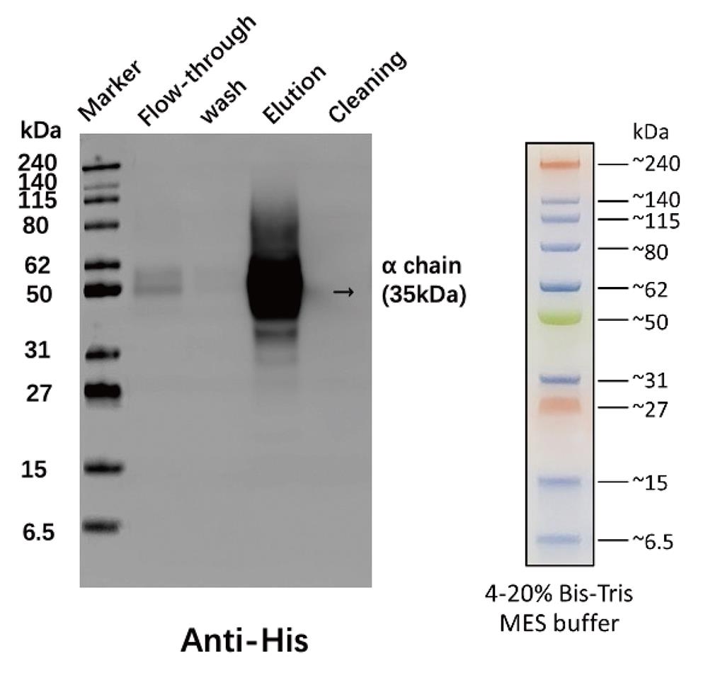
Figure 1. Evaluation of protein purification by Ni-NTA gravity chromatography. Samples collected during the purification process were analyzed by western blot using anti-His antibody to monitor protein recovery and binding efficiency. (Left) Western blot results. (Right) Broad Multi Color Pre-Stained Protein Standard.
D. Preparation of pMHC monomer
Note: To convert protein concentration from mg/mL to nmol/μL, use the following formula based on the molecular weight (MW) of the protein (for HLA-DR15, MW ≈ 76 kDa):
For example, a concentration of 1 mg/mL corresponds to approximately 0.0132 nmol/μL for a 76 kDa protein. To obtain 1 nmol of protein, add approximately 75.8 μL of a 1 mg/mL solution.
1. Calculate and transfer the volume containing 1 nmol of purified protein (based on the measured concentration and the conversion formula above) into a 0.2 mL PCR tube.
2. Add 2.5 μL of thrombin to the protein solution in the PCR tube.
3. Add 1× PBS to bring the total reaction volume to 100 μL.
4. Place the PCR tube in a thermocycler and incubate at 22 °C for 4 h.
5. After the 4-h incubation, transfer the 100 μL thrombin cleavage reaction mixture (containing the "empty" MHC complex) into a 1.5 mL centrifuge tube and add 900 μL of peptide exchange buffer.
6. Mix gently and incubate at 30 °C for 16 h in a metal bath.
7. Transfer the pMHC protein solution (peptide–MHC complex) into a 10 kDa (MWCO) ultrafiltration centrifugal tube.
8. Centrifuge the tube at 4,000× g for 5 min at 4 °C.
9. Repeat step D8 until the entire volume of protein supernatant is concentrated to the desired final volume (typically 50–200 μL).
10. Perform buffer exchange by adding 0.3–0.4 mL of 1× TBS buffer (pH 8.0) to the concentrated protein in the ultrafiltration tube. Centrifuge again at 4,000× g for 5 min at 4 °C.
11. Repeat the buffer exchange process for a total of 4–5 cycles to ensure complete exchange into 1× TBS.
12. Invert the ultrafiltration insert and place it into a clean collection tube. Centrifuge at 4,000× g for 3–5 min at 4 °C to recover the concentrated protein solution from the membrane surface.
13. Measure the final protein concentration using NanoDrop. Aliquot the concentrated pMHC complex into low-protein-binding microcentrifuge tubes. Figure 2 shows the efficiency of this method of biotinylation and thrombin cleavage using a western blot.
Pause point: The concentrated complex can be stored at 4 °C for immediate use (within 1 week) or flash-frozen in liquid nitrogen and stored at -80 °C for long-term preservation.
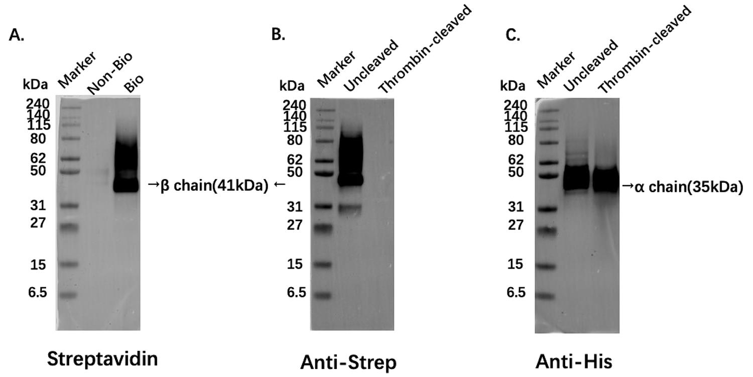
Figure 2. Western blot analysis of biotinylation and thrombin cleavage. (A) Biotinylation efficiency: Detected with streptavidin-HRP. Non-biotinylated controls show no signal; in vivo biotinylated samples show specific bands. (B) Thrombin cleavage efficiency: Detected with anti-Strep tag antibody. Strep tag signal is present in untreated samples and completely lost after thrombin cleavage. (C) Major histocompatibility complex (MHC) α chain integrity: Detected with anti-His tag antibody. His tag signal is present in both untreated and thrombin-cleaved samples, indicating the MHC heavy chain remains intact throughout the process.
E. Tetramer assembly
1. Calculate the required volumes based on the molecular weight and concentration of the pMHC protein solution. Add streptavidin conjugated to PE (SA-PE) into the pMHC protein solution. Maintain a precise molar ratio of SA-fluorophore to pMHC of 1:6.
2. Add PBS to the mixture to achieve a final pMHC concentration of 2.5 μM in the total reaction volume. Mix the components by gently pipetting up and down 10–15 times.
3. Protect the tube from light by wrapping it in aluminum foil. Incubate the reaction mixture at room temperature (22–25 °C) for 30 min in the dark.
Caution: PE-streptavidin is light-sensitive. Protect the conjugate from direct light to avoid photodegradation and signal loss.
4. After the initial incubation, gently flick the tube to mix the contents again.
5. Transfer the tube to a 4 °C refrigerator. Continue the incubation in the dark for 12–16 h (overnight).
6. Following the overnight incubation, the tetramer is ready for use.
Note: After assembly, store the tetramer at 4 °C in the dark and use it within one week.
F. Construction of over-expressed T-cell receptor (TCR) cell line
To generate over-expressed TCR cell lines for tetramer validation, we followed the strategy for T-cell receptor cloning and expression described by Xia et al. [7]. Briefly, TCRα and TCRβ chain sequences were synthesized and cloned into a lentiviral expression backbone using Gibson assembly. Lentiviral particles were produced by co-transfecting HEK-293T cells with the TCR expression plasmid and packaging plasmids (psPAX2 and pMD2.G). Viral supernatants were harvested 48 h post-transfection, filtered, and used to transduce TCRα/β-deficient Jurkat 76 cells. Transduced cells were expanded and sorted by flow cytometry for stable TCR surface expression using anti-human TCRα/β antibody. The resulting TCR-transduced cell lines were used for subsequent tetramer staining assays.
G. Tetramer staining of T cells
1. Resuscitate and culture the T-cell line expressing specific TCR. Harvest the cell suspension and add PBS for washing. Centrifuge at 300× g for 5 min at room temperature.
2. Suspend the cells with FACS buffer to achieve a final cell concentration of 5 × 10⁶ cells/mL
3. Transfer 100 μL of cells into 1.5 mL microcentrifuge tubes and add 2 μL of tetramer for staining.
Note: The CLIP-Tetramer serves as the essential negative control to define background staining and set the positive gate.
4. Mix gently and incubate the cells in the dark at 37 °C for 90 min.
Critical: Incubate at 37 °C for at least 90 min. Insufficient incubation time or lower temperature may result in weak staining signals and underestimation of tetramer-positive cells.
5. Add 1 mL of FACS buffer to each test tube, centrifuge at 300× g for 5 min, and carefully discard the supernatant.
6. Repeat the washing step again.
7. Resuspend the cells with 200 μL of FACS buffer for flow cytometry analysis.
Note: Before collection, keep the sample on ice and keep it away from light.
8. Collect data on flow cytometry. Use negative controls to set tetramer positive gating. A tetramer loaded with a specific peptide specifically recognizes and binds to cells expressing epitope-specific TCR, as shown in Figure 3.
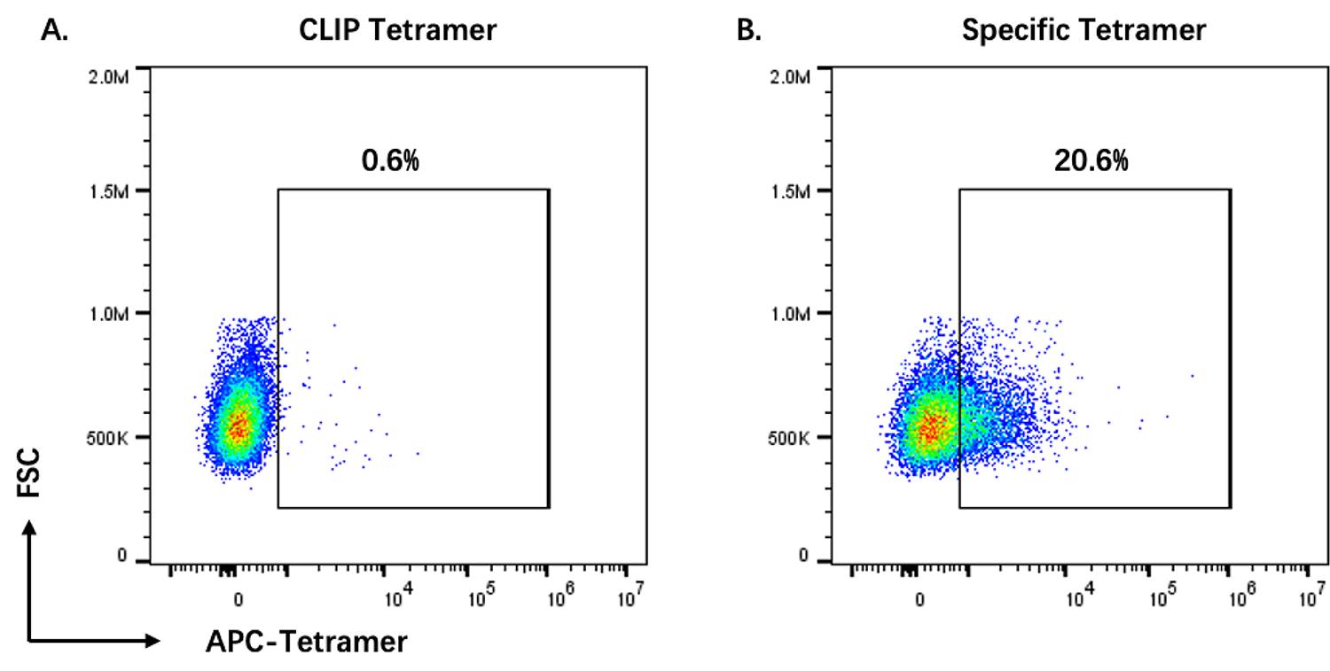
Figure 3. Flow cytometry analysis of T-cell receptor (TCR)-expressing T cells stained with peptide-loaded major histocompatibility complex (MHC) class II (pMHC) tetramers. Cells were stained with either CLIP-loaded tetramer or specific peptide-loaded tetramer. Tetramer-positive populations were gated based on the CLIP-tetramer control.
Data analysis
Flow cytometry data were analyzed with FlowJo v10.8.1.
Validation of protocol
Protein expression and purification steps were repeated three times (n = 3). Following purification, the presence of the His tag on the α chain was successfully detected in the elution fraction by western blot, appearing as a positive signal at an apparent molecular mass of approximately 35 kDa. A representative result is shown in Figure 1, and an additional independent replicate is presented in Figure 4A.
The preparation of pMHC monomers was also repeated three times (n = 3), encompassing assessments of biotinylation efficiency, thrombin cleavage, and peptide exchange. Biotinylation efficiency was assessed by western blot using HRP-conjugated streptavidin. Successful biotinylation of the Avi tag on the β chain was detected as a positive signal at an apparent molecular mass of approximately 41 kDa. Thrombin cleavage efficiency was assessed by western blot using an anti-Strep tag antibody. Upon thrombin-mediated removal of the CLIP peptide, the Strep tag on the β chain was removed along with the CLIP peptide, resulting in the loss of the Strep signal, while the His tag on the α chain remained unaffected. Representative data from these experiments are presented in Figure 2, and reproducibility across independent repeats (including additional samples) is shown in Figure 4B.
Tetramers prepared according to this method recognize and bind to cells expressing epitope-specific TCR. A representative result is shown in Figure 3, and an additional independent replicate is presented in Figure 4C. For functional validation of the tetramers, a CLIP-MHC tetramer was used as a negative control to define nonspecific background signals, providing a baseline reference for gating of tetramer-positive cells. CLIP peptide is a segment of the invariant chain (Ii chain) and is not recognized by any known CD4+ T cells in vivo, making it an ideal negative control.
Tetramer staining validation experiments comparing the specific tetramer with the CLIP negative control tetramer were performed at least six independent times. Statistical analysis using Welch's t-test (unpaired, unequal variance) showed a significant difference between the two groups (n = 6, ***p < 0.001), as shown in Figure 4D.
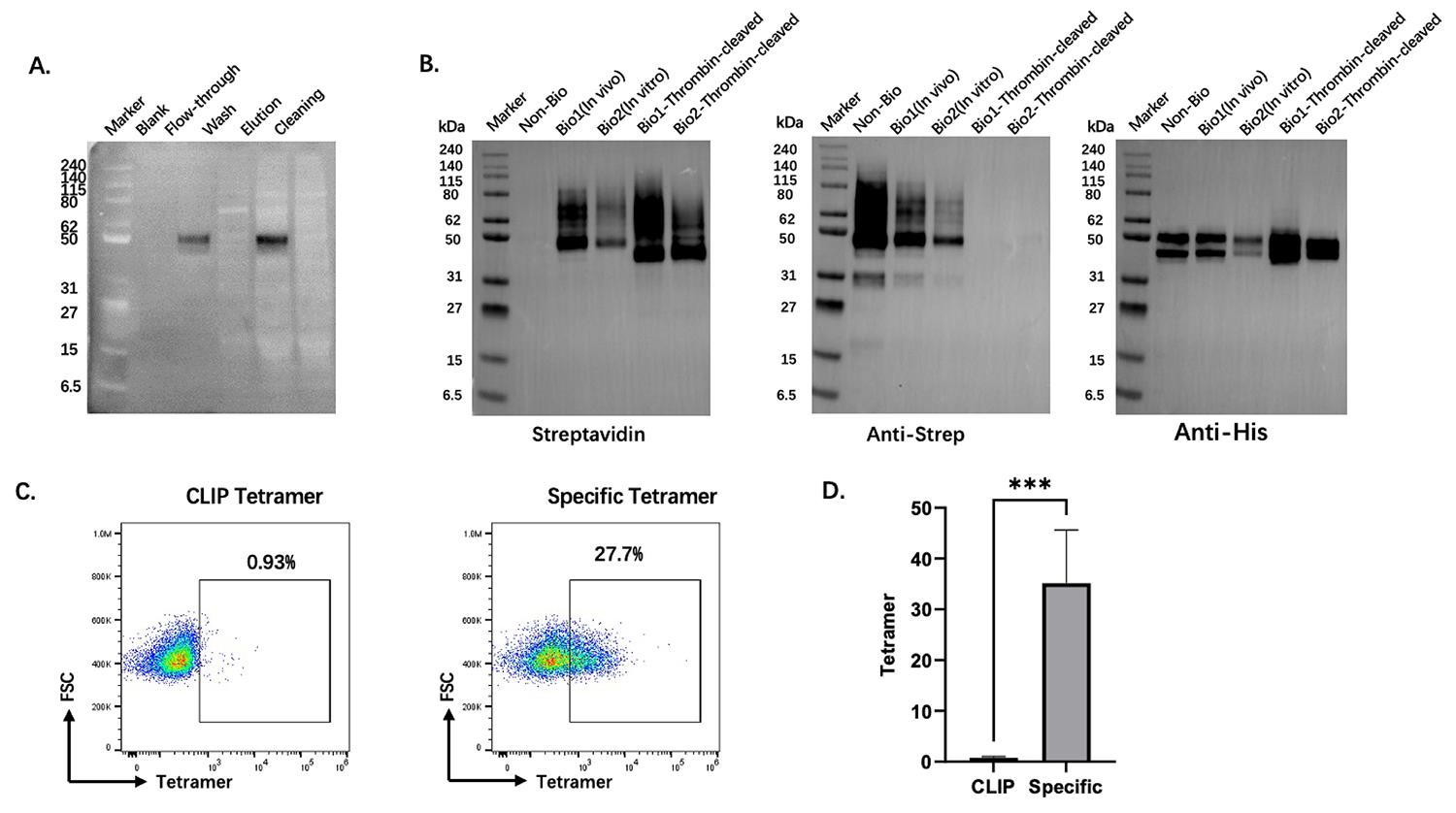
Figure 4. Independent replicate experiments and statistical validation. (A) Independent replicate of the protein purification and elution validation shown in Figure 1. Western blot detection of the His tag on the α chain confirmed a positive signal at approximately 35 kDa. (B) Independent replicate experiments for peptide-loaded major histocompatibility complex (MHC) class II (pMHC) monomer assessments. Western blots show biotinylation efficiency (HRP-streptavidin, ~41 kDa) and thrombin cleavage efficiency (anti-Strep tag antibody; loss of Strep signal upon CLIP removal, while the His tag on the α chain remained unaffected). (C) Independent replicate of tetramer staining shown in Figure 3, demonstrating specific binding of tetramers to cells expressing epitope-specific TCRs. (D) Statistical analysis of tetramer staining validation experiments comparing the specific tetramer with the CLIP-MHC negative control tetramer. Data were analyzed using Welch's t-test (unpaired, unequal variance), revealing a statistically significant difference between the two groups (n = 6, ***p < 0.001).
General notes and troubleshooting
General notes
1. This protocol was optimized for HLA-DRB1*15:01 in Expi293F cells. When adapting to other HLA-DR alleles, minor modifications may be necessary, including adjustment of protein harvest time based on cell viability and expression levels during the experiment. Protein yield may vary depending on the expression efficiency of different MHC alleles; therefore, pilot experiments are recommended when applying this protocol to new alleles.
2. Peptide exchange conditions should be empirically determined based on the specific peptide of interest. High-affinity peptides typically achieve efficient exchange under the standard conditions described here (100 μM peptide, 30 °C, 16 h). For low-affinity peptides or those with slow binding kinetics, higher peptide concentrations or extended incubation times may be required. For each new peptide, optimal exchange conditions may need to be determined experimentally.
3. This system is suitable for small-to-medium-scale protein preparation (e.g., 50–500 mL culture volume). For large-scale production, the current workflow relies on Ni-NTA gravity column purification and repeated ultrafiltration concentration steps using Amicon® Ultra centrifugal filters. Each concentration and buffer exchange cycle takes approximately 30–40 min, making the process labor-intensive and time-consuming when processing larger volumes. For scaling up to >500 mL of culture, automated chromatography systems (e.g., fast protein liquid chromatography, FPLC) and tangential flow filtration (TFF) may be required for more efficient processing.
Troubleshooting
Problem 1: Low protein yield after Ni-NTA purification.
Possible causes: Protein amount exceeds the binding capacity of the Ni-NTA column, or insufficient protein binding to the resin.
Solutions: Make sure the total protein amount does not exceed the manufacturer's recommended binding capacity of the Ni-NTA column. Load the sample at an appropriate flow rate (approximately 0.5–1 mL/min) to allow sufficient binding. Collect the flowthrough and reload it onto the column once more to improve binding efficiency and maximize protein recovery.
Problem 2: No tetramer staining or weak signal by flow cytometry.
Possible causes: Improper staining conditions; tetramer degradation.
Solutions: Store the tetramer at 4 °C in the dark and use it within one week. Always include CLIP-tetramer as a negative control to define background staining. If staining is weak, the incubation time may be appropriately extended (e.g., from 90 min to 2 h) to improve binding.
Supplementary information
The following supporting information can be downloaded here:
1. Figure S1. Design of plasmid for Class II CLIP/MHC protein expression.
Acknowledgments
X.-Y.Z. conceived the study, drafted the manuscript, and generated the figures and tables. H.-H. L. contributed to the conception of the study and participated in the experiments. L.C., Y.-W.W., and L.-J.L. supervised the study, provided critical revisions, and edited the manuscript. H.-Y. M., B.Y., and R.-Y.Q. participated in parts of the experiments. X.Z. edited the manuscript.
The work was funded by the Innovative Drug Research and Development National Science and Technology Major Project, the National Key Research and Development Program of China (No. 2023YFC2306401), the National Natural Science Foundation of China (No. 82471823), and the Major Program of the National Natural Science Foundation of China (No. 82293635). This work was supported in part by the Synergistic Teams for Industry-Education-Research Talent Development at Shanghai Higher Education Institutions.
Competing interests
The authors declare no conflicts of interest.
References
- Wragg, K. M., Lee, W. S., Koutsakos, M., Tan, H.-X., Amarasena, T., Reynaldi, A., Gare, G., Konstandopoulos, P., Field, K. R., Esterbauer, R., et al. (2022). Establishment and recall of SARS-CoV-2 spike epitope-specific CD4+ T cell memory. Nat Immunol. 23(5): 768–780. https://doi.org/10.1038/s41590-022-01175-5
- Farag, A. K., Roesner, L. M., Wieschowski, S., Heratizadeh, A., Eiz-Vesper, B., Kwok, W. W., Valenta, R. and Werfel, T. (2022). Specific T cells targeting staphylococcus aureus fibronectin-binding protein 1 induce a type 2/type 1 inflammatory response in sensitized atopic dermatitis patients. Allergy. 77(4): 1245–1253. https://doi.org/10.1111/all.15120
- Mori, S., Kohyama, M., Yasumizu, Y., Tada, A., Tanzawa, K., Shishido, T., Kishida, K., Jin, H., Nishide, M., Kawada, S., et al. (2024). Neoself-antigens are the primary target for autoreactive Tcells in human lupus. Cell. 187(21): 6071–6087.e20. https://doi.org/10.1016/j.cell.2024.08.025
- Poli, C., Raffin, C., Dojcinovic, D., Luescher, I., Ayyoub, M. and Valmori, D. (2013). MHC class II/ESO tetramer-based generation of in vitro primed anti-tumor T-helper lines for adoptive cell therapy of cancer. Haematologica. 98(2): 316–322. https://doi.org/10.3324/haematol.2012.071712
- Vollers, S. S. and Stern, L. J. (2008). Class II major histocompatibility complex tetramer staining: progress, problems, and prospects. Immunology. 123(3): 305–313. https://doi.org/10.1111/j.1365-2567.2007.02801.x
- Crawford, F., Kozono, H., White, J., Marrack, P. and Kappler, J. (1998). Detection of antigen-specific T cells with multivalent soluble class II MHC covalent peptide complexes. Immunity. 8(6): 675–682. https://doi.org/10.1016/S1074-7613(00)80572-5
- Xia, Q., Huang, H. and Davis, M. M. (2022). A High-Throughput Strategy for T-Cell Receptor Cloning and Expression. In H. Huang and M. M. Davis (Eds.), T-Cell Repertoire Characterization (Vol. 2574, pp. 251–264). New York, NY: Springer US. https://doi.org/10.1007/978-1-0716-2712-9_12
Article Information
Publication history
Received: Apr 29, 2026
Accepted: Jun 7, 2026
Available online: Jun 24, 2026
Published: Jul 20, 2026
Copyright
© 2026 The Author(s); This is an open access article under the CC BY-NC license (https://creativecommons.org/licenses/by-nc/4.0/).
How to cite
Zhao, X. Y., Li, H. H., Ma, H. Y., Yang, B., Qian, R. Y., Zhang, X., Luo, L. J., Wu, Y. W. and Chen, L. (2026). A Streamlined and Time-Saving Approach to Generate HLA-DR15 MHC Class II Tetramers via In Vivo Biotinylation. Bio-protocol 16(14): e5755. DOI: 10.21769/BioProtoc.5755.

 Download Citation in RIS Format
Category
Immunology > Immune cell staining > Flow cytometry
Immunology > Immune cell function > Antigen-specific response
Biochemistry > Protein > Labeling
Do you have any questions about this protocol?
Post your question to gather feedback from the community. We will also invite the authors of this article to respond.
 Post a Question


