


- Protocols

- Articles and Issues
- For Authors
- About
- Become a Reviewer


A Simplified Langendorff-Based Method for Mouse Cardiac Myocyte Isolation

Published: Vol 16, Iss 14, Jul 20, 2026 DOI: 10.21769/BioProtoc.5743 Views: 334
Reviewed by: Olga KopachRishith RavindranKarthik Amudhala Hemanthakumar
 PDF
PDF  Ask a question
Original research article
The authors used this protocol in:

Sep 2025
Advertisement


Protocol Collections
Comprehensive collections of detailed, peer-reviewed protocols focusing on specific topics




Related protocols
Abstract
Isolation of adult mouse ventricular myocytes is essential for studying cardiac physiology and cellular function. Traditional methods commonly rely on Langendorff perfusion systems, which provide continuous retrograde coronary perfusion but require specialized equipment and can be complex to operate. Here, we describe a simplified Langendorff-based protocol that uses a syringe pump–driven system to achieve constant-flow retrograde aortic perfusion during enzymatic digestion. The setup incorporates an inline heater for precise temperature control and uses widely available laboratory components, enabling consistent delivery of digestion enzymes. This approach maintains stable perfusion despite changes in coronary resistance and reduces variability associated with conventional gravity-driven systems. The protocol yields high-quality adult ventricular myocytes suitable for downstream functional analyses, including electrophysiology, contractility, and calcium imaging. Compared with traditional systems, this method is more accessible, reduces technical complexity, and improves reproducibility, facilitating adoption in laboratories without dedicated isolated-heart perfusion infrastructure.
Key features
• Accessible cardiomyocyte isolation without dedicated Langendorff apparatus, suitable for laboratories with limited perfusion infrastructure.
• Constant-flow perfusion overcomes enzyme delivery variability caused by changing coronary resistance during tissue digestion.
• Inline heating enables rapid, precise temperature control without water-jacket systems, reducing setup complexity and contamination risk.
• Optimized for producing calcium-tolerant adult ventricular myocytes for electrophysiology, contractility, and calcium imaging studies. Alternative methods may be more appropriate for non-cardiomyocyte populations.
Keywords: Langendorff perfusion Cardiomyocyte isolation
Mouse ventricular myocytes
Retrograde aortic perfusion
Constant-flow perfusion
Cardiomyocyte isolation
Mouse ventricular myocytes
Retrograde aortic perfusion
Constant-flow perfusion
Graphical overview
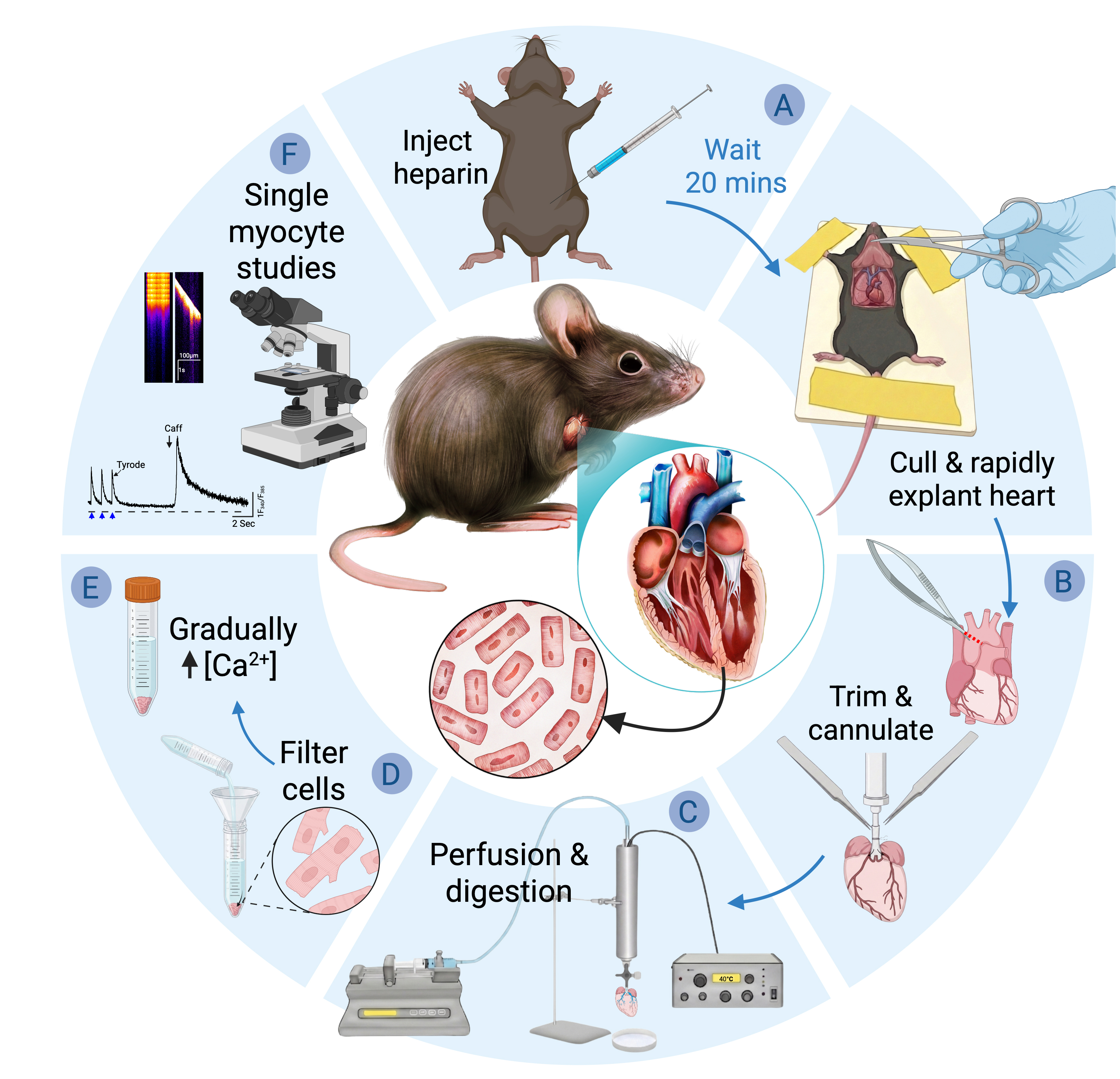
Simplified Langendorff-based mouse cardiac myocyte isolation protocol. Schematic overview of the workflow for isolating adult mouse ventricular cardiomyocytes. (A) Animal preparation and heart excision. (B) Cannulation and setup. (C) Perfusion and enzymatic digestion. (D) Tissue dissociation and cell isolation. (E) Gradual Ca2+ reintroduction. (F) Isolated cardiomyocytes can be used for downstream single-cell functional analyses, including calcium imaging and electrophysiological measurements.
Background
The isolation of intact adult cardiac myocytes is a foundational technique in cardiac research, enabling direct investigation of cellular structure and function under controlled experimental conditions. The isolated heart preparation described by Oscar Langendorff in 1897 [1], and methodologically refined by others [2,3], forms the basis of the modern “Langendorff” method [4]. In this preparation, the excised heart is rapidly cannulated via the ascending aorta and perfused ex vivo while suspended in a temperature-controlled chamber. Retrograde perfusion through the aorta allows controlled delivery of physiological solutions through the intact coronary vasculature while preserving myocardial architecture [1]. Its further adaptation in the 1970s for enzymatic dissociation established the Langendorff method as the standard approach for isolating viable cardiac myocytes [5,6], a status it has maintained for more than five decades [7–9]. Langendorff perfusion enables enzymatic delivery through the coronary circulation while minimizing mechanical trauma to the myocardium by retrograde aortic perfusion. In this configuration, flow delivered through the cannulated proximal aorta fills the aortic root, generating pressure that causes coaptation of the aortic cusps and closure of the valve. Aortic valve closure prevents retrograde ventricular filling and directs perfusate into the coronary circulation via the coronary ostia at the base of the root [4]. This technique is particularly well-suited to rodents and other small-animal models, in which the aorta is sufficiently large for reliable cannulation yet small enough to allow economical use of dissociation enzymes [10]. For larger hearts, perfusing a coronary branch and excising the region is more practical than perfusing the whole heart [10].
Langendorff systems operate under either constant-pressure or constant-flow perfusion regimes [8]. Constant-pressure setups are typically gravity-fed from a reservoir of perfusate and exhibit variable coronary flow as vascular resistance changes during digestion. As enzymatic dissociation reduces resistance, coronary flow increases, providing a useful indicator of digestion-progression that is commonly monitored by changes in drip rate [10,12–15]. Because coronary vascular resistance dynamically influences the pressure–flow relationship [16], variations in flow under constant-pressure perfusion may result in variable delivery of enzyme, particularly as resistance drops during digestion [10,15]. Variability in extracellular matrix composition and fibrosis degree across strain, age, and pathological conditions ostensibly will also influence myocardial resistance and thereby perfusion flow dynamics [15].
In contrast, constant-flow systems employ a fixed volumetric perfusion rate while perfusion pressure varies in response to downstream resistance. Careful selection of the flow rate is important: if set too low, inadequate coronary perfusion may occur; if excessive, supraphysiological perfusion pressures can invert the aortic valve cusps and divert perfusate into the ventricular cavity rather than the coronary circulation. Many traditional constant-flow Langendorff setups rely on peristaltic pumps as well as dedicated perfusion rigs incorporating water-jacketed glassware, bubble traps, recirculation loops, and continuous carbogen or oxygen gassing of bicarbonate-buffered solutions [5,6,9,12]. The complexity of specialized apparatus, combined with the technical expertise required for rapid aortic cannulation, has been criticized as limiting accessibility. In response, simplified “Langendorff-free” methods have been developed [17–20].
Langendorff-free methods, sometimes referred to as “injection methods,” are based on direct injection of perfusate into the ventricular lumen [18–21]. In these approaches, the aorta is clamped, and buffer is forced into the coronary circulation in an antegrade manner, flowing through the aortic valve into the coronary ostia. Although injection-based methods are touted as requiring less technical skill than aortic cannulation, they nonetheless depend on accurate clamp placement and precise ventricular injections into the right ventricular lumen, followed by multiple injections into the left ventricular lumen. These procedures demand operator precision and introduce distinct risks. Repeated ventricular punctures, particularly in the small and thin-walled mouse heart, introduce the risk of rupture or buffer leakage. Preservation of aortic integrity is also critical, as damage can compromise effective coronary perfusion. These risks may be exacerbated in structurally fragile or diseased hearts or those exhibiting pathological remodeling.
Moreover, injection methods rely on non-physiological alkaline buffers (pH ~7.8) for optimal cell yield [17–19], the basis for which is speculative rather than mechanistically established [18]. Cardiomyocyte function is sensitive to pH, and extracellular alkalinization directly influences intracellular pH via established acid–base transport mechanisms, including Na+/H+ exchange and lactate/H+ transport [22,23]. Because intracellular pH must be tightly maintained within a narrow physiological range (~7.15–7.25) to preserve excitation–contraction coupling and calcium homeostasis, even modest deviations can alter Ca2+ handling and myofilament sensitivity [24–27]. Furthermore, extracellular pH fluctuations regulate autophagy, a process central to cellular viability and metabolic control [28]. Exposure to alkaline conditions during isolation may influence post-isolation cellular physiology and subsequent functional readouts. For studies requiring transcriptomic or functional characterization of the full spectrum of cardiac cell types, including fibroblasts, endothelial cells, and immune cells, non-perfusion enzymatic digestion approaches have been developed that enable isolation of viable cells across all major cardiac populations, as demonstrated by single-cell sequencing studies of both healthy and diseased myocardium [29,30].
Taken together, these considerations highlight the need for a method that preserves the physiological fidelity and controlled delivery afforded by constant-flow coronary perfusion while eliminating the specialized equipment and technical expertise required by traditional Langendorff systems. The protocol described here addresses this need through a simplified, syringe pump–driven Langendorff approach. It retains the critical advantages of aortic cannulation and continuous retrograde perfusion while implementing them within a minimal and accessible apparatus optimized for the isolation of adult mouse ventricular myocytes.
Materials and reagents
Biological materials
1. Adult C57BL/6N mice, obtained from Janvier Labs (Le Genest-Saint-Isle, France)
Reagents
1. Collagenase II (Worthington Biochemical, catalog number: LS004177)
Note: Store as a lyophilized powder at 2–8 °C. Stable for long-term storage when kept dry and protected from moisture. Reconstitute fresh prior to use.
2. Collagenase IV (Worthington Biochemical, catalog number: LS004189)
Note: Store as a lyophilized powder at 2–8 °C. Stable for long-term storage when kept dry and protected from moisture. Reconstitute fresh prior to use.
3. Protease XIV (Merck, catalog number: P5147)
Note: Store as a solid at -20 °C. Stable for long-term storage when kept dry and protected from moisture. Reconstitute fresh prior to use.
4. Calcium chloride (CaCl2) (Merck/Sigma-Aldrich, catalog number: 21115)
5. Magnesium chloride hexahydrate (MgCl2·6H2O) (Merck/Sigma-Aldrich, CAS number: 7791-18-6)
6. Bovine serum albumin (BSA) (Merck/Sigma-Aldrich, CAS number: 9048-46-8)
7. Sodium chloride (NaCl) (Merck/Sigma-Aldrich, CAS number: 7647-14-5)
8. HEPES (Merck/Sigma-Aldrich, CAS number: 7365-45-9)
9. MES (Merck/Sigma-Aldrich, CAS number: 4432-31-9)
10. Glucose (Merck/Sigma-Aldrich, CAS number: 50-99-7)
11. Taurine (Merck/Sigma-Aldrich, CAS number: 107-35-7)
12. 2,3-butanedione monoxime (BDM) (Merck/Sigma-Aldrich, CAS number: 57-71-6)
13. EDTA (Merck/Sigma-Aldrich, CAS number: 60-00-4)
14. Potassium chloride (KCl) (Merck/Sigma-Aldrich, CAS number: 7447-40-7)
15. Sodium phosphate monohydrate (NaH2PO4·H2O) (Merck/Sigma-Aldrich, CAS number: 10049-21-5)
16. Sodium hydroxide (NaOH) (Merck/Sigma-Aldrich, CAS number: 1310-73-2)
17. (Optional) Fura-2 AM (Thermo Fisher Scientific, InvitrogenTM, catalog number: F1221); store at -20 °C and protect from light
Solutions
1. EDTA buffer (see Recipes)
2. Perfusion buffer (see Recipes)
3. Enzyme solution (see Recipes)
4. Stop buffer (see Recipes)
5. Ca2+-free Tyrode’s solution (see Recipes)
Recipes
1. EDTA buffer
| Reagent | Final concentration | Quantity for 100 mL |
|---|---|---|
| NaCl | 125 mM | 730.5 mg |
| KCl | 5 mM | 37.3 mg |
| NaH2PO4·H2O | 0.5 mM | 6.9 mg |
| HEPES | 10 mM | 238.3 mg |
| BDM | 10 mM | 101.1 mg |
| Taurine | 10 mM | 125.2 mg |
| EDTA | 5 mM | 146.1 mg (free acid) |
| Glucose | 10 mM | 180.2 mg |
Prepare using ultrapure (Milli-Q) water. Adjust pH to 7.4 at 37 °C using NaOH and bring to a final volume of 100 mL.
Note: Reagent masses are calculated for the specific hydration states listed above. Adjust quantities accordingly if using hydrated salts or alternative chemical forms.
2. Perfusion buffer
| Reagent | Final concentration | Quantity for 100 mL |
|---|---|---|
| NaCl | 130 mM | 759.7 mg |
| KCl | 5 mM | 37.3 mg |
| NaH2PO4·H2O | 0.5 mM | 6.9 mg |
| HEPES | 10 mM | 238.3 mg |
| BDM | 10 mM | 101.1 mg |
| Taurine | 10 mM | 125.2 mg |
| MgCl2·6H2O | 1 mM | 20.3 mg |
| Glucose | 10 mM | 180.2 mg |
Prepare using ultrapure (Milli-Q) water. Adjust pH to 7.4 at 37 °C using NaOH and bring to a final volume of 100 mL.
Note: Reagent masses are calculated for the specific hydration states listed above. Adjust quantities accordingly if using hydrated salts or alternative chemical forms.
3. Enzyme solution
Perfusion buffer supplemented with collagenase II (3,250 U/25 mL), collagenase IV (26,25 U/25 mL), and protease XIV (1.25 mg/25 mL). Prepare fresh immediately prior to use.
4. Stop buffer
Perfusion buffer supplemented with 5% (w/v) BSA. Minimum volume: 10 mL.
5. Ca2+-free Tyrode’s solution
| Reagent | Final concentration | Quantity for 100 mL |
|---|---|---|
| NaCl | 136 mM | 794.8 mg |
| KCl | 4 mM | 29.8 mg |
| HEPES | 5 mM | 119.2 mg |
| MES | 5 mM | 97.6 mg |
| MgCl2·6H2O | 0.8 mM | 16.3 mg |
| Glucose | 10 mM | 180.2 mg |
Prepare using ultrapure (Milli-Q) water. Adjust pH to 7.4 at 37 °C using NaOH and bring to a final volume of 100 mL.
Laboratory supplies
Note: For any item, equivalent products meeting the same specifications are acceptable. Listed brands and catalog numbers are examples only.
1. Beakers (Corning/PYREX®, catalog numbers: 1395-25, 1395-100)
2. Volumetric flask, 50 mL (Corning/PYREX®, catalog number: CLS564050FO)
3. Petri dishes, 60–100 mm (any supplier)
4. Braided silk sutures, USP size 5-0 (Galerie Vömel GmbH, catalog number: 14739)
5. Cell strainer, 300 μm (pluriSelect Life Science, catalog number: 43-50300)
6. Analytical funnel (Semadeni, catalog number: 224)
7. Conical tubes, 15 mL (Sarstedt, catalog number: 62.554.502)
8. Pasteur pipettes, 3 mL (Auxilab, catalog number: DLB009)
9. Syringes, 1 mL (BD Plastipak Luer-Slip, catalog number: 303172)
10. Syringes, 10 mL (B. Braun, catalog number: 4617100V-02)
11. 26G Luer-lock needle (BD, catalog number: 305110)
12. Flexible tubing (compatible with syringe pump and cannula)
Equipment
Note: For any item, equivalent products meeting the same specifications are acceptable. Listed brands and catalog numbers are examples only.
1. Analytical balance (Mettler Toledo, model: AT261 DeltaRange)
2. Magnetic stirrer (IKA, model: COMBIMAG RCO)
3. Water bath (Techne, model: Tempette TE-8A)
4. Syringe pump (KD Scientific, model: 100)
5. Inline heater and temperature controller (Warner Instruments, models: SH-27B and TC-324B)
6. Dissecting microscope (ZEISS, model: Stemi 305)
7. Cannulation/dissection chamber (custom or standard)
8. Benchtop centrifuge (capable of low-speed centrifugation)
9. Aortic metal cannula for mouse heart perfusion (Harvard Apparatus, catalog number: 73-2800; outer diameter 1.3 mm)
Note: Equivalent cannulas compatible with mouse aortic cannulation (approximately 23–25G) may also be used. Cannulas may be modified by creating a shallow circumferential ridge/groove near the tip to improve suture retention and secure the aorta during perfusion.
10. Tissue forceps (Aesculap, catalog number: BD557R)
11. Iris scissors (Aesculap, catalog number: BC110R)
12. Spring scissors (Vannas-Tübingen, catalog number: 15003-08)
13. Operating scissors (KRUUSE, catalog number: 130460)
14. Hemostatic forceps (Aesculap, catalog number: BH111R)
15. Curved iris forceps (Graefe/CHIRU+, catalog number: 19-7013.01)
16. Fine forceps No. 5 (Dumont, catalog number: F6521)
Procedure
Note: Total time of the procedure is ~1.5–2 h, including 20 min of heparin pre-treatment.
A. Animal preparation and heart excision
Note: Steps A2–5 take <3 min; perform as rapidly as possible.
1. Inject 0.2 mL of anticoagulant heparin sodium at 1,000 U/mL intraperitoneally (200 U per mouse; ~10 U/g for a 20 g mouse) 20 min prior to euthanasia.
2. Euthanize the mouse by cervical dislocation with or without prior anesthesia, in accordance with applicable institutional and national animal welfare regulations. Quickly tape the forelimbs and tail to immobilize the animal in the supine position. Following respiratory arrest, interruption of oxygen delivery rapidly initiates myocardial ischemia and metabolic depletion [31]. Therefore, perform all subsequent steps rapidly until consistent coronary perfusion is established (step C2).
3. Lift the skin at the sternum with tissue (rat tooth) forceps and cut using blunt-end operating scissors. Reflect the skin caudally and cranially to expose the abdominal cavity.
4. Make an incision under the ribcage, cutting the diaphragm; bilaterally cut to retroflect the thoracic cage with hemostatic forceps, exposing the heart.
5. Elevate the heart gently with curved iris forceps and excise, keeping the dissection with iris scissors close to the dorsal thoracic wall to preserve aortic length. Sever the inferior vena cava and descending aorta first to facilitate rapid mobilization of the heart while minimizing tension on the ascending aorta. Remove the lungs, thymus, and pericardial fat pads, ensuring that the heart, including the ascending aorta and atria, remains intact.
B. Cannulation and setup
Note: Section B takes <5 min; perform as rapidly as possible.
1. Transfer the heart to ice-cold EDTA buffer in the cannulation chamber under the dissecting microscope. Trim using spring micro scissors (Figure 1). There is no need to rinse the heart to remove blood, as this unnecessarily extends the period of ischemia. In sufficiently heparinized animals, latent blood will be washed out after the onset of perfusion. Any thrombi formed in the arterial coronary vasculature will not be removed by rinsing anyway.
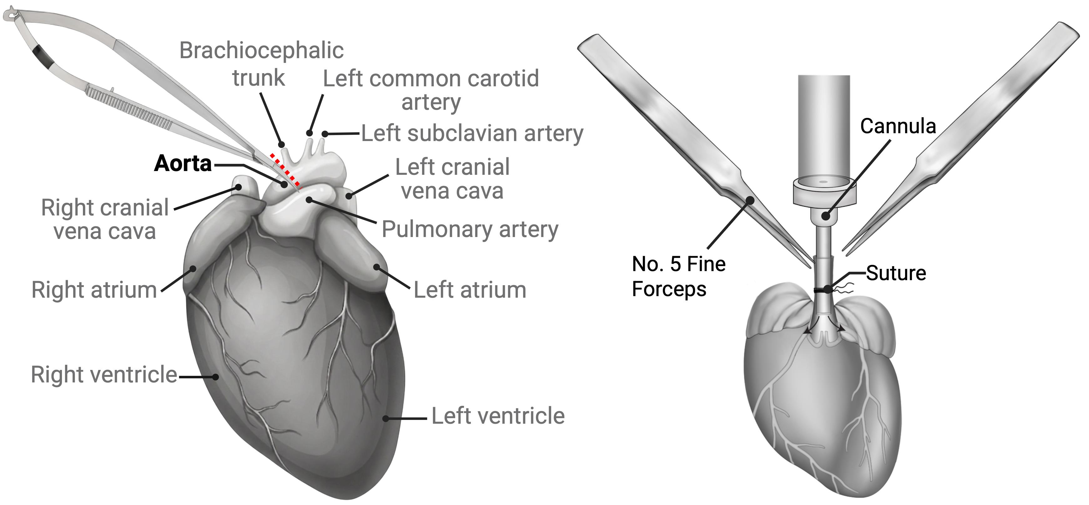
Figure 1. Preparation to cannulate aorta. (Left) The ascending aorta is trimmed using micro scissors beneath the brachiocephalic trunk to preserve a sufficient length of aorta for cannulation. (Right) The aorta is guided onto the cannula using fine forceps and fixed in place with a suture.
2. Insert the cannula into the aorta and gently advance it toward the aortic root, positioning the tip just above the aortic valve. Using fine forceps, guide the aorta over the cannula. See Video 1 for an example. The cannula and perfusion line should be pre-filled with EDTA buffer and be free of air bubbles. Secure the aorta in place with a double suture (reef knot) and perfuse gently with EDTA until the coronary veins are cleared of blood (see Figure 2).

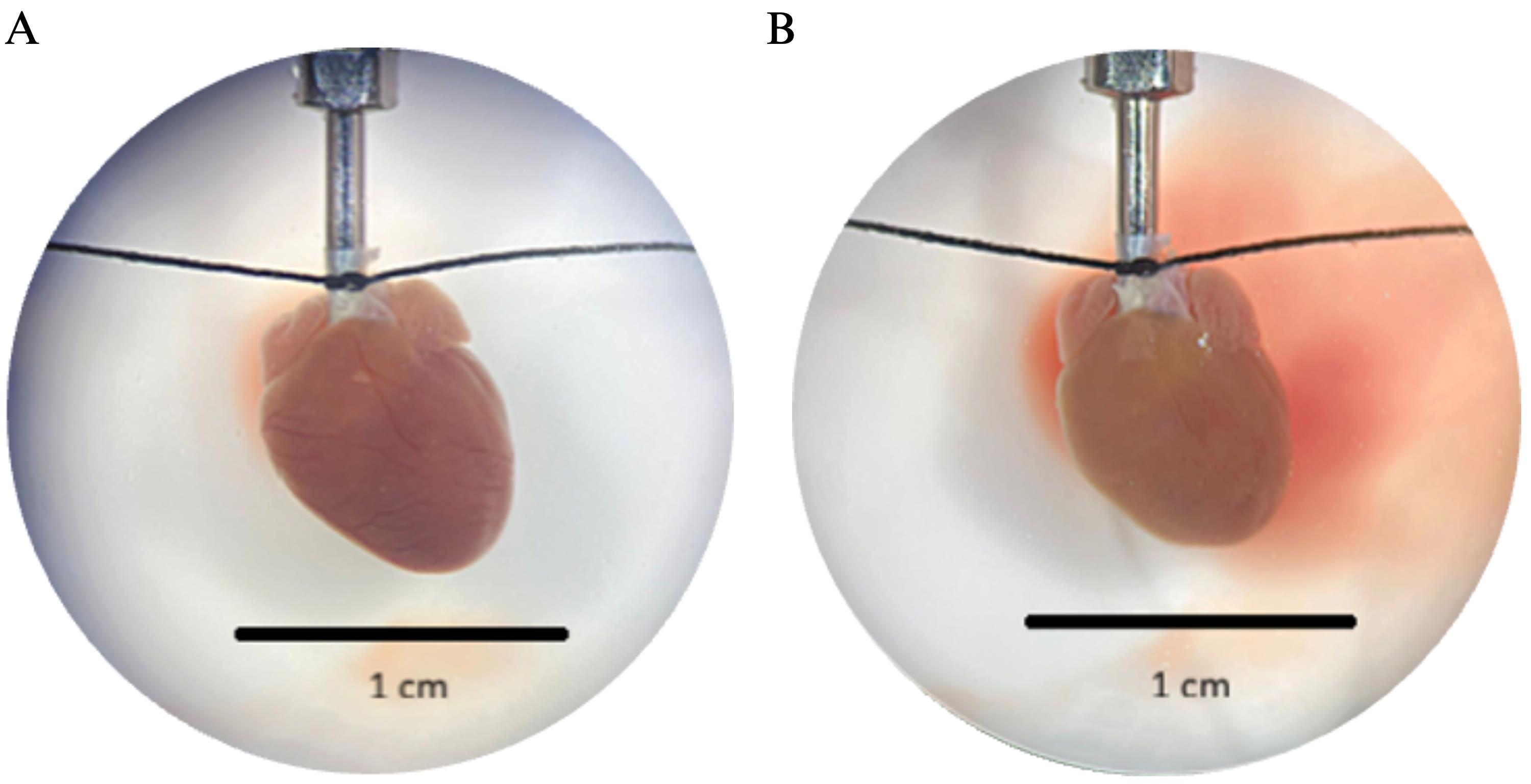
Figure 2. Mouse heart cannulation under the microscope (10×). (A) The aorta is secured on the cannula using silk suture thread. (B) After flushing with EDTA buffer, the coronary veins are cleared of blood.
C. Perfusion and digestion
Note: This section takes ~12–18 min, being endpoint-dependent.
1. Transfer the cannulated heart to the perfusion setup. To do this, carefully lift the cannula (not the heart tissue) and connect the cannula inlet to the outflow tubing of the syringe pump via the inline heater. Ensure the connection is secure and free of air bubbles before initiating flow. Confirm that the tubing and inline heater are prewarmed and that the syringe pump (e.g., KD Scientific Model 100) is loaded and ready prior to transfer (see Figure 3).
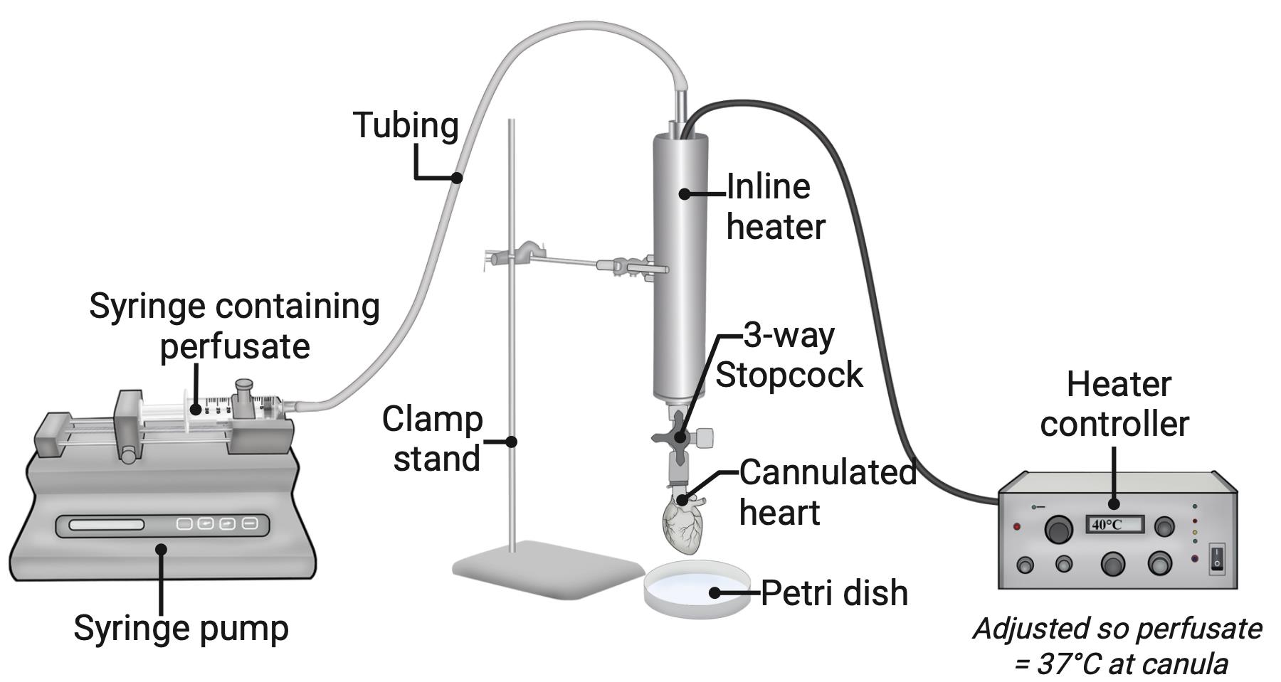
Figure 3. Syringe pump setup. The cannulated heart is transferred to the setup with a syringe pump and an inline heater.
2. Perfuse EDTA buffer at 1 mL/min for 5 min. Meanwhile, dissolve enzyme aliquots to prepare enzyme solution.
3. Switch to perfusion buffer at 1.5 mL/min for 2 min.
4. Perfuse enzyme solution at 2 mL/min for ~5 min. Monitor digestion progress continuously. The heart should become progressively swollen, softer, and flaccid. A slight shift from a reddish/dark pink to pale pink is expected during proper digestion. As digestion proceeds, the tissue should feel soft and spongy when gently pinched with forceps, and the surface may show loosening or separation of muscle fibers.
Note: A pale pink coloration during digestion should not be confused with a white or yellowish appearance, which may indicate poor coronary perfusion (e.g., incorrect cannulation or air embolism).
D. Tissue dissociation and cell isolation
Note: This section takes ~15–20 min.
1. Endpoint: Stop perfusion when the heart is clearly soft, flaccid, and spongy, while maintaining a light-pink (not white) appearance. See Video 2 for an example. Transfer the heart to a new dish containing ~5 mL of stop buffer.
2. Gently tease the tissue with forceps. Well-digested tissue is pale, soft, and tears easily between forceps to release cells (see Figure 4). If tissue appears firm and dark, this indicates ineffective coronary perfusion, insufficient enzymatic digestion, or both. Optionally use scissors in a feathering motion and a cut-tip Pasteur pipette for gentle trituration (avoid bubbles, as shear stress can damage cell membranes).
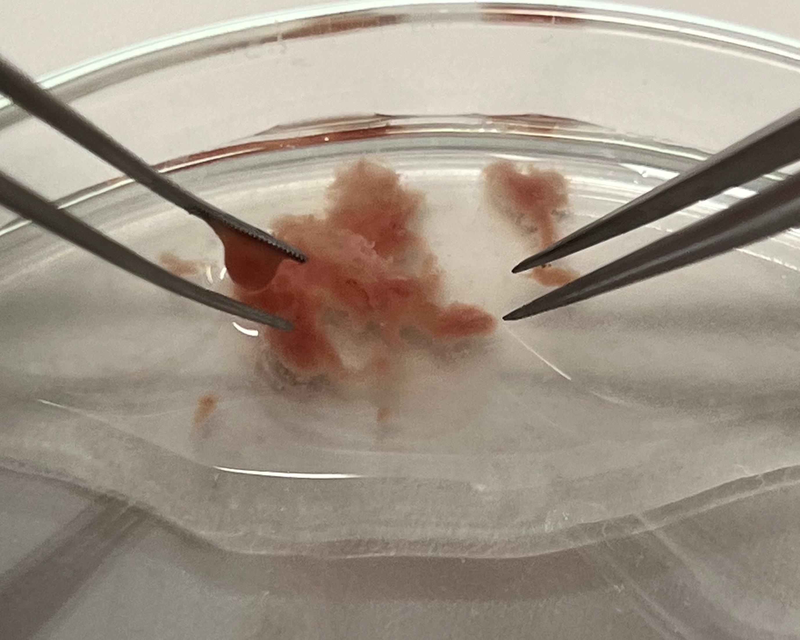
Figure 4. Heart tissue after digestion. Tissue readily dissociates with gentle forceps manipulation.
3. Filter suspension through a 300 μm mesh into a 15 mL conical tube. Allow cells to pellet by gravity (~12 min) (see Figure 5). Aspirate supernatant to discard and resuspend the cell pellet in Ca2+-free Tyrode’s solution at room temperature.
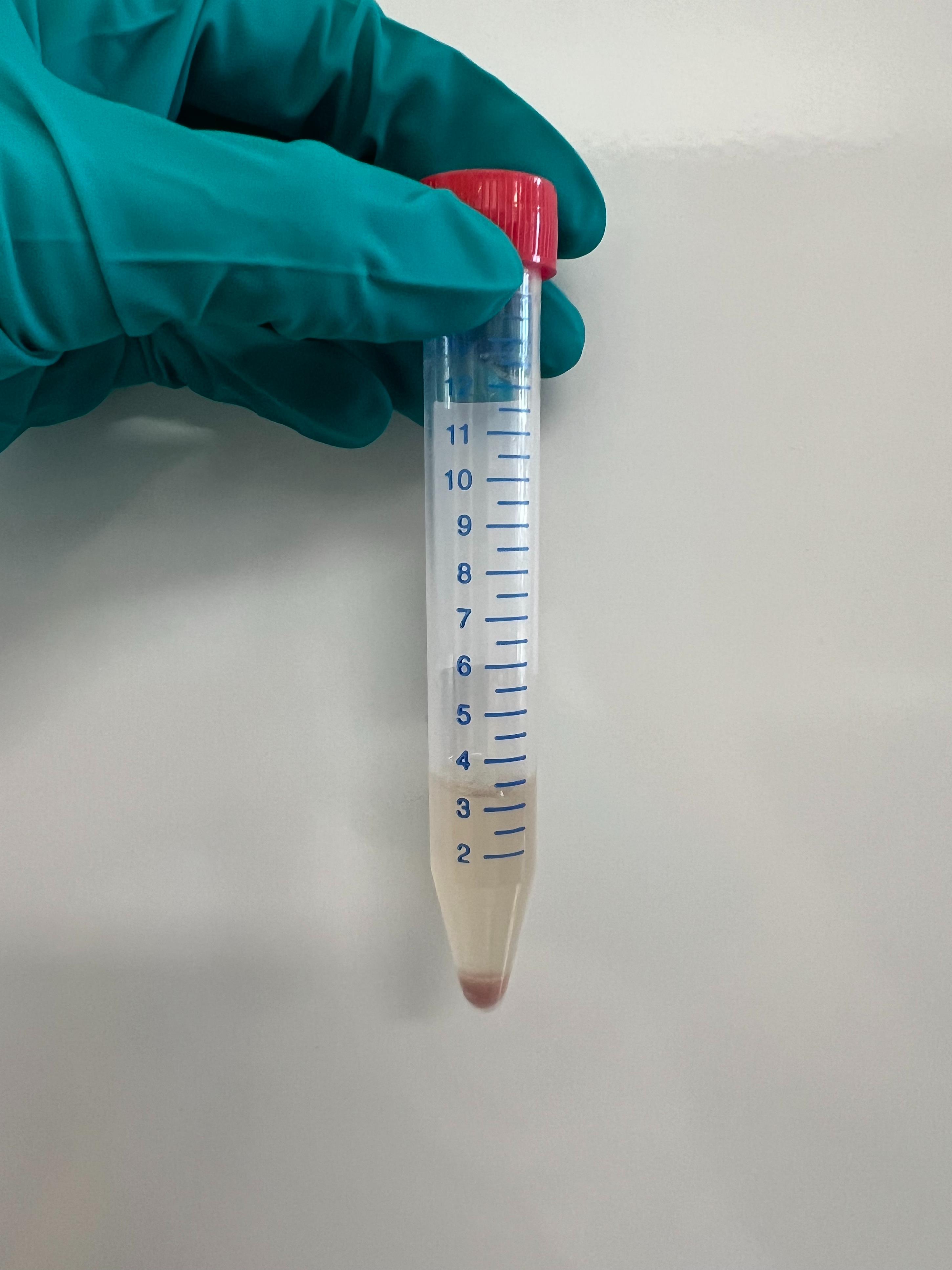
Figure 5. Cell pellet exhibiting a distinct pink color formed after gravity sedimentation. An overly pale or colorless pellet may indicate poor cell viability.
4. Assess cell viability under the microscope (10×). Cell viability is considered acceptable if >70% of cells exhibit a rod-shaped morphology.
E. Calcium reintroduction
Note: This section takes ~30–40 min.
1. Aspirate the supernatant and resuspend the myocyte pellet in 4 mL of Ca2+-free Tyrode’s solution.
2. Reintroduce Ca2+ gradually in a stepwise manner to prevent calcium overload. The final experimental [Ca2+] is 1.8 mM.
Increase extracellular Ca2+ concentration sequentially in 5–6 steps (4 min incubation at room temperature between steps), for example:
Stop buffer (0 mM Ca2+) → ~100 μM → ~250 μM → ~500 μM → ~750 μM → ~1.5 mM
Mix gently after each addition.
3. Allow cells to settle by gravity, then resuspend in Tyrode’s solution containing 1.8 mM Ca2+.
F. Representative downstream applications
For calcium imaging, cardiomyocytes were loaded with 1–5 μM Fura-2 AM for 20 min (protected from light), followed by transfer to dye-free Tyrode’s solution prior to analysis. Fura-2 AM was selected for experiments because its ratiometric properties provide a robust assessment of intracellular Ca2+ handling while minimizing variability arising from dye loading, photobleaching, and cell size. When selecting a calcium indicator, key considerations include the imaging system available, the temporal resolution required, and whether ratiometric correction for loading variability is needed. Alternative indicators (e.g., Fluo-4, Rhod-2, or Indo-1) may be preferable depending on the research question and microscope configuration. Notably, Fura-2 AM requires a dual-excitation imaging system, whereas single-wavelength indicators such as Fluo-4 may be more suitable for higher-speed imaging applications.
Data analysis
Cell viability is assessed by light microscopy based on morphology, with viable cardiomyocytes identified as rod-shaped cells with clear striations. Preparations are considered acceptable when >70% of cells exhibit this morphology. For functional validation, calcium handling may be assessed using established fluorescence-based methods (e.g., Fura-2 AM), with analysis performed using standard imaging software (such as FIJI/ImageJ, Clampfit, and IonWizard). No specific statistical analyses are required to perform or validate the isolation procedure. However, where quantitative comparisons are made, appropriate statistical tests should be selected based on experimental design.
Validation of protocol
This protocol has been used to reproducibly isolate viable adult mouse ventricular myocytes across multiple independent preparations (>60 hearts). Preparations typically yield >70% rod-shaped, calcium-tolerant cells, indicating high cell viability and structural integrity. The absolute number of viable cardiomyocytes per preparation was not systematically quantified in this study; however, preparations consistently yielded sufficient cell numbers for downstream single-cell functional analyses, including calcium imaging and electrophysiology. The remaining cardiomyocytes (typically <30%) comprise rounded, hypercontracted, or fragmented cells that have not survived the isolation process. These non-rod-shaped cells can be largely excluded by allowing cells to settle by gravity sedimentation prior to experiments or by selecting rod-shaped cells for analysis based on morphological criteria during imaging. Isolated cardiomyocytes exhibit consistent, stimulus-evoked Ca2+ transients and preserved functional responses to β-adrenergic stimulation and caffeine upon reintroduction to physiological Ca2+ concentration (1.8 mM), demonstrating maintained excitation–contraction coupling.
Representative results are shown in Figure 6. For Figure 6B, Fura-2 AM–loaded cardiomyocytes (1 μM, 20 min, protected from light) were transferred to a superfusion chamber and continuously superfused with Tyrode’s solution containing 1.8 mM Ca2+ at 37 °C. Cells were field-stimulated at 1 Hz using platinum electrodes. Ca2+ transients were recorded as the ratio of fluorescence emission at 510 nm upon alternating excitation at 340 and 380 nm (340/380 ratio). To assess β-adrenergic responsiveness, isoprenaline (50 nM) was applied by superfusion, and Ca2+ transients were recorded during steady-state stimulation. Sarcoplasmic reticulum (SR) Ca2+ content was assessed by rapid application of 10 mM caffeine under non-stimulated conditions, which triggers SR Ca2+ release via ryanodine receptors. The amplitude of the caffeine-evoked transient provides an estimate of SR Ca2+ load.
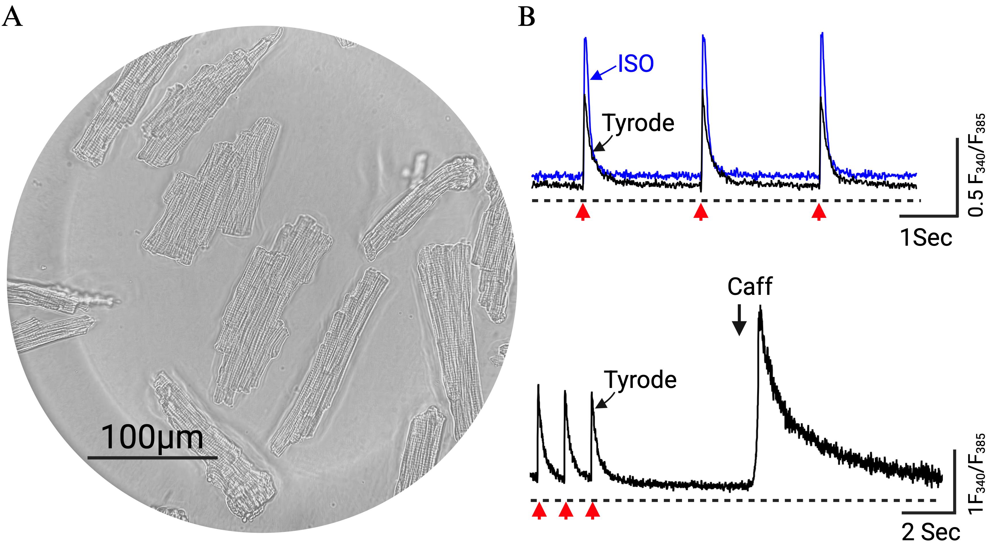
Figure 6. Isolated ventricular myocytes in 1.8 mM Ca2+ Tyrode’s solution. (A) Representative phase-contrast image of isolated cardiomyocytes. Scale bar = 100 μM. (B) Representative traces of Fura-2 AM–loaded cells treated with 50 nM isoprenaline (ISO) (top, blue) and 10 mM caffeine (Caff) (bottom). Red arrows indicate field stimulation.
General notes and troubleshooting
General notes
1. To prevent contamination, microbial growth, salt precipitation, and flow obstruction, all tubing should be properly maintained. After each use, flush the tubing with 70% ethanol followed by Milli-Q or ultrapure water. Inspect tubing regularly and replace at defined intervals or immediately if discoloration, increased back pressure, reduced flow, or visible residue is observed.
2. Solutions should be prepared fresh on the day of use when possible, particularly enzyme-containing buffers, to ensure optimal activity and reproducibility. Prolonged storage may lead to reduced enzyme activity, pH drift, or precipitation, which can negatively affect digestion efficiency and cell viability.
3. Cells were collected using gravity sedimentation to minimize mechanical stress and preserve viability. Alternatively, low-speed centrifugation (e.g., 300× g for 1 min) may be used if required; however, excessive centrifugation can increase cell damage and reduce viability.
4. Although this method offers advantages in accessibility and simplicity, several limitations related to traditional Langendorff systems should be considered. Traditional constant-pressure Langendorff systems allow coronary flow to vary naturally in response to changes in vascular resistance during digestion, and the resulting increase in drip rate serves as a useful real-time indicator of digestion progression. In the constant-flow approach described here, this indirect readout is absent, and the digestion endpoint must be assessed entirely by visual and tactile evaluation of tissue compliance, a skill that requires operator experience. Traditional systems also commonly incorporate recirculating perfusion loops, bubble traps, and carbogen-gassed bicarbonate-buffered solutions, which provide superior oxygenation and pH stability over extended perfusion periods. The simplified setup described here uses HEPES-buffered, non-recirculating solutions without active oxygenation, which may be less suitable for longer perfusion durations or for hearts with high metabolic demand, such as those from hypertrophied or failing models. Additionally, syringe volume limits the maximum perfusion duration without interruption to reload. For standard adult mouse hearts, this is unlikely to be restrictive, but may require planning for larger hearts or extended digestion protocols.
5. This protocol has been optimized for adult C57BL/6N mice and may require adjustment of enzyme concentrations, flow rates, and digestion duration when applied to other strains, ages, or disease models.
Troubleshooting
Problem 1: No or low perfusion flow (little or no solution exiting the heart).
Possible causes: Cannula incorrectly positioned; knot too loose; blockage; cannula inserted too deep (into ventricle) or not deep enough (causing leakage).
Solution: Re-seat the cannula, ensuring correct positioning in the ascending aorta (tip must be above the aortic valve), and secure firmly with a suture.
Problem 2: Heart remains dark pink/red with no color change.
Possible causes: Poor or absent coronary perfusion due to incorrect cannulation; coronary vessels remain filled with blood.
Solution: Re-cannulate, ensuring a tight seal and correct positioning. Confirm that the perfusate flows through the coronary circulation (coronaries should clear as blood is flushed out). Ensure there is no external leakage.
Problem 3: Heart becomes white or yellowish early during perfusion.
Possible cause: Air embolism blocking coronary microcirculation.
Solution: Remove all air bubbles from the tubing before cannulation. Monitor continuously and stop immediately if bubbles enter the system. Flush bubbles from the system before continuing.
Problem 4: Low yield; tissue remains firm despite good coronary perfusion.
Possible causes: Under-digestion; low enzyme activity (batch variability); insufficient perfusion; incorrect buffer pH or temperature.
Solutions: Extend digestion time; verify enzyme activity (test new batches where possible); ensure effective coronary perfusion; verify buffer temperature and pH.
Problem 5: Low cell yield despite having soft tissue.
Possible cause: Incomplete coronary perfusion leading to uneven digestion.
Solutions: Ensure correct heparin dose and timing prior to isolation. Check cannula positioning and flow; confirm perfusion throughout digestion.
Problem 6: Cell fragility or low viability.
Possible causes: Over-digestion; prolonged enzyme exposure; delays after isolation; incorrect buffer pH or temperature.
Solutions: Reduce digestion time; stop perfusion promptly at the endpoint; minimize delays before downstream processing; verify buffer temperature and pH.
Problem 7: Inconsistent digestion between experiments.
Possible causes: Variability in enzyme activity; differences in animal age or condition; timing variability.
Solutions: Adjust digestion time based on tissue response (softness/sponginess); standardize enzyme batches or test activity prior to use.
Problem 8: Perfusate leaks from the heart.
Possible causes: Cannula not inserted sufficiently into the aorta; insufficient suture seal; aorta cut too short.
Solutions: Ensure adequate ascending aortic length during excision and trimming; insert cannula securely and tie firmly to prevent leakage.
Problem 9: No or poor digestion despite correct timing.
Possible cause: Cannulation of the incorrect vessel (e.g., pulmonary artery).
Solution: Confirm correct identification and cannulation of the aorta before perfusion. Coronary vessels should be clear of blood during initial perfusion; failure indicates incorrect cannulation.
Problem 10: Low viability despite good yield.
Possible causes: Ischemic damage due to slow excision or delayed perfusion; mechanical stress during handling.
Solutions: Perform excision rapidly and establish coronary perfusion as soon as possible. Use gentle trituration with wide-bore or cut tips; minimize pipetting steps and avoid foaming or shear stress.
Acknowledgments
Conceptualization, T.Z.; Investigation, T.Z. and M.L.; Formal analysis, T.Z. and M.L.; Writing—Original Draft, T.Z.; Writing—Review & Editing, T.Z., M.L., and M.T.; Visualization, M.L. and T.Z.; Supervision, T.Z and M.T. Cardiac myocyte isolations were performed by T.Z. and M.L. Representative images were acquired by M.L. Aortic cannulation shown in Video 1 was performed by M.T. and recorded by T.Z.
This work was supported by the Independent Research Fund Denmark (grant number 2034-00073B), the Carlsberg Foundation (grant number CF19-0067), and the Novo Nordisk Foundation (grant number NNF18OC0032728) to M.T.
This protocol has been used and validated in the following research article:
• Zawadzki et al. bioRxiv (2025). https://doi.org/10.1101/2025.09.10.675427
This protocol has also been applied in ongoing, unpublished work.
The following figures were created using BioRender: Graphical overview, biorender.com/37jkfp3; Figure 1, biorender.com/c5udss6; Figure 3, biorender.com/m6fmw9a.
Competing interests
The authors declare no competing financial or non-financial interests.
Ethical considerations
All procedures described in this protocol were approved by the Danish National Animal Experiments Inspectorate under the Danish Ministry of Environment and Food (license no. 2023-15-0201-01576) and conducted in accordance with European Parliament Directive 2010/63/EU on the protection of animals used for scientific purposes. All experiments complied with applicable institutional and national animal welfare regulations and were performed in accordance with the ARRIVE guidelines. Mice were euthanized by cervical dislocation performed by trained personnel in accordance with the approved project license. The study design adhered to the principles of the 3Rs (Replacement, Reduction, and Refinement), with efforts made to minimize animal use and suffering. Investigators implementing this protocol must obtain appropriate ethical approval and ensure that all animal procedures comply with relevant institutional and national regulations prior to initiating experiments.
References
- Langendorff, O. (1895). Untersuchungen am überlebenden Säugethierherzen. Pflüger, Archiv für die Gesammte Physiologie des Menschen und der Thiere 61(6): 291–332. https://doi.org/10.1007/bf01812150
- Gottlieb, R. and Magnus, R. (1903). Digitalis und Herzarbeit. Archiv für Experimentelle Pathologie und Pharmakologie 51(1): 30–63. https://doi.org/10.1007/bf01840739
- Heymans, J. F., Kochmann, M. (1904). Une nouvelle méthode de circulation artificielle à travers le coeur isolé de mammifère. Archives Internationales de Pharmacodynamie et de Thérapie. 13: 531–538.
- Skrzypiec-Spring, M., Grotthus, B., Szeląg, A. and Schulz, R. (2007). Isolated heart perfusion according to Langendorff—Still viable in the new millennium. J Pharmacol Toxicol Methods. 55(2): 113–126. https://doi.org/10.1016/j.vascn.2006.05.006
- Powell, T. and Twist, V. (1976). A rapid technique for the isolation and purification of adult cardiac muscle cells having respiratory control and a tolerance to calcium. Biochem Biophys Res Commun. 72(1): 327–333. https://doi.org/10.1016/0006-291x(76)90997-9
- Berry, M. N., Friend, D. S. and Scheuer, J. (1970). Morphology and Metabolism of Intact Muscle Cells Isolated from Adult Rat Heart. Circ Res. 26(6): 679–687. https://doi.org/10.1161/01.res.26.6.679
- Liao, R. and Jain, M. (2007). Isolation, Culture, and Functional Analysis of Adult Mouse Cardiomyocytes. Methods in Molecular Medicine™. 251–262. https://doi.org/10.1007/978-1-59745-571-8_16
- Louch, W. E., Sheehan, K. A. and Wolska, B. M. (2011). Methods in cardiomyocyte isolation, culture, and gene transfer. J Mol Cell Cardiol. 51(3): 288–298. https://doi.org/10.1016/j.yjmcc.2011.06.012
- MacLeod, K. T. and Harding, S. E. (1991). Effects of phorbol ester on contraction, intracellular pH and intracellular Ca2+ in isolated mammalian ventricular myocytes.. J Physiol. 444(1): 481–498. https://doi.org/10.1113/jphysiol.1991.sp018889
- Chen, X., O’Connell, T. D., Xiang, Y. K. (2016). With or without Langendorff: a new method for adult myocyte isolation to be tested with time. Circ Res. 119 (8): 888–890. https://doi.org/10.1161/CIRCRESAHA.116.309630
- Cerbai, E., Sartiani, L., De Paoli, P. and Mugelli, A. (2000). Isolated cardiac cells for electropharmacological studies. Pharmacol Res. 42(1): 1–8. https://doi.org/10.1006/phrs.1999.0654
- Wolska, B. M. and Solaro, R. J. (1996). Method for isolation of adult mouse cardiac myocytes for studies of contraction and microfluorimetry. Am J Physiol Heart Circ Physiol. 271(3): H1250–H1255. https://doi.org/10.1152/ajpheart.1996.271.3.h1250
- Hintz, M. O., Zellhuber-McMillan, U. and Jones, P. P. (2024). Simple Method to Isolate Individual Mouse Cardiomyocytes Using Enzymatic Digestion via Retrograde Perfusion. Methods Mol Biol. 7–20. https://doi.org/10.1007/978-1-0716-4342-6_2
- Page, M. T., Quintana, P. J., Ligutti, J. A. and Sabbadini, R. A. (2000). Cell death in cultured adult rat cardiomyocytes: use of the comet assay to distinguish apoptosis from necrosis. BAM-PADOVA. 10: 159–170.
- Li, H., Liu, C., Bao, M., Liu, W., Nie, Y., Lian, H. and Hu, S. (2020). Optimized Langendorff perfusion system for cardiomyocyte isolation in adult mouse heart. J Cell Mol Med. 24(24): 14619–14625. https://doi.org/10.1111/jcmm.15773
- Goodwill, A. G., Dick, G. M., Kiel, A. M. andTune, J. D. (2017). Regulation of coronary blood flow. Compr. Physiol. 7 (2): 321–382. https://doi.org/10.1002/cphy.c160016
- Ackers-Johnson, M. and Foo, R. S. (2019). Langendorff-Free Isolation and Propagation of Adult Mouse Cardiomyocytes. Methods Mol Biol. 193–204. https://doi.org/10.1007/978-1-4939-9086-3_14
- Ackers-Johnson, M., Li, P. Y., Holmes, A. P., O’Brien, S. M., Pavlovic, D. and Foo, R. S. (2016). A Simplified, Langendorff-Free Method for Concomitant Isolation of Viable Cardiac Myocytes and Nonmyocytes From the Adult Mouse Heart. Circ Res. 119(8): 909–920. https://doi.org/10.1161/circresaha.116.309202
- Nikouee, A., Yap, J. Q., Rademacher, D. J., Kim, M. and Zang, Q. S. (2024). An optimized Langendorff-free method for isolation and characterization of primary adult cardiomyocytes. BMC Cardiovasc Disord. 24(1): 649. https://doi.org/10.1186/s12872-024-04256-5
- Myachina, T. A., Butova, X. A. and Khohlova, A. D. (2019). A modified Langendorff-free method for isolation of cardiomyocytes from adult rat heart. AIP Conf Proc. 2174: 020140. https://doi.org/10.1063/1.5134291
- Butova, X., Myachina, T. and Khokhlova, A. (2021). A combined Langendorff-injection technique for simultaneous isolation of single cardiomyocytes from atria and ventricles of the rat heart. MethodsX. 8: 101189. https://doi.org/10.1016/j.mex.2020.101189
- Vaughan‐Jones, R. D. and Wu, M. L. (1990). Extracellular H+ inactivation of Na(+)‐H+ exchange in the sheep cardiac Purkinje fibre. J Physiol. 428(1): 441–466. https://doi.org/10.1113/jphysiol.1990.sp018221
- Dennis, S. C., McCarthy, J., Keding, B. and Opie, L. H. (1986). Lactate efflux from the ischaemic myocardium: The influence of pH. J Mol Cell Cardiol. 18: 119–119. https://doi.org/10.1016/s0022-2828(86)80835-5
- Orlowski, A., Di Mattia, R. A. and Aiello, E. A. (2025). Intracellular pH Regulation in Ventricular Myocytes: Implications for Cardiac Health and Disease. Circ Res. 136(12): 1636–1656. https://doi.org/10.1161/circresaha.125.325386
- Choi, H. S., Trafford, A. W., Orchard, C. H. and Eisner, D. A. (2000). The effect of acidosis on systolic Ca2+ and sarcoplasmic reticulum calcium content in isolated rat ventricular myocytes. J Physiol. 529(3): 661–668. https://doi.org/10.1111/j.1469-7793.2000.00661.x
- Fabiato, A. and Fabiato, F. (1978). Effects of pH on the myofilaments and the sarcoplasmic reticulum of skinned cells from cardiace and skeletal muscles.. J Physiol. 276(1): 233–255. https://doi.org/10.1113/jphysiol.1978.sp012231
- Orchard, C. H., Cingolani, H. E. (1994). Acidosis and arrhythmias in cardiac muscle. Cardiovasc Res. 28 (9): 1312–1319. https://doi.org/10.1093/cvr/28.9.1312
- Zhao, L., Cui, L., Jiang, X., Zhang, J., Zhu, M., Jia, J., Zhang, Q., Zhang, J., Zhang, D., Huang, Y., et al. (2016). Extracellular pH regulates autophagy via the AMPK–ULK1 pathway in rat cardiomyocytes. FEBS Lett. 590(18): 3202–3212. https://doi.org/10.1002/1873-3468.12359
- Wehrens, M., de Leeuw, A. E., Wright-Clark, M., Eding, J. E., Boogerd, C. J., Molenaar, B., van der Kraak, P. H., Kuster, D. W., van der Velden, J., Michels, M., et al. (2022). Single-cell transcriptomics provides insights into hypertrophic cardiomyopathy. Cell Rep. 39(6): 110809. https://doi.org/10.1016/j.celrep.2022.110809
- Gladka, M. M., Molenaar, B., de Ruiter, H., van der Elst, S., Tsui, H., Versteeg, D., Lacraz, G. P., Huibers, M. M., van Oudenaarden, A., van Rooij, E., et al. (2018). Single-Cell Sequencing of the Healthy and Diseased Heart Reveals Cytoskeleton-Associated Protein 4 as a New Modulator of Fibroblasts Activation. Circulation. 138(2): 166–180. https://doi.org/10.1161/circulationaha.117.030742
- Döring, H. J., Dehnert, H. (1988). The Isolated Perfused Warm-Blooded Heart According to Langendorff. Biomessetechnik-Verlag March GmbH.
Article Information
Publication history
Received: Apr 22, 2026
Accepted: Jun 2, 2026
Available online: Jun 11, 2026
Published: Jul 20, 2026
Copyright
© 2026 The Author(s); This is an open access article under the CC BY-NC license (https://creativecommons.org/licenses/by-nc/4.0/).
How to cite
Larsen, M. S., Thomsen, M. B. and Zawadzki, T. (2026). A Simplified Langendorff-Based Method for Mouse Cardiac Myocyte Isolation. Bio-protocol 16(14): e5743. DOI: 10.21769/BioProtoc.5743.

 Download Citation in RIS Format
Category
Cell Biology > Cell isolation and culture > Cell isolation
Cell Biology > Cell-based analysis > Electrophysiological technique
Medicine > Cardiovascular system > Heart tissue culture techniques
Do you have any questions about this protocol?
Post your question to gather feedback from the community. We will also invite the authors of this article to respond.
 Post a Question


