


- Protocols

- Articles and Issues
- For Authors
- About
- Become a Reviewer


Stepwise Differentiation of Mouse Embryonic Stem Cells Into Murine Blood Vessel Organoids With Endothelial Lineage Tracing for Quality Control

(§ Technical contact) Published: Vol 16, Iss 13, Jul 5, 2026 DOI: 10.21769/BioProtoc.5727 Views: 337
Reviewed by: Jessica DavisThirupugal GovindarajanNeha Saxena
 PDF
PDF  Ask a question
Original research article
The authors used this protocol in:
Oct 2025
Advertisement


Protocol Collections
Comprehensive collections of detailed, peer-reviewed protocols focusing on specific topics




Abstract
In vitro vascular models are most informative when they recapitulate endothelial assembly within a 3D microenvironment. Blood vessel organoids (BVOs) enable the study of vascular heterogeneity, function, and organ-instructive cues in development, homeostasis, and disease. Here, we present a robust stepwise method to generate murine blood vessel organoids (mBVOs) from feeder-dependent mouse embryonic stem cells (mESCs) of common genetic backgrounds. Embryoid bodies (EBs) are formed using strain-specific seeding densities (day 0–3), followed by mesoderm induction (day 3–6) and vascular induction (day 6–8). Induced EBs are embedded in collagen I with Geltrex to drive sprouting and network formation (day 8–13). Vascular networks are microdissected and grown in suspension to yield mature mBVOs (day 21–30). The inclusion of a Cre-inducible VE-cadherin-GFP reporter line enables a quantitative quality control, reducing variability by excluding poorly differentiated organoids. The protocol reliably produces ~100 mBVOs per differentiation and is compatible with engineered mouse strains for gain- and loss-of-function studies, functional assays of vascular plasticity, and syngeneic grafting to assess perfusion. Thus, mBVOs provide a scalable and traceable 3D platform that bridges endothelial assays, mouse models, and human organoid systems.
Key features
• A detailed timeline to differentiate feeder-dependent mESCs into mBVOs, with key success readouts and troubleshooting.
• Efficient across three genetic backgrounds with strain-specific EB seeding densities and typical yields of ~100 mBVOs per differentiation.
• The inducible VE-cadherin-GFP lineage tracing/reporter system provides an endothelial quality control to quantify efficiency and exclude poorly differentiated organoids.
• Compatible with engineered mouse strains for gain/loss-of-function, with in vitro assays of vascular plasticity/remodeling, and with syngeneic in vivo validation.
Keywords: Murine blood vessel organoids (mBVOs) Mouse embryonic stem cells (mESCs)
Feeder-dependent culture
Embryoid bodies (EBs)
Vascular differentiation
Sprouting angiogenesis
VE-cadherin (Cdh5) lineage tracing
Quality control of differentiation
Mouse embryonic stem cells (mESCs)
Feeder-dependent culture
Embryoid bodies (EBs)
Vascular differentiation
Sprouting angiogenesis
VE-cadherin (Cdh5) lineage tracing
Quality control of differentiation
Graphical overview
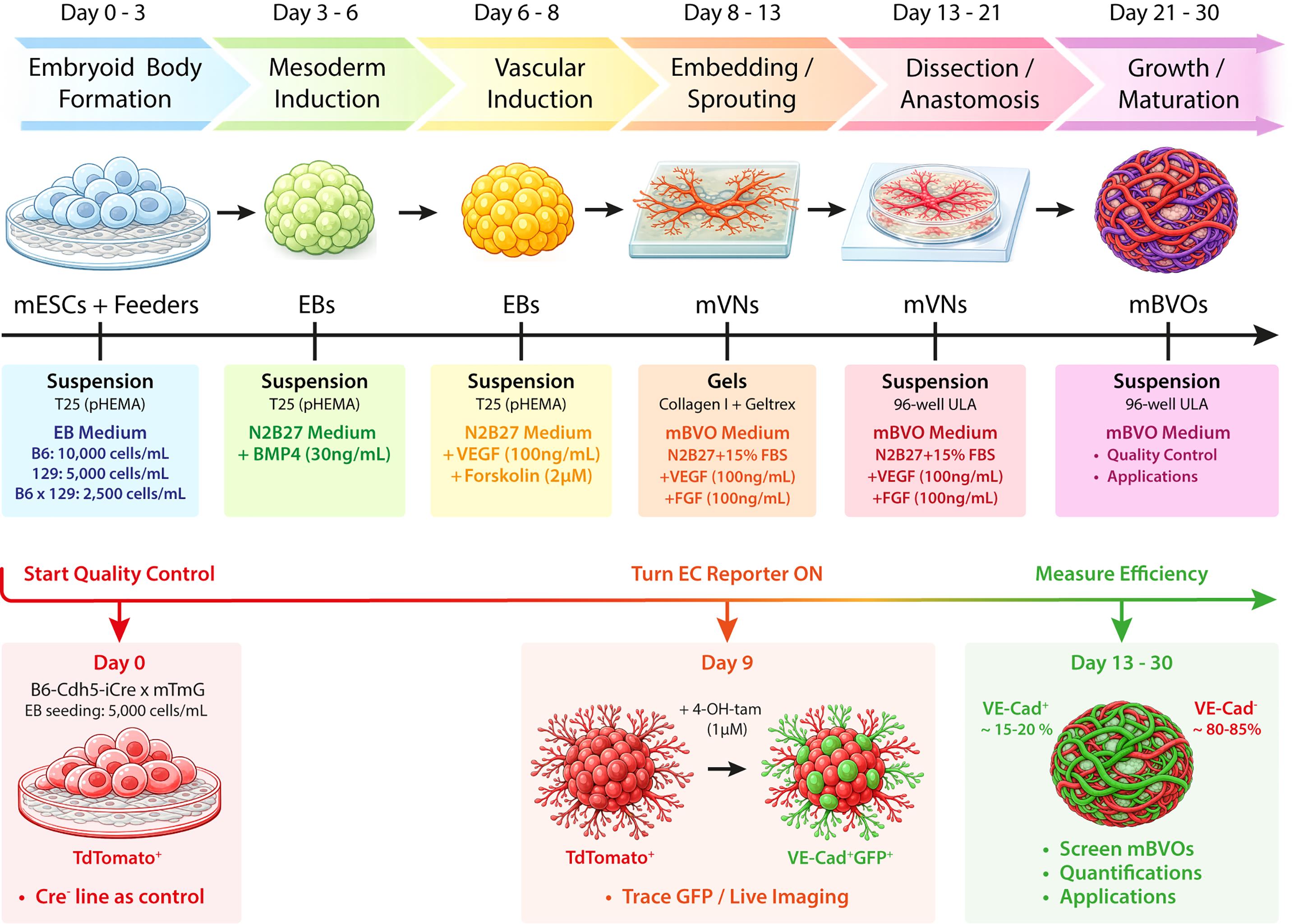
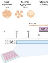
Workflow for the differentiation of mESCs into mBVOs, including the B6-Cdh5-iCre × mTmG reporter-based quality control. 129, 129S6 wild-type mouse strain; 4-OH-tam, 4-hydroxytamoxifen; B6, C57BL/6 wild-type mouse strain; B6x129, C57BL/6x129S6 F1 hybrid mouse strain; BMP, bone morphogenic protein 4; EB, embryoid body; EC, endothelial cell; FBS, fetal bovine serum; FGF, fibroblast growth factor; GFP, green fluorescent protein; mBVO, murine blood vessel organoid; mESC, murine embryonic stem cell; mVN, murine vascular network; pHEMA, poly(2-hydroxyethyl methacrylate); T25, cell culture flasks (25 cm2); ULA, ultra-low attachment; VE-Cad, vascular endothelial cadherin; VEGF, vascular endothelial growth factor.
Background
Modeling blood vessels in vitro has long been technically challenging because a functional vasculature is not solely defined by endothelial cells. In vivo, endothelial tubes are supported by mural/perivascular cells, a basement membrane, and the surrounding stroma, altogether maintaining barrier function and stability [1–3].
As a result, 2D endothelial cultures or simple sprouting assays are well-suited for endothelial-centered studies, but they provide limited insight into how multicellular vascular units self-organize and adapt within vascular beds [4]. More complex co-culture and matrix-based systems increase physiological relevance, yet they are typically optimized to answer narrow questions rather than capturing a broader, self-organizing vascular architecture that can be compared across experiments [5,6].
Human blood vessel organoids (hBVOs), initially described by Wimmer and colleagues, marked a major step forward. Taking advantage of the multilineage potential of pluripotent stem cells, they self-assemble into complex vascular structures in 3D, including endothelial, mural, mesenchymal, and immune compartments [7–9]. Since the original reports, hBVOs have been incorporated into a variety of organ-instructed assembloid systems. Notable examples include their integration with neural tissues [10,11] and their combination with pancreatic islets [12]. Beyond these applications, hBVOs have been widely used for disease modeling, including studies of diabetes [7], CNS malformations [13], neurodegeneration [14,15], aging [16], sepsis [17], and cancer metastasis [18].
Like other organoid systems [19], hBVOs offer clear advantages but also carry important limitations. A major strength is their ability to recapitulate human vascular organization and tissue-context effects. However, the system has practical limits. Human pluripotent stem cells are not always readily available, and hBVOs lack a substantial immune component, which restricts their use in vascular-immune and inflammation-focused studies [20]. Moreover, although perfusion can be engineered in vitro [21], many long-term perfusion and remodeling assays rely on transplantation into immunocompromised mice, which complicates the interpretation of immune-related processes [7,22].
Vascular studies have long been performed in mice because of their genetic traceability and physiological relevance [23]. Yet, there are significant differences between mouse and human vascular gene-expression programs [24,25]. Importantly, there is a strong push to replace, reduce, and refine animal experiments when possible [26], in accordance with European legislation (Directive 2010/63/EU; available at https://eur-lex.europa.eu/eli/dir/2010/63/oj/eng).
In this protocol, we describe the stepwise differentiation of mouse embryonic stem cells (mESCs) into murine blood vessel organoids (mBVOs) following a defined timeline [27]. We adapted the hBVO method logic to the requirements of feeder-dependent mESC cultures grown on mitotically inactivated mouse embryonic fibroblasts (MEFs). The protocol is reproducible with distinct genetic backgrounds and uses strain-specific embryoid body (EB) seeding densities to enhance reproducibility across lines. During routine differentiations, we typically obtain a consequent number of mBVOs (~100), enabling sufficient replicates for quantitative assays and statistical analyses [27].
A common challenge in organoid differentiation is achieving—and accurately quantifying—differentiation efficiency. To address this, we include an endothelial lineage-tracing module using B6-Cdh5-iCre × mTmG mESCs. After 4-hydroxytamoxifen (4-OH-tam) induction in vitro, VE-cadherin-expressing cells switch from membrane tdTomato to membrane GFP, allowing identification and exclusion of poorly differentiated organoids. Cre-negative controls define the tomato-only baseline [27,28]. This approach offers a practical advantage over strategies that rely mostly on endpoint marker staining or morphology, and it provides a standardized method for batch selection [27].
Because the system is based on mESCs, it is compatible with genetically engineered mouse strains, enabling gain- or loss-of-function studies targeting the endothelial compartment, as well as mural and immune cells, if suitable Cre tools are available. Finally, mBVOs provide a platform to study vascular plasticity and remodeling in vitro. In our original study, we used mBVOs to assess responses to pro-inflammatory cues, anti-angiogenic perturbations, and tumor–vasculature interactions using glioblastoma assembloids. We also demonstrated perfusion after syngeneic implantation in mice [27].
Together, mBVOs bridge the gap between mouse models, endothelial assays, and human BVO platforms. They provide a scalable murine 3D vascular system that is genetically traceable, with a quantitative quality control step to measure differentiation efficiency. We expect this protocol to enhance the reproducibility of BVO generation and to facilitate mechanistic studies that connect murine genetics with questions rooted in human vascular biology and disease.
Materials and reagents
Biological materials
1. Mouse embryonic stem cells (mESCs), C57BL/6 × 129S6 F1 hybrid (B6x129)
2. mESCs, C57BL/6 wild type (B6)
3. mESCs, 129S6 wild type (129)
4. mESCs, B6-Cdh5-iCre × mTmG (VE-cadherin lineage tracing line)
5. mESCs, B6-Cdh5-iCre × mTmG Cre-negative (tdTomato-only control)
6. Mouse embryonic fibroblasts (MEFs), isolated from mouse embryos and expanded as primary cultures before mitomycin C (MMC) inactivation and cryo-storage as feeder stocks
Note: mESC strains and primary MEFs were previously isolated and provided by the laboratories of Prof. Pieter Carmeliet and Prof. Kian Peng Koh, following previously described procedures [29–37].
Reagents
mESC/MEF culture
1. KnockOut DMEM (Thermo Fisher Scientific, catalog number: 10829-018)
2. ESC-qualified fetal bovine serum (FBS) (Thermo Fisher Scientific, catalog number: 10270106)
3. L-glutamine (Thermo Fisher Scientific, catalog number: 25030-024)
4. Sodium pyruvate (Thermo Fisher Scientific, catalog number: 11360-039)
5. Non-essential amino acids (Thermo Fisher Scientific, catalog number: 11140-035)
6. 2-mercaptoethanol (Sigma-Aldrich, catalog number: M6250); store at 4 °C
7. Penicillin-streptomycin (Thermo Fisher Scientific, catalog number: 15140-122)
8. Mouse leukemia inhibitory factor (mLIF) (PeproTech, catalog number: 250-02)
9. DMEM high glucose (Thermo Fisher Scientific, catalog number: 41965-062)
10. Trypsin-EDTA 0.25% (Thermo Fisher Scientific, catalog number: 25200056)
11. Gelatin, from porcine skin (Sigma-Aldrich, catalog number: G1890)
12. Distilled water, cell culture grade (Thermo Fisher Scientific, catalog number: 15230162)
13. D-PBS without calcium and magnesium (Thermo Fisher Scientific, catalog number: 14190094)
14. Mitomycin C (MMC) (Sigma-Aldrich, catalog number: M0503)
Differentiation (N2B27 medium + growth factors)
15. DMEM/F-12 (Thermo Fisher Scientific, catalog number: 11320-033)
16. Neurobasal medium (Thermo Fisher Scientific, catalog number: 21103-049)
17. N-2 supplement (Thermo Fisher Scientific, catalog number: 17502-048)
18. B-27 supplement (Thermo Fisher Scientific, catalog number: 17504-044)
19. Bovine albumin fraction V, 7.5% (Thermo Fisher Scientific, catalog number: 15260-037)
20. Monothioglycerol (Sigma-Aldrich, catalog number: D6145); store at 4 °C
21. Recombinant murine BMP4 (PeproTech, catalog number: 315-27)
22. Recombinant murine VEGF165 (PeproTech, catalog number: 450-32)
23. Forskolin (Bio-Techne, catalog number: 1099)
Embedding/matrices
24. MEM (10×), no glutamine (Thermo Fisher Scientific, catalog number: 21430020)
25. Ham’s F-12 (Thermo Fisher Scientific, catalog number: 11765054)
26. HEPES, 1 M (Thermo Fisher Scientific, catalog number: 15630080)
27. NaHCO3, 7.5% (Sigma-Aldrich, catalog number: S8761)
28. NaOH, 1.0 N (Sigma-Aldrich, catalog number: S2770)
29. Bovine collagen I (PureCol, 3 mg/mL) (Advanced BioMatrix, catalog number: 5005)
30. Geltrex LDEV-free reduced growth factor basement membrane matrix (Gibco, catalog number: A1413202)
mBVO maturation
31. Recombinant bFGF (PeproTech, catalog number: 450-33)
32. Fetal bovine serum (FBS), batch-tested (Biowest, catalog number: S1810)
Endothelial tracing for quality control
32. 4-hydroxytamoxifen (4-OH-tamoxifen) (Sigma-Aldrich, catalog number: SML1666)
Coating for embryoid body (EB) culture
33. Poly(2-hydroxyethyl methacrylate) (polyHEMA) (Sigma-Aldrich, catalog number: P3932)
34. Ethanol, 95% (VWR Chemicals, catalog number: 85829.360)
Solutions
1. ESC medium (see Recipes)
2. MEF medium (see Recipes)
3. Mitomycin C (MMC) (see Recipes)
4. Embryoid body (EB) medium (see Recipes)
5. N2B27 medium (N2B27) (see Recipes)
6. Mesoderm induction medium (N2B27 + BMP4) (see Recipes)
7. Vascular induction medium (N2B27 + VEGF + forskolin) (see Recipes)
8. Collagen I/Geltrex embedding mix (Embedding mix) (see Recipes)
9. mBVO maturation medium (mBVO medium) (see Recipes)
10. 0.1% gelatin coating solution (see Recipes)
11. 4-hydroxytamoxifen (4-OH-tamoxifen) working solution (QC module) (see Recipes)
12. polyHEMA coating solution (see Recipes)
Recipes
1. ESC medium (for feeder-dependent mESC maintenance)
| Reagent | Final concentration | Quantity or volume |
|---|---|---|
| KnockOut DMEM | n/a | to 500 mL |
| ESC-qualified FBS | 20% (v/v) | 100 mL |
| L-glutamine | 2 mM | 5 mL of 200 mM stock |
| Sodium pyruvate | 1 mM | 5 mL of 100 mM stock |
| Non-essential amino acids | 0.1 mM each | 5 mL of 100× stock |
| 2-mercaptoethanol | 0.1 mM | 6 μL |
| Penicillin-streptomycin | 100 U/mL (or 100 μg/mL) | 5 mL of 100× stock |
| mLIF | 20 U/mL (or 10 ng/mL) | 100 μL |
Prepare to a 500 mL total volume, filter-sterilize using a 0.22 μm PES membrane vacuum filter, and store at 4 °C for up to 2 weeks. Store mLIF stocks at -80 °C following your lab practices. ESC-qualified FBS has been previously batch tested and validated for mESC maintenance.
2. MEF medium
| Reagent | Final concentration | Quantity or volume |
|---|---|---|
| DMEM high glucose | n/a | to 500 mL |
| ESC-qualified FBS | 10% (v/v) | 50 mL |
| L-glutamine | 2 mM | 5 mL of 200 mM stock |
| Penicillin-streptomycin | 100 U/mL (or 100 μg/mL) | 5 mL of 100× stock |
Store at 4 °C for up to 4 weeks. Use the same batch of ESC-qualified FBS for this medium.
3. Mitomycin C (MMC)
Resuspend one vial of MMC powder in 4 mL of sterile D-PBS without calcium and magnesium to prepare a 0.5 mg/mL stock solution. Using a sterile 5 mL syringe, filter-sterilize the solution through a 0.22 μm PES syringe filter into a sterile 15 mL tube. Aliquot the filtered MMC stock, protect from light, and store at 4 °C for up to 1 week once resuspended.
Safety note: MMC is toxic. Prepare the stock solution under a biological safety cabinet, wear appropriate protection, and discard liquid and solid waste according to institutional chemical waste procedures.
4. EB medium
Prepare 250 mL of ESC medium (see Recipe 1) without mLIF, filter-sterilize using a 0.22 μm PES membrane vacuum filter, and store at 4 °C up to 2 weeks.
5. N2B27 medium (base medium used throughout differentiation)
| Reagent | Final concentration | Quantity or volume |
|---|---|---|
| DMEM/F-12 | 50% (v/v) | to 250 mL |
| Neurobasal medium | 50% (v/v) | to 250 mL |
| N-2 supplement | 0.5% (v/v) | 2.5 mL |
| B-27 supplement | 1% (v/v) | 5 mL |
| Bovine albumin fraction V, 7.5% | 0.5 mg/mL | 3.33 mL |
| L-glutamine | 2 mM | 5 mL of 200 mM stock |
| Penicillin-streptomycin | 100 U/mL (or 100 μg/mL) | 5 mL of 100× stock |
| Monothioglycerol | 150 μM | 6.3 μL |
Prepare to a 500 mL total volume, filter-sterilize using a 0.22 μm PES membrane vacuum filter, and store at 4 °C for up to 2 weeks.
6. Mesoderm induction medium
| Reagent | Final concentration | Quantity or volume |
|---|---|---|
| N2B27 medium | n/a | 30 mL |
| Recombinant BMP4 | 30 ng/mL | 9 μL (100 μg/mL stock) |
Prepare fresh on the day of use. Store BMP4 stocks at -80 °C following your lab practices.
7. Vascular induction medium
| Reagent | Final concentration | Quantity or volume |
|---|---|---|
| N2B27 medium | n/a | 50 mL |
| Recombinant VEGF165 | 100 ng/mL | 50 μL (100 μg/mL stock) |
| Forskolin | 2 μM | 10 μL (10 mM stock) |
Prepare fresh on the day of use. Store VEGF165 and forskolin stocks at -80 °C following your lab practices.
8. Collagen I/Geltrex embedding mix, pH 7.4
| Reagent | Final concentration | Quantity or volume |
|---|---|---|
| MEM (10×), no glutamine | n/a | 313 μL |
| Ham’s F-12 | n/a | 460 μL |
| HEPES, 1 M | n/a | 63 μL |
| NaHCO3, 7.5% | n/a | 49 μL |
| L-glutamine | n/a | 31 μL |
| Penicillin-streptomycin | n/a | 25 μL |
| Bovine collagen I, 3 mg/mL | n/a | 3.33 mL |
| NaOH, 1.0 N | adjust pH to 7.4 | 500 μL |
| Geltrex | n/a | 750 μL |
Prepare fresh on ice and use immediately. Mix by gentle inversion and slow pipetting to minimize bubble formation and to prevent premature gelation/precipitation.
Note: The first components listed (up to and excluding collagen I) can be combined in any order. After adding collagen I, adjust the pH to ~7.4 with 1.0 N NaOH, and add Geltrex last. This mix is used to prepare layer 1 and then layer 2 (~5.5 mL per layer) and is sufficient to cover an entire 12-well plate during the embedding step.
9. mBVO maturation medium
| Reagent | Final concentration | Quantity or volume |
|---|---|---|
| N2B27 medium | n/a | to 100 mL (84.8 mL) |
| FBS | 15% (v/v) | 15 mL |
| Recombinant VEGF165 | 100 ng/mL | 100 μL (100 μg/mL stock) |
| Recombinant bFGF | 100 ng/mL | 100 μL (100 μg/mL stock) |
Prepare fresh on the day of use. Store bFGF stocks at -80 °C following your lab practices.
Note: Use batch-tested FBS. ESC-qualified FBS is required for feeder-dependent mESC maintenance but is not required at this stage.
10. 0.1% gelatin coating solution
| Reagent | Final concentration | Quantity or Volume |
|---|---|---|
| Gelatin (porcine skin) | 0.1% (w/v) | 0.5 g |
| Distilled water | n/a | to 500 mL |
Dissolve, filter-sterilize using a 0.22 μm PES membrane vacuum filter, and store at 4 °C up to 1 month.
11. 4-hydroxytamoxifen (4-OH-tamoxifen) working solution
Prepare a 1 mM working solution by diluting the commercial 13 mM stock in ethanol. Aliquot (5 μL) and store at -20 °C protected from light (≤1 month). Avoid repeated freeze-thaw cycles and use fresh aliquots for each induction. Dilute in mBVO medium to a 1 μM final concentration and treat overnight on day 9.
12. polyHEMA 1% coating solution for EB suspension culture
| Reagent | Final concentration | Quantity or volume |
|---|---|---|
| polyHEMA | 1% (w/v) | 5 g |
| Ethanol, 95% | n/a | to 500 mL |
Dissolve with stirring and warming at 75 °C, filter-sterilize using a 0.22 μm PES membrane vacuum filter, and store at 4 °C for up to 1 month.
Laboratory supplies
1. Cell culture flasks, T25 (Corning, catalog number: 430639)
2. Cell culture plates, 6-well (Corning, catalog number: 3516)
3. Cell culture plates, 12-well (Corning, catalog number: 3513)
4. Cell culture dishes, 60 mm (Corning, catalog number: 430166)
5. Cell culture plates, 96-well round-bottom ultra-low attachment (ULA) (Corning, catalog number: 7007)
6. 15 mL conical sterile tubes (Corning, catalog number: 430790)
7. 50 mL conical sterile tubes (Corning, catalog number: 430828)
8. 1.5 mL microcentrifuge tubes (Eppendorf, catalog number: 0030120086)
9. Serological pipettes (5 mL) (Corning, catalog number: 4487)
10. Serological pipettes (10 mL) (Corning, catalog number: 4492)
11. Serological pipettes (25 mL) (Corning, catalog number: 4251)
12. Filter pipette tips, sterile, 10/20 μL (Corning Axygen, catalog number: TF-420-L-R-S)
13. Filter pipette tips, sterile 200 μL (Corning Axygen, catalog number: TF-200-L-R-S)
14. Filter pipette tips, sterile 1,000 μL (Corning Axygen, catalog number: TF-1000-L-R-S)
15. Pipette tips, non-filtered, sterile, 200 μL (Corning Axygen, catalog number: T-200-C-R-S
16. pH indicator strips (Merck, catalog number: 1.09543)
17. Vacuum filter bottles [500 mL, 0.22 μm, polyethersulfone (PES)] (Corning, catalog number: 431097)
18. Syringes, 5 mL, sterile, disposable (VWR, catalog number: 613-0917)
19. Sterile syringe filters (0.22 μm, PES) (VWR, catalog number: 514-4122)
20. Reagent reservoirs, sterile (25 mL) (VWR Collection, catalog number: 613-1175)
21. Plastic forceps (Fine Science Tools, catalog number: 11700-00)
22. Noyes spring scissors (straight) (Fine Science Tools, catalog number: 15012-12)
23. Double spatula (straight, flat/rounded ends, 9 mm blade width) (VWR, catalog number: 231-1034)
Equipment
1. Class II biological safety cabinet (Clean Air, model: EF/S 4)
2. Open biological cabinet with horizontal laminar flow (Telstar, model: Aeolus H5)
3. CO2 incubator, humidified, 37 °C, 5% CO2 (Binder, model: C170-230V-R)
4. Inverted phase-contrast microscope for routine culture (Leica, model: DM IL)
5. Inverted phase-contrast microscope for imaging (Zeiss, model: Axiovert S100)
6. Hemocytometer for cell counting (Blaubrand, model: Neubauer Improved Bright-Line)
7. Fluorescence microscope (for mTmG tdTomato/GFP readout) (Leica, model: Thunder)
8. Stereomicroscope (2×–10×) (Zeiss, model: Stemi 508)
9. LED Lamp, cold light source (Schott, model: KL1600 LED)
10. Refrigerated centrifuge with swinging-bucket rotor (Eppendorf, model: 5702R)
11. Water bath (room temperature to 37 °C) (Grant, model: SUB Aqua Pro 12L)
12. Analytical balance (Sartorius, model: Entris 124-1S)
13. Pipette, P10, 1–10 μL (Gilson, catalog number: F144802)
14. Pipette, P20, 2–20 μL (Gilson, catalog number: F123600)
15. Pipette, P200, 20–200 μL (Gilson, catalog number: F123601)
16. Pipette, P1000, 100–1,000 μL (Gilson, catalog number: F123602)
17. Multichannel pipette (30–300 μL) (Eppendorf, model: 3125000052)
18. Vacuum aspiration system with disinfectant trap (Integra Biosciences, Vacusafe 158320)
19. Aspiration pipette system with accessories (Integra Biosciences, Vacuboy hand operator set 155500)
20. Refrigerator (4 °C) and freezer (-20 °C) (Liebherr)
21. -80 °C freezer (for long-term reagent storage) (Thermo, model: UXF70086 ULT)
22. Dry incubator (room temperature to 60 °C) (VWR, model: VWI1296B03)
Software and datasets
1. Fiji (ImageJ) (NIH, version 1.54p), free
2. GraphPad Prism (GraphPad Software, version 10), license required
3. LasX Office (Leica, version 1.4.7.28982), license required
Procedure
This method builds on the available human blood vessel organoid (hBVO) differentiation protocols from Wimmer and colleagues [7,8] with key adaptations to the specifics of murine embryonic stem cells (mESCs). The procedure spans over 3–4 weeks, from the initiation of mESC differentiation up to the generation of mBVOs at the growth spike between day 21 and day 30 [27].
Overview and timing:
• Day -6 to day 0: mESC recovery and expansion on inactivated MEFs
• Days 0–3: EB formation
• Days 3–6: Mesoderm induction with BMP4
• Days 6–8: Vascular induction with VEGF + forskolin
• Days 8–13: Embedding and sprouting in collagen I/Geltrex
• Days 13–21: Dissection and anastomosis of vascular networks (mVNs)
• Days 21–30: mBVO maturation
A. Feeder preparation and maintenance of mESCs
Timing: MEF expansion and mitomycin C inactivation are performed in advance. Mitomycin C (MMC)-inactivated MEFs are thawed and plated one day before mESC thawing or passaging. mESCs are passaged every 48 h on fresh feeder layers and are used for differentiation only after at least two recovery passages after thawing.
A1. Prepare mitomycin C–inactivated MEFs stocks (in advance)
1. Expand primary MEFs in MEF medium (see Recipes) until passage 3–4. They should be 80%–90% confluent at the time of inactivation with mitomycin C (MMC).
2. Prepare MMC stock solution at 0.5 mg/mL in D-PBS (see Recipes).
3. Aspirate the MEF medium and add fresh MEF medium containing 10 μg/mL MMC.
4. Incubate for 2 h at 37 °C.
5. Wash the cells twice with D-PBS.
6. Add fresh MEF medium and allow the cells to recover for 1 h.
7. Use MMC-inactivated MEFs directly or freeze them as feeder stocks at approximately 3,000,000 cells per cryovial.
Safety note: MMC is toxic. Handle under a biosafety cabinet, protect from light, and dispose of waste according to institutional safety procedures.
Note: MEFs are expanded before MMC inactivation to generate enough feeder stock for routine mESC maintenance. In our workflow, MMC-inactivated MEFs are frozen in aliquots that allow one cryovial to be used for one 6-well feeder plate.
A2. Thaw and plate mitomycin-inactivated MEFs (day -2)
1. Coat one 6-well plate with 0.1% gelatin using 1 mL per well.
2. Incubate for 20–30 min at room temperature in a biosafety cabinet.
3. Aspirate gelatin.
4. Thaw one cryovial of MMC-inactivated MEFs.
5. Gently resuspend the cells in 9 mL of prewarmed MEF medium.
6. Centrifuge at 300× g for 5 min.
7. Discard the supernatant and resuspend in 12 mL of fresh MEF medium.
8. Plate 2 mL per well of the gelatin-coated 6-well plate. This corresponds to ~400,000 cells per well (200,000 cells/mL).
9. Incubate overnight at 37 °C before mESC thawing or passaging (Figure 1A).
Note: Plated MMC-inactivated MEFs can be maintained at 37 °C and used sequentially for routine mESC passaging for up to 10 days after plating. Refresh MEF medium every 48 h until the feeder wells are used. Do not use feeder wells that show poor attachment, contamination, or obvious deterioration.
A3. Recovery and maintenance of mESCs on feeder layers (day -6 to day 0)
1. Thaw one cryovial of mESCs and seed the cells directly onto a freshly prepared MMC-inactivated MEF feeder well in 3 mL of mESC medium (see Recipes).
2. Refresh the mESC medium the next day and then daily thereafter.
3. Passage mESCs every 48 h onto MMC-inactivated MEFs.
4. For routine passaging from one well of a 6-well plate, aspirate the mESC medium and rinse once with PBS.
5. Add 0.5 mL of 0.25% trypsin-EDTA and incubate for 3 min at 37 °C.
6. Quench with an equal volume of mESC medium and gently triturate 3–5 times to obtain a single-cell suspension; avoid bubbles.
7. Collect the cell suspension and centrifuge at 200× g for 5 min.
8. Resuspend in fresh mESC medium and plate on fresh MMC-inactivated MEFs. Plate 300,000 cells per well of a 6-well plate in a final volume of 3 mL (100,000 cells/mL).
9. Maintain mESCs for at least two passages after thawing before initiating mBVO differentiation.
Critical: mESC colonies should be compact and present sharp and bright edges. Spontaneous mESC differentiation should be kept to a minimum, typically less than 10% of the total number of colonies (Figure 1B–D).
Note: This procedure is applicable to all mESC lines used in this protocol.
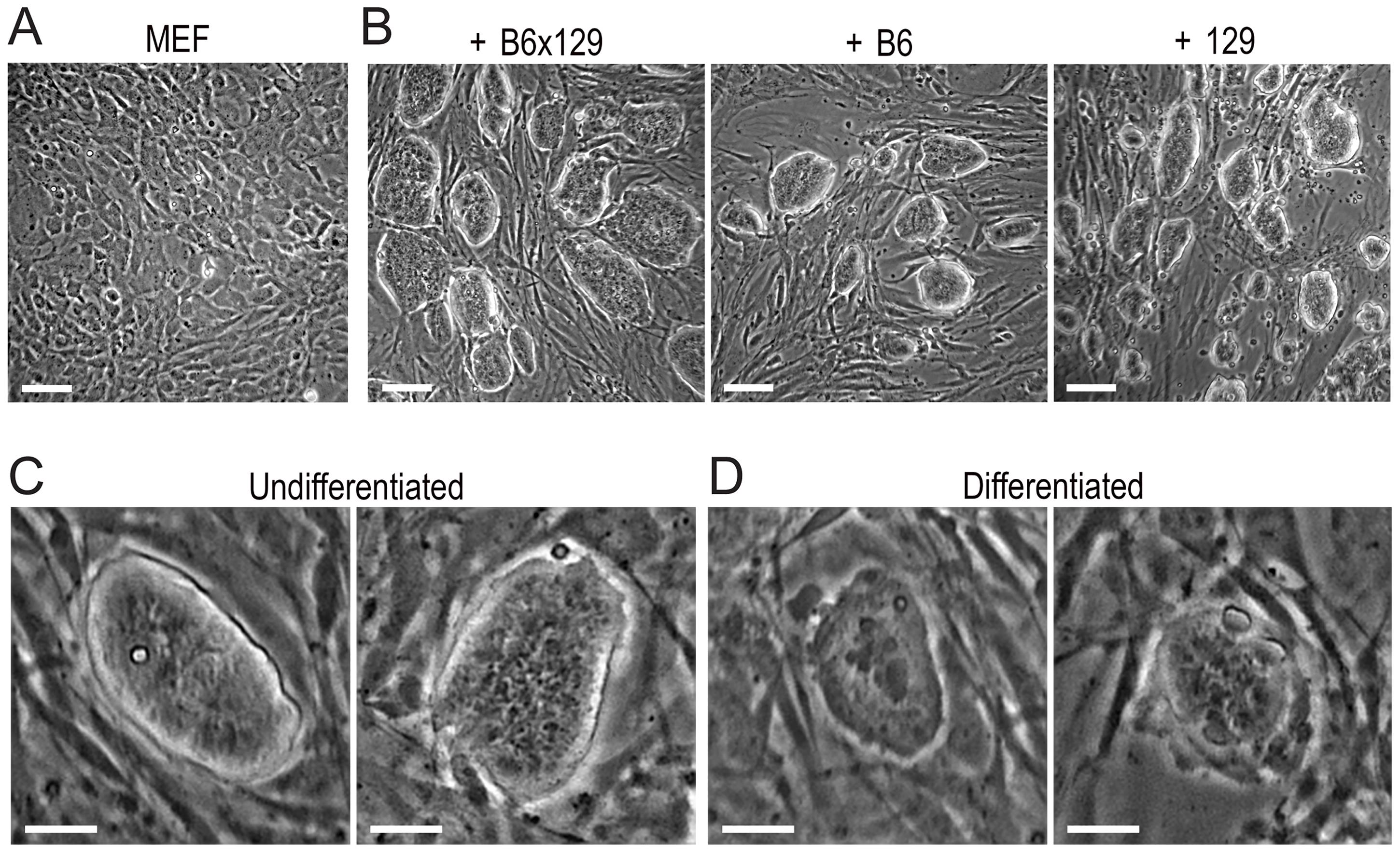
Figure 1. Maintenance of mouse embryonic stem cells (mESCs) under feeder-dependent culture conditions. (A) Monolayer of MEFs before plating. (B) mESC cultures on mouse embryonic fibroblast (MEF) feeders, including B6x129, B6, and 129 strains. (C) Examples of undifferentiated B6x129 colonies, with shiny/defined borders and round shapes. (D) Examples of differentiated B6x129 colonies, with undefined borders and flat cells. Scale bars: 50 μm (A, B), 20 μm (C, D).
B. Preparation of polyHEMA-coated T25 flasks
Timing: Overnight (day -1) or earlier.
1. Prepare 1% polyHEMA solution (see Recipes).
2. Add ~3 mL of polyHEMA stock to each T25 flask.
3. Tilt/rotate to coat the full growth surface evenly.
4. Allow complete ethanol evaporation in a dry incubator until a uniform film forms (typically overnight at 60 °C).
5. Before use, rinse twice with sterile D-PBS.
Critical: Do not use flasks with patchy coating. Uneven coating increases EB attachment and fusion.
Note: PolyHEMA-coated T25 flasks can be prepared in advance and stored sealed at room temperature for up to one week before use.
C. Formation of embryoid bodies (EBs) in suspension
Timing: 3 days (day 0 to day 3).
C1. Prepare EB medium and flasks
1. Rinse polyHEMA-coated T25 flasks twice with sterile D-PBS.
2. Add 5 mL of EB medium (see Recipes) to each T25 flask.
C2. Prepare a single-cell suspension of mESCs
1. Detach mESCs from feeders using 0.25% trypsin-EDTA as described in step A3.
2. After trypsinization, collect the full cell suspension containing both mESCs and MMC-inactivated MEFs.
3. Count viable mESCs.
Note: In this feeder-dependent workflow, both mESCs and MMC-inactivated MEFs detach during trypsinization and are collected. When counting cells for EB seeding, count mESCs only based on their smaller size and distinct morphology. Residual MMC-inactivated MEFs can be carried over, but because they are mitotically inactivated and do not expand in EB suspension conditions, they do not overtake the culture or interfere with EB formation.
C3. Seed strain-specific EB densities
1. B6x129: 2,500 cells/mL
2. B6: 10,000 cells/mL
3. 129: 5,000 cells/mL
4. Seed the required number of mESCs into each T25 flask containing 5 mL of EB medium and gently rock to distribute.
C4. EB culture (day 0 to day 3)
1. Incubate for 3 days at 37 °C and 5% CO2.
2. Once daily, gently shake the flask (x and y axes motions) to reduce EB fusion.
Critical: On day 3, EBs should be relatively uniform with limited fusion and an average diameter of ~100 μm (Figure 2A). Inaccurate and excessive seeding density can promote EB fusion (Figure 2B).
Note: Differentiation is initiated by culturing mESCs in suspension and by removing mLIF from the ESC medium (EB medium, see Recipes).
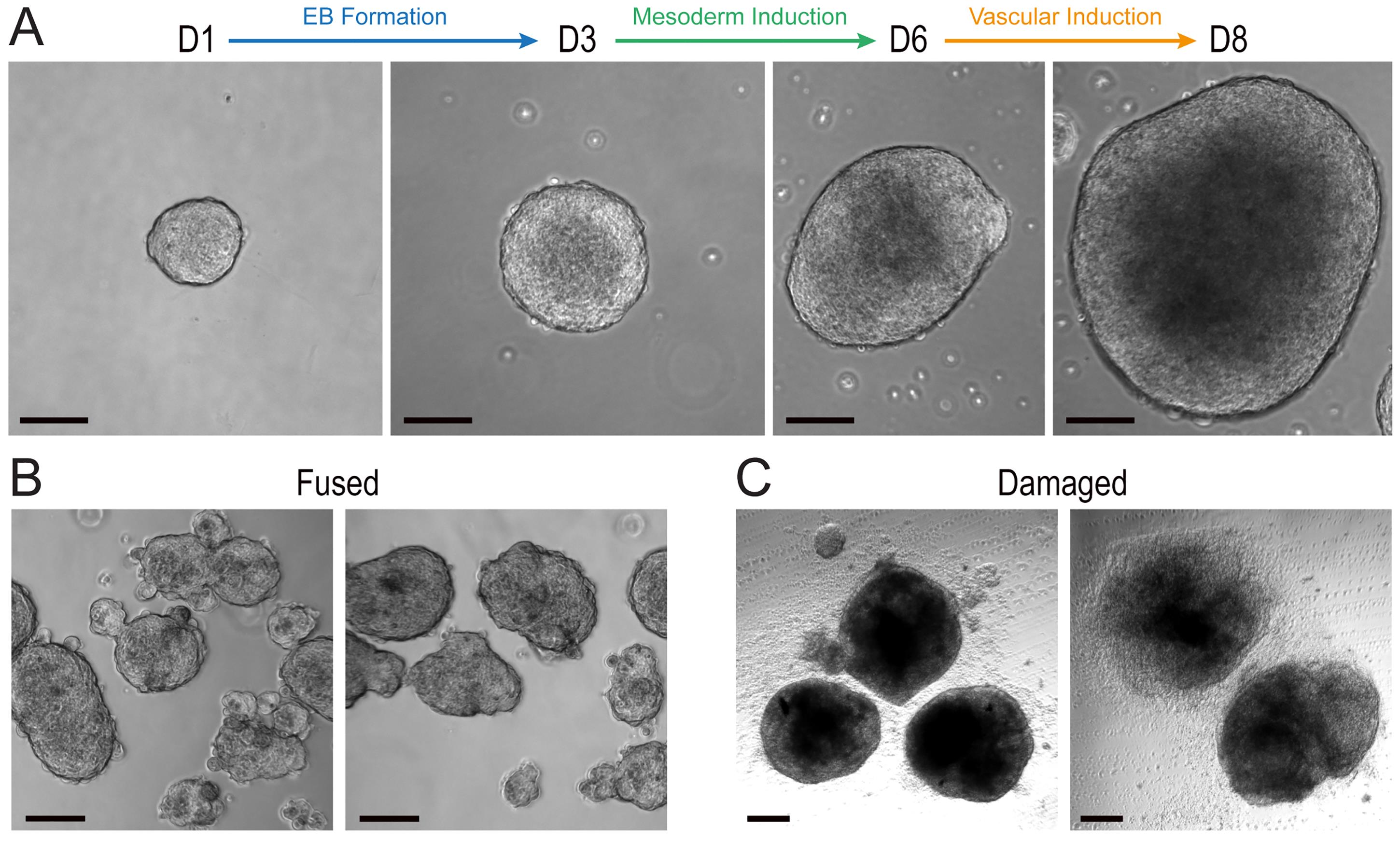
Figure 2. Examples of suitable and suboptimal embryoid bodies (EB). (A) Optimal sizes and morphologies of EBs during EB formation and mesoderm and vascular induction steps, shown on day 1 (D1, ~50 μm), day 3 (~100 μm), day 6 (~150–200 μm), and day 8 (~200–300 μm). EBs should have shiny borders and, at later stages, the core should not be too dense/dark. (B) Two examples of fused EBs on day 3, seeded too densely and too large. (C) Two examples of damaged EBs on day 8 as a consequence of centrifugation during media changes. B6x129 mESCs were used here for all images of the panels. Scale bars: 50 μm.
D. Mesoderm induction with BMP4
Timing: 3 days (day 3 to day 6).
1. Prepare mesoderm induction medium: N2B27 basal medium supplemented with BMP4 at 30 ng/mL (see Recipes).
2. Gently collect EBs into a 15 mL tube using a 5 mL serological pipette and allow to settle by gravity for 15–20 min (do not centrifuge).
3. While waiting, prefill T25 flasks with 5 mL of fresh mesoderm induction medium.
4. Carefully aspirate the supernatant, leaving a small volume to avoid EB loss (~100–200 μL).
5. Gently resuspend EBs in 1 mL of fresh mesoderm induction medium using a P1000 micropipette, then return to the same T25 flask.
6. Culture for 3 days. Gently shake daily to limit fusion. Do not perform additional medium changes during this window.
Critical: On day 6, EBs should be relatively uniform with an average diameter of ~150–200 μm (Figure 2A). To minimize EB fusion, resuspend them using the pipetting method described above (Figure 2B). Avoid centrifugation of EBs to prevent compaction and damage (Figure 2C).
Note: A minimum volume of 6 mL is required at this stage to sustain EB growth. When using 5 mL serological pipettes, use the lowest speed of aspiration/dispensing. Gentle trituration with a P1000 micropipette provides an optimal balance between minimizing mechanical stress on EBs and efficiently dispersing them to prevent clumping.
E. Vascular induction with VEGF and forskolin
Timing: 2 days (day 6 to day 8).
1. Prepare vascular induction medium: N2B27 supplemented with VEGF at 100 ng/mL and forskolin at 2 μM (see Recipes).
2. Gently collect EBs into a 15 mL tube using a 5 mL serological pipette and allow to settle by gravity for 15–20 min (do not centrifuge).
3. While waiting, prefill T25 flasks with 6.5 mL of fresh vascular induction medium.
4. Carefully aspirate the supernatant, leaving a small volume to avoid EB loss (~100–200 μL).
5. Gently resuspend EBs in 1 mL of fresh vascular induction medium using a P1000 micropipette, then return to the same T25 flask.
6. Culture for 2 days. Gently shake daily to reduce fusion. No additional medium change is required during this window.
Critical: On day 8, EBs should be relatively uniform with an average diameter of ~200–300 μm (Figure 2A). To minimize EB fusion, resuspend EBs using the pipetting method described above (Figure 2B). Avoid centrifugation of EBs to prevent compaction and damage (Figure 2C).
Note: A minimum volume of 7.5 mL is required at this stage to sustain EB growth. A medium color change to orange-yellow is normal and is caused by forskolin-induced increase in EB growth.
F. Embedding of EBs and induction of vascular sprouting
Timing: 5 days (day 8 to day 13).
Critical: Keep collagen I and Geltrex cold and prepare mixes on ice to control gelation.
F1. Prepare layer 1 gel base (day 8)
1. Prepare collagen I mix on ice and adjust the pH to approximately 7.4 using NaOH and pH strips (see Recipes) (Figure 3A).
2. Add Geltrex to the collagen I mix (typical collagen I:Geltrex ratio is 8:1). Gently swirl and rotate the tube to homogenize the layer 1 mix (Figure 3B, Video 1A).
3. Dispense 400–500 μL per well of a 12-well plate, avoiding bubbles. The volume of layer 1 (~5.5 mL) is sufficient to cover an entire 12-well plate (Figure 3C, Video 1B).
4. Gently tilt the plate for uniform coverage (Video 1C).
5. Polymerize for at least 2 h at 37 °C.

F2. Prepare layer 2 with EBs (day 8)
1. Prepare layer 2 similar to layer 1.
2. Collect induced EBs into a 15 mL tube using a 5 mL serological pipette and allow to settle by gravity for 15–20 min (do not centrifuge).
3. Carefully aspirate the supernatant, leaving a small volume to avoid EB loss (~100–200 μL).
4. Rinse once with PBS, allow to settle, and repeat the aspiration step.
5. Gently resuspend all EBs in layer 2 gel using a P1000 micropipette. Avoid introducing bubbles (Figure 3D, Video 1D).
6. Overlay the EB-containing layer 2 onto polymerized layer 1, dispensing 400–500 μL per well. The volume of layer 2 (~5.5 mL) is sufficient to cover the entire 12-well plate (Figure 3E, Video 1E).
7. Gently tilt the plate for uniform coverage (Video 1F).
8. Polymerize at 37 °C for at least another 2 h; gels will appear slightly opaque (Figure 3F).
9. Slowly add mBVO medium (see Recipes) on top of gels using a 10 mL serological pipette and dispense 1 mL per well (Figure 3G, Video 1G).
Critical: A practical target is approximately 15–30 EBs per well of a 12-well plate. Using the optimized EB seeding densities for each mESC line detailed in section C, all EBs grown in one T25 are resuspended with layer 2 to cover an entire 12-well plate. This is crucial for efficient sprouting and for the dissection of networks.
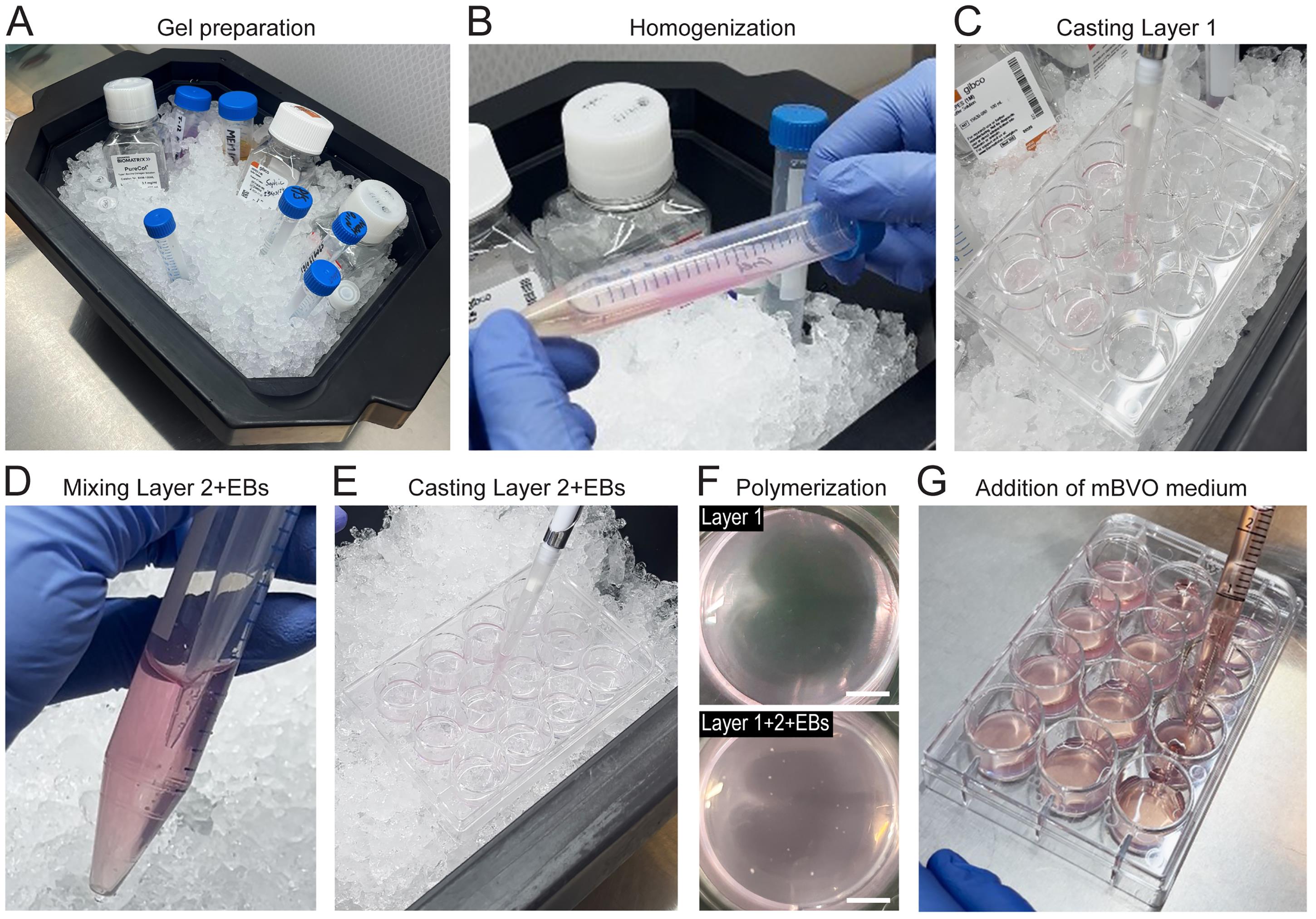
Figure 3. Step-by-step procedure for embedding embryoid bodies (EBs) in collagen I/Geltrex on day 8. (A) Gel preparation with all reagents kept on ice; add collagen I, measure the pH, and finish by adding Geltrex. (B) Gently swirl and rotate the tube for homogenization. (C) Casting of layer 1 gel onto the 12-well plate (400–500 μL per well). (D) Pipetting EBs into layer 2 gel using a P1000 micropipette and gentle mixing. (E) Casting of layer 2 + EBs gel onto the 12-well plate (400–500 μL per well). (F) Images of wells after gel polymerization, showing layer 1 (upper panel) and layer 1 topped with layer 2 and EBs (lower panel). ~15–30 EBs should be plated in each well. Once polymerized, gels appear slightly opaque. Scale bars: 5 mm. (G) Addition of murine blood vessel organoid (mBVO) medium on top of gels (1 mL per well), using a 10 mL serological pipette and the lowest speed of dispensing.
F3. Feeding EBs during vascular sprouting (day 10 to day 13)
1. Refresh mBVO medium after 48 h, then daily until day 13.
Critical: Slowly aspirate the media from the bottom edge of wells, leaving a minimal volume of media (~100 μL), while tilting the plate to avoid aspirating or damaging the gels. Carefully add mBVO medium using a 10 mL serological pipette to slowly dispense 1 mL per well and to avoid detaching the gels (Video 2).
Note: Sprouting typically starts within 24–48 h.
G. Dissection of murine vascular networks (mVNs)
Timing: Day 13.
1. Prepare a sterile dissection area with a stereomicroscope, an LED lamp, and sterile instruments, including plastic forceps, Noyes spring scissors, and a flat double spatula. Perform the procedure in an open sterile biological cabinet under horizontal laminar flow (Figure 4A).
2. Sterilize instruments and the dissection area with 70% ethanol.
3. Prefill an ultra-low attachment (ULA) 96-well plate with 200 μL of mBVO medium per well.
4. Select wells with well-branched networks for dissection and optimal network density (Figure 4B).
5. Aspirate the medium on top of the gels as described in section F3.
6. Using the rounded edge of the flat double spatula, gently lift the gel from the edge of the well and transfer it into a sterile 60 mm cell culture dish prefilled with 1mL of mBVO medium (Figure 4C, Video 3).
7. Using the stereomicroscope, identify well-sprouted vascular networks and cut them out using Noyes spring scissors (Figure 4D, Video 4A–C).
8. Transfer single networks (mVNs) into the prepared 96-well ULA plate, using plastic forceps and placing one mVN per well.
Critical:
1. Dissect only mVNs that show multiple radially distributed sprouts with clear branching. Avoid compact and poorly sprouted structures (Figure 4D, E).
2. Remove as much surrounding gel as technically possible, but keep a thin rim around the mVN so that it remains embedded to further grow into an mBVO (Video 4).
3. During transfer, hold the surrounding gel with the plastic forceps rather than gripping the vascular network directly.
4. Keep gels and dissected mVNs hydrated with mBVO medium throughout the procedure. Drying makes the gel fragile and increases the risk of damaging the networks.
Notes:
1. Minor variation in the amount of residual gel surrounding each mVN is acceptable, as long as it remains intact and embedded. If a few peripheral sprouts are accidentally cut, the mVN can still be transferred, provided that the central network remains intact.
2. Dissection is easier when the embedding density on day 8 is not too high. Wells with too many overlapping networks are more difficult to dissect cleanly and may lead to lower recovery of usable mVNs.
3. Variability at this stage is due to differences in sprouting efficiency between wells or mESC lines, rather than loss of sprouts during cutting.
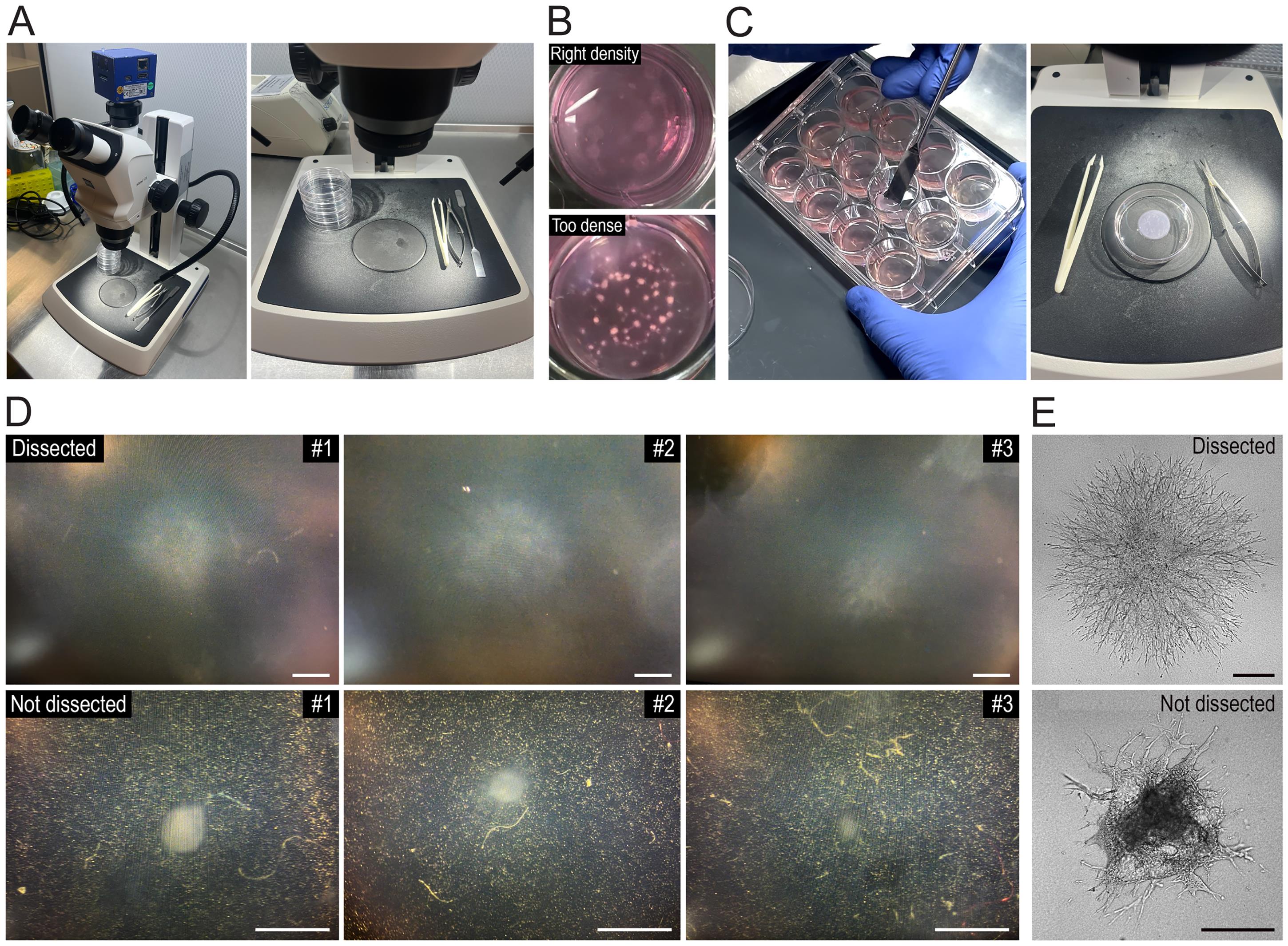
Figure 4. Sterile dissection of murine vascular networks (mVNs) on day 13. (A) Experimental setup for the dissection of mVNs, including a stereomicroscope, an LED lamp (left), sterile instruments and 60 mm dishes (right), all manipulated in an open sterile biological cabinet (horizontal laminar flow). (B) Representative wells containing sprouting mVNs within gels on day 13, showing optimal network density for dissection (upper well), and excessive network density (lower well). (C) Transfer of a gel into a 60 mm dish using the rounded end of a flat double spatula (left), followed by dissection under the stereomicroscope using plastic forceps and Noyes spring scissors (right). (D) Representative examples of mVNs selected or not selected for dissection, as seen under the stereomicroscope during the procedure. See Video 4 for the cutting procedure. (E) Phase contrast images of B6 mVNs on day 13, showing an efficiently sprouted mVN selected for dissection and a poorly sprouted mVN not selected for dissection. Scale bars: 500 μm (D); 200 μm (E).
H. Single-organoid culture and maturation
Timing: 18 days (day 13 to day 30)
1. Grow individual mVNs in suspension in 96-well ULA plates until their maturation into mBVOs (Figure 5A, B).
2. Refresh the mBVO medium every 48 h until day 21 at least and up until day 30 (Figure 5B). To change medium, tilt the plate and carefully aspirate the medium from the upper edge of each well using sterile non-filtered 200 μL pipette tips connected to the aspiration system. Leave a minimal volume of residual medium (~20 μL) to allow mBVOs to settle by gravity and to avoid aspirating them (Figure 5C, Video 5A). Add 200 μL of fresh mBVO medium per well using a multichannel micropipette and sterile reservoirs (Figure 5D, Video 5B).
Critical: Aspirate slowly and keep the tip away from the bottom of the well and from the mBVOs. Dispense fresh medium gently against the wall of the well to avoid disturbing the organoids.
Endpoint: Mature mBVOs are typically obtained between day 21 and day 30 and can be used for subsequent analyses and manipulations [27].
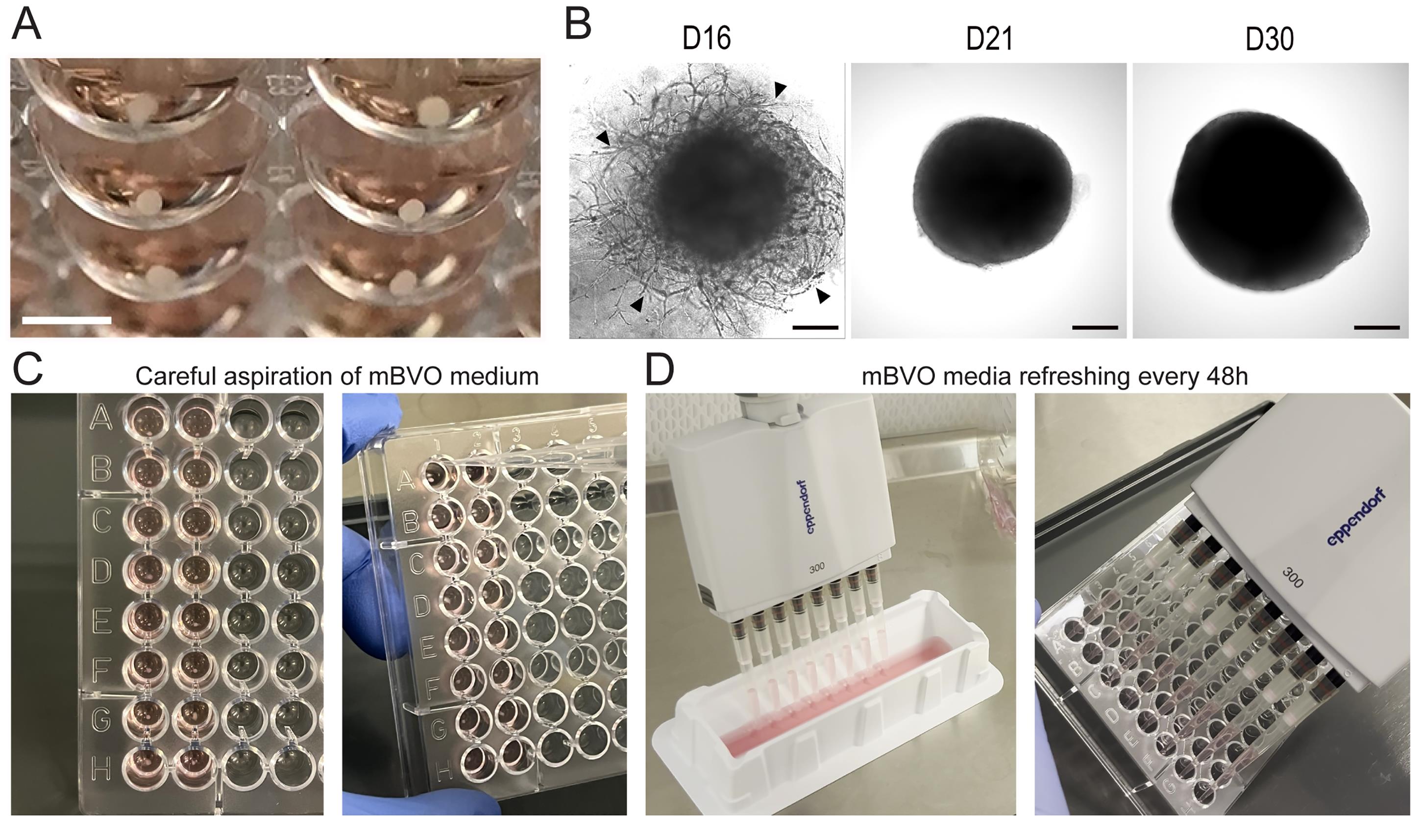
Figure 5. Growth and maturation of murine blood vessel organoids (mBVOs). (A) Six mBVOs (day 30) cultured individually in 96-well ULA plates. Scale bar: 2 mm. (B) Anastomosis of murine vascular networks (mVNs) (day 16) (left), and growth and maturation into mBVOs from day 21 (middle) until day 30 (right). Arrowheads indicate anastomosis of mVNs. 129 mESCs were used here for differentiation. Scale bars: 200 μm. Images for this figure were adapted from Figure S1C, S1E of our original study [27]. (C) Images of how to aspirate media from mBVOs grown individually in 96-well ULA plates. Use a vacuum system and a P200 tip to carefully aspirate media from the upper part of each well, leaving a minimal volume (~20 μL), while tilting the plate. mBVOs settle by gravity in the bottom part of the rounded well and thus are not aspirated. (D) mBVO medium refreshing with 200 μL per well, using a multichannel micropipette and sterile reservoirs.
I. B6-Cdh5-iCre × mTmG endothelial tracing for quality control of differentiation
Timing: Overnight 4-OH-tamoxifen treatment on day 9.
1. Maintain B6-Cdh5-iCre × mTmG mESCs in feeder-dependent conditions as described in section A; include both Cre-negative and Cre-positive lines.
2. On day 0, seed EBs at a density of 5,000 cells/mL in 5 mL of EB medium for both lines, as described in section B.
3. Follow the stepwise differentiation timeline as previously until day 9.
4. On day 9, add 4-OH-tamoxifen to the sprouting cultures to a final concentration of 1 μM (see Recipes).
5. Incubate overnight (Figure 6A).
6. The next day, replace with standard mBVO medium without 4-OH-tamoxifen.
7. Proceed with mBVO differentiation until the desired time point.
8. Perform live imaging (brightfield plus tdTomato and GFP channels) (Figure 6B) to assess endothelial differentiation efficiency and screen poorly differentiated organoids (Figure 6C–E).
Safety: Carefully handle 4-OH-tamoxifen in compliance with the safety procedures of your laboratory.
Critical: Always include a Cre-negative (tdTomato-only) control line processed in parallel to set imaging parameters and confirm specificity (Figure 6C, D).
Note: 4-OH-tamoxifen can be added at any time point. For quality control (QC) of differentiation, a treatment on day 9 overnight is optimal as it targets the sprouting phase of EBs (Figure 6B).
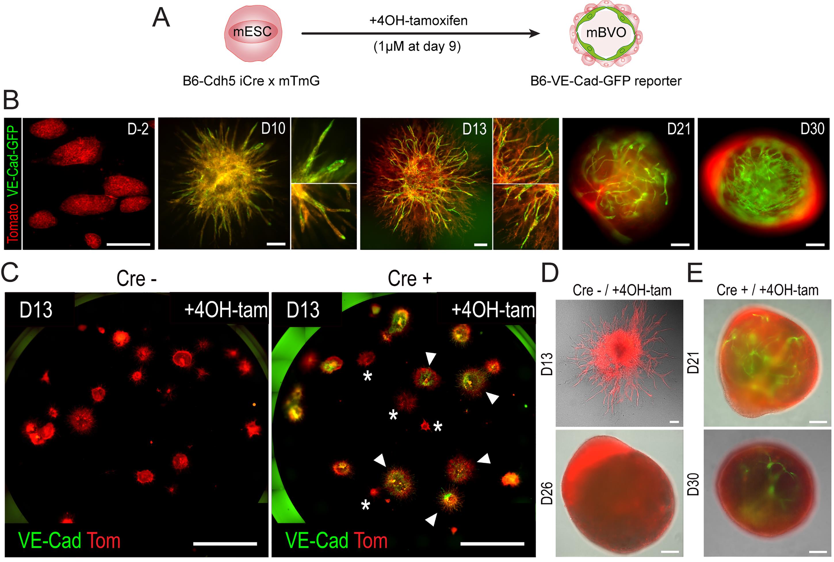
Figure 6. Lineage tracing to follow murine blood vessel organoid (mBVO) differentiation. (A) Diagram of the differentiation of B6-Cdh5-iCre × mTmG mESCs into B6-VE-Cad-GFP reporter mBVOs. 4-OH tamoxifen (4OH-tam) is added on day 9 overnight (1 μM) to induce recombination. (B) Live fluorescent imaging on day -2 (D-2), day 10, day 13, day 21, and day 30. Scale bars: 200 μm. (C) Live fluorescent imaging of sprouting vascular networks (VNs) from Cre-negative controls (left panel) and B6-VE-Cad-GFP reporters (right panel) on day 13; 4-OH-tam is added in both conditions. Arrowheads indicate differentiated mVNs; asterisks indicate poorly differentiated mVNs. Scale bars: 5 mm. (D) Live fluorescent imaging of Cre-negative control tomato+ mBVO on day 13 and day 26. Scale bars: 200 μm. (E) Live fluorescent imaging of a B6-VE-Cad-GFP reporter mBVO on day 21 and day 30, showing poor differentiation efficiency. Scale bars: 200 μm. Images for this figure were adapted from Figure 1H, 1I, S2A, S2B, S2C, and S2D of our original study [27].
Data analysis
1. Recommended replicates
• Biological replicate (n): One independent differentiation experiment starts from a separate mESC culture and is carried through all steps until mBVO maturation.
• Technical replicates: Multiple organoids/networks measured within the same differentiation.
• Typical yield per differentiation: Starting from EB formation, one independent differentiation typically generates approximately one full 96-well plate of mature organoids; after dissection on day 13, this corresponds to approximately one full 12-well plate of dissected vascular networks placed into maturation, although the final yield varies depending on the mESC line and differentiation quality.
• Recommended minimum: Perform at least n ≥ 3 independent differentiations per mESC line or condition. When feasible, analyze at least 10–20 individual mVNs or mBVOs per independent differentiation for image-based analyses. For FACS or single-cell RNA sequencing, multiple mBVOs may need to be pooled, depending on the required cell number.
2. Day 13 sprouting efficiency and morphology-based selection
On day 13, morphology is used as an early screening step to evaluate sprouting and to select mVNs suitable for dissection. This readout should be considered a measure of sprouting/selection efficiency, not a formal measurement of endothelial differentiation efficiency.
For each well or biological replicate:
• Count the total number of EB-derived structures or mVNs present in the gel on day 13 (N_total).
• Count the number of mVNs selected and dissected based on sprouting morphology (N_dissected).
• Calculate sprouting/selection efficiency as:
Sprouting efficiency (%) = (N_dissected/N_total) × 100
mVNs selected for dissection should show clear radial sprouting and vascular network-like morphology. Structures with poor or absent sprouting, extensive fusion, collapse, necrosis, or contamination should be excluded (Figure 7A).
This morphology-based step is useful to compare early protocol performance between mESC strains or differentiations (Figure 7B), but it does not by itself determine whether the resulting mBVOs are well differentiated at the endothelial level.
3. Endothelial differentiation efficiency in non-reporter mESC lines
To assess vascular differentiation efficiency in non-reporter mESC lines, quantify endothelial marker expression in day-21 individual mBVOs. The recommended minimal readout is the percentage of CD31-positive endothelial cells or CD31-positive area, measured by FACS or immunofluorescence, depending on sample format and available material (Figure 7C).
This analysis allows discrimination between well-differentiated mBVOs and poorly differentiated mBVOs.
In the validation dataset shown in Figure 7, individual mBVOs with CD31 positivity ≥7% were classified as well differentiated, whereas mBVOs with CD31 positivity ≤5% were classified as poorly differentiated (Figure 7C). If intermediate values occur, these samples should be considered borderline and excluded from downstream experiments requiring strict endothelial differentiation quality control.
A more complete characterization of mBVO differentiation can include additional endothelial, mesenchymal, mural, and myeloid markers by IF, FACS, or single-cell RNA sequencing [27] (see the validation of the protocol section), but CD31 quantification provides the essential vascular differentiation QC for routine use.
4. Quality control (QC) of differentiation based on endothelial lineage tracing
For the B6-Cdh5-iCre × mTmG reporter line, endothelial differentiation can also be monitored by induction of Cre recombination with 4-OH-tamoxifen on day 9. This turns Cdh5/VE-cadherin-expressing endothelial cells GFP-positive, while non-recombined or non-endothelial cells remain tdTomato-positive.
Because dissecting GFP-positive networks precisely at the early sprouting stage is technically challenging, mVNs are dissected on day 13 as in the standard protocol and followed during maturation. Fluorescence imaging of GFP and tdTomato can then be used to rapidly assess endothelial differentiation quality in live mVNs or mBVOs.
Acquire GFP and tdTomato images using identical acquisition settings within each experiment and compare them with Cre-negative tomato-only controls. GFP positivity provides a convenient qualitative and quantitative QC readout for endothelial differentiation in the reporter line.
For individual day-21 B6-Cdh5-iCre × mTmG reporter mBVOs, GFP positivity can be quantified by live imaging. In the validation dataset shown in Figure 7, mBVOs with GFP positivity ≥7% were classified as well differentiated, whereas mBVOs with GFP positivity ≤5% were classified as poorly differentiated (Figure 7D).
The reporter line is useful for direct visualization and live tracking of endothelial differentiation, but it is not required to be differentiated in parallel with every non-reporter mESC line. Non-reporter lines should be QCed independently by CD31 quantification.
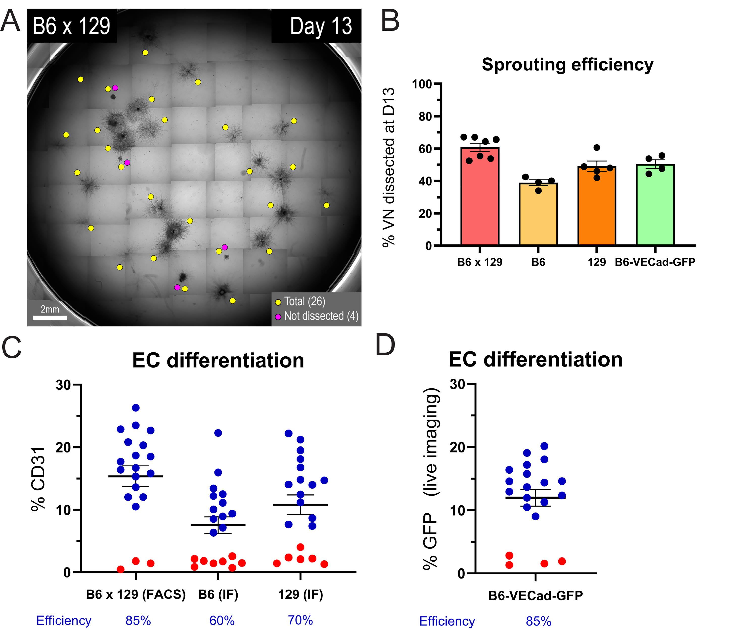
Figure 7. Quantification of sprouting and endothelial differentiation efficiency. (A) Representative day 13 gel containing sprouting murine vascular networks (mVNs) differentiated from B6x129 mouse embryonic stem cells (mESCs). Yellow dots indicate the total embryoid body (EB)-derived structures/mVNs scored in the well, and magenta dots indicate structures that were not selected for dissection. Scale bar: 2 mm. (B) Sprouting/selection efficiency on day 13, calculated as the percentage of mVNs selected for dissection among the total EB-derived structures/mVNs scored in the gel. Quantifications are shown for each mESC strain. n ≥ 4 independent murine blood vessel organoid (mBVO) differentiations per mESC strain; mean ± SEM. (C, D) Quantification of endothelial cell (EC) differentiation efficiency in individual day-21 mBVOs. CD31-positive endothelial cells were quantified by FACS in B6x129-derived mBVOs and by immunofluorescence (IF) in B6- and 129-derived mBVOs (C). For the B6-Cdh5-iCre × mTmG reporter line, VE-cadherin-GFP signal was quantified by live fluorescence imaging (D). Each dot represents one mBVO; n = 20 mBVOs per mESC strain; mean ± SEM. Blue dots indicate well-differentiated mBVOs (CD31+ or GFP+ ≥7%), and red dots indicate poorly differentiated mBVOs (CD31+ or GFP+ ≤5%). Values below the graphs indicate the percentage of well-differentiated mBVOs for each strain.
5. Recommended software
• Leica LasX Office for brightfield/fluorescence image acquisition (xyz planes).
• Fiji/ImageJ for fluorescence quantification and network metrics.
• GraphPad Prism for statistics/plots (optional; R/Python can be used as free alternatives).
Validation of protocol
This protocol has been used and validated in the following research article:
Guelfi et al. [27]. Murine vascular organoids are responsive and adaptable 3D systems with cellular heterogeneity and dynamic plasticity. Science Advances (2025), doi:10.1126/sciadv.ady4738.
Based on this work, we provide a summary of key assays to validate the generation of mBVOs:
1. Reproducible formation of sprouting vascular networks and mBVOs
Stage-specific whole-mount immunostaining confirms the formation of CD31+ endothelial networks associated with mural/perivascular cells and a basement membrane consistent with organized and lumenized vascular structures at the expected stages (Figure 8A–C).
2. Validation of expected cellular composition
We performed flow cytometry and immunophenotyping on day 21 mBVOs and identified three main lineages (Figure 8C, D):
• Endothelial cells (ECs): ~15% of total live cells.
• Mural/mesenchymal populations: ~80% of total live cells, dominant compartment.
• Myeloid cells: ~3% of total live cells, classified as macrophages.
These results provide an internal validation that the protocol yields the expected cell composition in vitro.
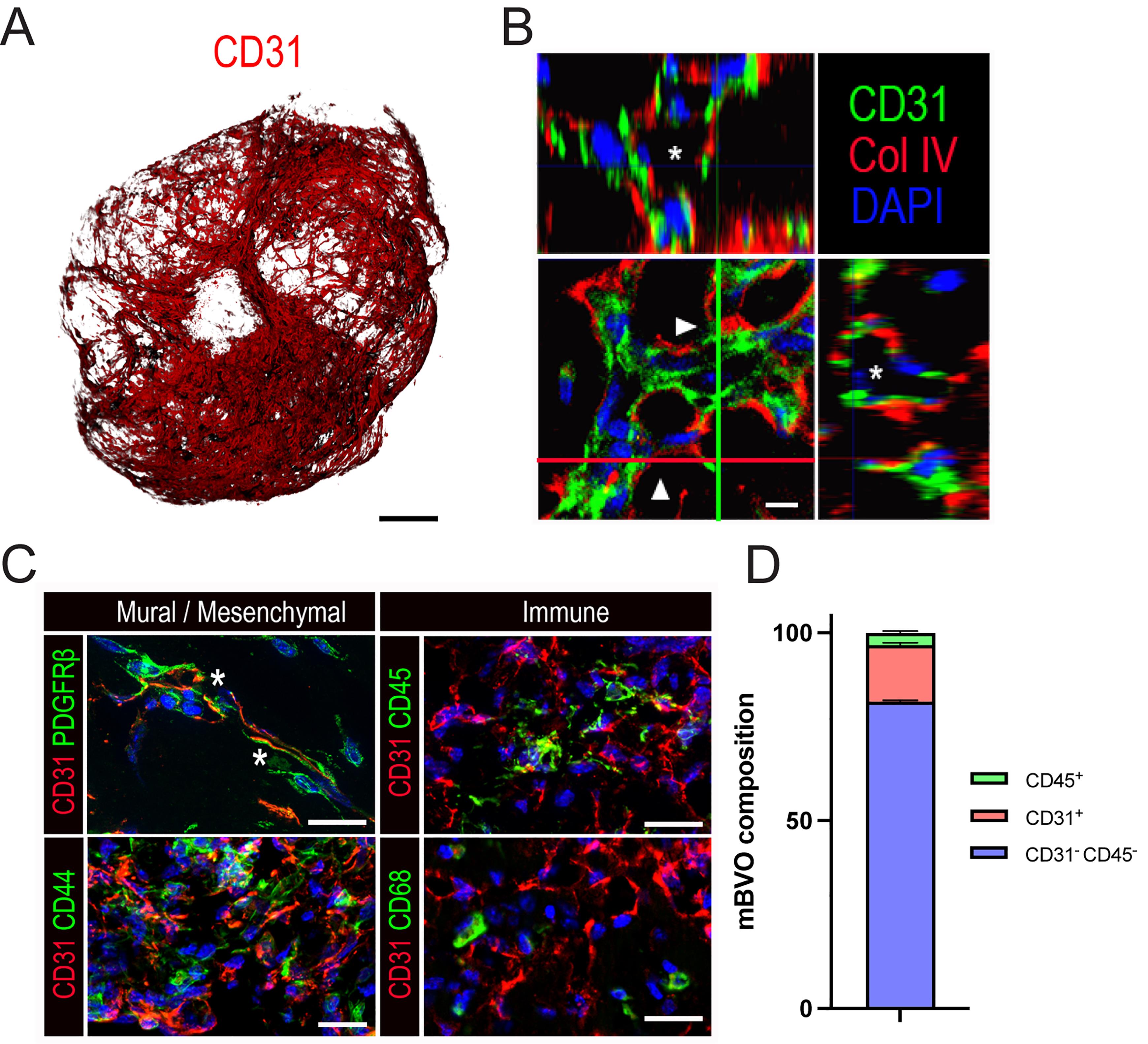
Figure 8. Readouts for validation of murine blood vessel organoid (mBVO) differentiation. (A) 3D image of CD31+ networks within a mBVO (day 21), anterior view. Scale bar: 100 μm. (B) Orthogonal projections of CD31+ColIV+ (Collagen IV) vessels within a mBVO (day 21) differentiated from B6x129 mESCs, showing lumenized vascular networks. The x projection (red line and arrowhead) is shown on the upper panel, the y projection (green line and arrowhead) is shown in the right panel, and asterisks indicate lumens. Scale bar: 5 μm. (C) Cryosections of mBVOs (day 21) co-stained for CD31 with PDGFRβ and CD44 to identify mural/mesenchymal cells, and with CD45 and CD68 to identify immune cells. Asterisks indicate interactions of CD31+ cells with PDGFRβ+ cells. Scale bars: 20 μm. (D) FACS quantifications of the percentage of CD31-CD45-, CD31+, and CD45+ live cell populations composing mBVOs (day 21). n = 3 mBVO differentiations; mean ± SEM. Images for this figure were adapted from Figure 1D, 1F, 3A, and 3D of our original study [27].
General notes and troubleshooting
General notes
1. Strain-dependent variability in mESC differentiation
mESC lines can differ in their baseline pluripotent state, germline competence, EB formation, and lineage differentiation capacity, as previously reported for different mouse genetic backgrounds [38–41]. Therefore, EB seeding density should not be assumed to be interchangeable between mESC lines. In this protocol, seeding densities were empirically optimized for each strain: 2,500 cells/mL for B6x129, 10,000 cells/mL for B6, and 5,000 cells/mL for 129 mESCs. Under these optimized conditions, EB numbers were broadly comparable by day 8, but vascular differentiation efficiency still differed between strains, with B6 mESCs showing lower efficiency in our hands. Users should therefore re-optimize EB seeding density for each new mESC line using EB morphology, EB number, sprouting efficiency, and endothelial differentiation readouts.
2. Feeder-dependent vs. feeder-free mESC culture
This protocol was optimized and validated using mESCs maintained on MMC-inactivated MEF feeders in serum-containing ESC medium supplemented with LIF. These conditions provided the most robust mBVO differentiation efficiency in our hands. Feeder-free conditions, including 2i conditions, may be compatible with mBVO differentiation and have been used in a recent mESC-derived vascular organoid study [42]. However, this adaptation was not validated in our original study [27]. Users starting from feeder-free mESCs should re-optimize EB seeding density, EB size, differentiation timing, sprouting efficiency, and endothelial readouts such as CD31 expression or VE-cadherin-reporter positivity.
3. Starting mESC quality is a key determinant of success
Begin EB formation only from healthy, compact colonies with minimal spontaneous differentiation. If cultures look heterogeneous, passage once more on fresh feeders and reassess morphology before initiating differentiation.
4. Feeder layer quality affects mESC maintenance and thus differentiation
Use appropriately dense, freshly prepared mitotically inactivated MEFs. Over-aged or sparse feeders can promote differentiation or slow growth. Do not grow primary MEFs further than passage 4 before MMC inactivation and freezing of stocks. Transfer MMC-inactivated MEFs cryovials rapidly to the liquid nitrogen for optimal preservation.
5. Strain-specific EB seeding density must be applied exactly
Use the fixed densities (B6x129: 2,500 cells/mL; B6: 10,000 cells/mL; 129: 5,000 cells/mL; B6-Cdh5-iCre x mTmG: 5,000 cells/mL) and ensure accurate counting and high cell viability at seeding to reduce EB size variability.
6. Batch effects (growth factors and matrices) are a common source of run-to-run variability
Minimize freeze–thaw cycles with single-use aliquots for mLIF, BMP4, VEGF, bFGF, and forskolin. Record lot numbers for collagen I and Geltrex, and consider pre-testing new lots if performance changes.
7. Gentle handling reduces loss of differentiation potential
Throughout EB culture, embedding, and dissection, avoid excessive trituration, rapid pipetting, or drying of gels. Mechanical stress often manifests later as weak sprouting or fragile vascular networks.
8. Matrix preparation is sensitive to temperature and pH
Keep collagen I/Geltrex components cold during preparation and cast promptly. Incorrect pH or warming during preparation can reduce polymerization quality and impair sprouting.
9. For EC tracing, always include the Cre-negative (tdTomato-only) control for interpretation
This control is essential to set imaging parameters and confirm the specificity of GFP induction in the reporter line.
10. Quality control of mBVO differentiation across mESC lines
Sprouting morphology on day 13 provides an early checkpoint for selecting mVNs suitable for dissection, but it should not be used as the only readout of vascular differentiation efficiency. Because differentiation potential can vary between mESC strains, each mESC line should be assessed independently. To reduce batch-to-batch variability, compare mESC lines using the same lots of serum, matrix, growth factors, and basal media whenever possible.
Troubleshooting
Problem 1: mESC colonies look differentiated or grow slowly before starting EB formation.
Possible causes: Over-confluency, suboptimal feeder layer (too sparse/too old), stressed cells after thaw, or inconsistent daily medium changes.
Solutions: Start differentiations only from healthy cultures with compact colonies. Use freshly prepared inactivated feeders at appropriate density, avoid overgrowth, and passage onto fresh feeders if differentiation is evident before starting EB formation.
Problem 2: EBs are too small on day 3 (slow growth) or very heterogeneous in size.
Possible causes: Inaccurate cell counting, incorrect strain-specific seeding density, reduced viability, or uneven distribution in the T25.
Solutions: Verify counting accuracy and apply the fixed strain-specific seeding densities exactly. Mix thoroughly immediately before seeding and distribute evenly. Start from high-viability cultures.
Problem 3: EB fusion (large aggregates), followed by poor differentiation.
Possible causes: Inadequate polyHEMA coating, too many EBs per flask, insufficient gentle agitation or centrifugation during media changes.
Solutions: Confirm polyHEMA coating quality. Reduce EB density per vessel (split across additional flasks) if fusion is frequent, gently agitate daily to keep EBs suspended, and do not centrifuge EBs.
Problem 4: Collagen I/Geltrex gel does not polymerize properly (soft/uneven) or polymerizes too quickly during embedding.
Possible causes: Incorrect pH, warming of components, slow handling, or lot variability.
Solutions: Keep all components cold and work quickly. Ensure the pH of the collagen I mix is appropriate before adding Geltrex and casting. Document lot numbers and switch lots if polymerization is affected.
Problem 5: Gel detaches from the well or is disrupted during medium changes (sprouts damaged).
Possible causes: Aspiration too close to the gel, forceful dispensing, insufficient polymerization time, or temperature shock.
Solutions: Allow full polymerization before feeding. Aspirate from the edge, add prewarmed medium slowly down the sidewall, and avoid directing flow onto the gel surface.
Problem 6: Poor sprouting between day 8 and 13 (few EBs sprout; sprouts are short) and poor yield of ECs within growing mBVOs.
Possible causes: EB quality issues (fusion/heterogeneity), matrix preparation problems (pH/temperature/ratio), or reduced growth factor activity.
Solutions: Use early quality control (EB size distribution on day 3; fusion) to exclude poor batches. Re-check matrix preparation (temperature and pH control) and replace suspect growth factor aliquots.
Problem 7: Low or absent GFP induction after 4-OH-tamoxifen (day 9 overnight).
Possible causes: 4-OH-tamoxifen degradation (light/temperature, repeated freeze–thaw), incorrect dilution/timing, or reporter line handling issues.
Solutions: Use light-protected single-use aliquots, avoid repeated freeze–thaw, and confirm the induction (day 9 overnight, 1 μM 4-OH-tamoxifen). Verify line identity and imaging timing.
Problem 8: Degradation of mBVOs during the growing phase in ULA 96-well plates.
Possible cause: Degradation of the ULA coating of 96-well plates over time (from Corning).
Solutions: Transfer individual mBVOs into a fresh plate. As an alternative, use polyHEMA-coated 96-well plates for in-house production and cost reduction.
Problem 9: Bacterial contamination of individual mBVOs grown in ULA 96-well plates.
Possible causes: Problem with aseptic techniques during the dissection of networks and media changes of ULA 96-well plates, misuse of the open cabinet.
Solutions: Review aseptic techniques, autoclave instruments, transfer non-contaminated mBVOs to fresh plates, carefully change tips during aspiration/media changes.
Problem 10: Low or variable sprouting/differentiation efficiency between mESC lines or between experiments.
Possible causes: Differentiation efficiency can vary depending on mESC strain, passage number, colony quality, EB size, EB fusion, feeder quality, matrix lot, growth factor activity, serum lot, or handling during embedding and dissection.
Solutions: Start differentiation from healthy, undifferentiated mESC colonies and use the strain-specific EB seeding densities indicated in the protocol. Avoid EB fusion during suspension culture and keep the number of embedded EBs per well consistent. Use the same lots of serum, collagen I/Geltrex, growth factors, and differentiation media when comparing different mESC lines or experimental conditions.
Acknowledgments
S.G.: Conceptualization, Methodology, Investigation, Data curation, Formal analysis, Visualization, Writing—Original Draft, Writing—Review & Editing. S.B: Investigation, Data curation, Formal analysis and Visualization. G.B.: Conceptualization, Supervision, Funding acquisition, Validation, Writing—Original Draft, Writing—Review & Editing.
This work was funded by Fonds Wetenschappelijk Onderzoek Vlaanderen (FWO), project numbers G072021N (S.G) and G0A1122N (G.B).
We thank Els Gils, Luc Schoonjans, Pieter Carmeliet, Ben van der Veer, and Kian Peng Koh for providing C57BL/6 × 129S6, C57BL/6 Cdh5-iCre × mTmG, C57BL/6 and 129S mESC strains, MEF feeders, and related reagents. We thank all collaborators who co-authored our initial study regarding mBVOs. We are grateful to the VIB Flow Core (Leuven), VIB Single Cell Core (Leuven), and VIB Nucleomics Core (Leuven) for support.
This protocol builds on previously described human blood vessel organoid methods, including Wimmer et al. [8], and is used in [27]: Science Advances (2025), doi:10.1126/sciadv.ady4738.
Competing interests
The authors declare no conflicts of interest.
Ethical considerations
This protocol describes in vitro culture and differentiation of established mouse embryonic stem cell lines and the use of mitotically inactivated feeder cells. No new human samples, clinical data, or in vivo animal experiments are performed as part of the protocol.
References
- Aird, W. C. (2005). Spatial and temporal dynamics of the endothelium. J Thromb Haemost. 3(7): 1392–1406. https://doi.org/10.1111/j.1538-7836.2005.01328.x
- Aird, W. C. (2007). Phenotypic heterogeneity of the endothelium: I. Structure, function, and mechanisms. Circ Res 100(2): 158–173. https://doi.org/10.1161/01.RES.0000255691.76142.4a
- Rajendran, P., Rengarajan, T., Thangavel, J., Nishigaki, Y., Sakthisekaran, D., Sethi, G. and Nishigaki, I. (2013). The vascular endothelium and human diseases. Int J Biol Sci. 9(10): 1057–1069. https://doi.org/10.7150/ijbs.7502
- Pries, A. R., Secomb, T. W. and Gaehtgens, P. (1995). Design principles of vascular beds. Circ Res. 77(5): 1017–1023. https://doi.org/10.1161/01.res.77.5.1017
- Bogseth, A., Ramirez, A., Vaughan, E. and Maisel, K. (2023). In Vitro Models of Blood and Lymphatic Vessels-Connecting Tissues and Immunity. Adv Biol (Weinh) 7(5): e2200041. https://doi.org/10.1002/adbi.202200041
- Nwokoye, P. N. and Abilez, O. J. (2024). Blood vessels in a dish: the evolution, challenges, and potential of vascularized tissues and organoids. Front Cardiovasc Med. 11: 1336910. https://doi.org/10.3389/fcvm.2024.1336910
- Wimmer, R. A., Leopoldi, A., Aichinger, M., Wick, N., Hantusch, B., Novatchkova, M., Taubenschmid, J., Hämmerle, M., Esk, C., Bagley, J. A., et al. (2019). Human blood vessel organoids as a model of diabetic vasculopathy. Nature. 565(7740): 505–510. https://doi.org/10.1038/s41586-018-0858-8
- Wimmer, R. A., Leopoldi, A., Aichinger, M., Kerjaschki, D. and Penninger, J. M. (2019). Generation of blood vessel organoids from human pluripotent stem cells. Nat Protoc. 14(11): 3082–3100. https://doi.org/10.1038/s41596-019-0213-z
- Nikolova, M. T., He, Z., Seimiya, M., Jonsson, G., Cao, W., Okuda, R., Wimmer, R. A., Okamoto, R., Nikoloff, J. M., Dittrich, P. S., et al. (2025). Fate and state transitions during human blood vessel organoid development. Cell. 188(12): 3329–3348.e31. https://doi.org/10.1016/j.cell.2025.03.037
- Sun, X. Y., Ju, X. C., Li, Y., Zeng, P. M., Wu, J., Zhou, Y. Y., Shen, L. B., Dong, J., Chen, Y. J. and Luo, Z. G. (2022). Generation of vascularized brain organoids to study neurovascular interactions. eLife. 11: e76707. https://doi.org/10.7554/eLife.76707
- Sun, X. Y., Ju, X. C., Zhao, H. F., You, Z. W., Han, R. R. and Luo, Z. G. (2023). Generation of Human Blood Vessel and Vascularized Cerebral Organoids. Bio Protoc. 13(21): e4870. https://doi.org/10.21769/BioProtoc.4870
- Tubbs, E., Mehanović, M., Lopes, M., Quintard, C., Combe, S., Pirlian, C., Armanet, M., Domet, T., Sabatier, J., Granziera, S., et al. (2025). Human assembloid of human blood vessel organoids with pancreatic islets improves insulin secretion over time ex vivo. Cell Rep. 44(10): 116378. https://doi.org/10.1016/j.celrep.2025.116378
- Dao, L., You, Z., Lu, L., Xu, T., Sarkar, A. K., Zhu, H., Liu, M., Calandrelli, R., Yoshida, G., Lin, P., et al. (2024). Modeling blood-brain barrier formation and cerebral cavernous malformations in human PSC-derived organoids. Cell Stem Cell. 31(6): 818–833.e811. https://doi.org/10.1016/j.stem.2024.04.019
- Kong, D., Park, K. H., Kim, D. H., Kim, N. G., Lee, S. E., Shin, N., Kook, M. G., Kim, Y. B. and Kang, K. S. (2023). Cortical-blood vessel assembloids exhibit Alzheimer's disease phenotypes by activating glia after SARS-CoV-2 infection. Cell Death Discov. 9(1): 32. https://doi.org/10.1038/s41420-022-01288-8
- Sun, X., Kofman, S., Ogbolu, V. C., Karch, C. M., Ibric, L. and Qiang, L. (2024). Vascularized Brain Assembloids With Enhanced Cellular Complexity Provide Insights Into the Cellular Deficits of Tauopathy. Stem Cells. 42(2): 107–115. https://doi.org/10.1093/stmcls/sxad086
- Sun, X., Che, S., Wang, H., Fan, Y., Ding, Y., Tan, A., Yang, K., Hu, J., Zhang, Y., Ma, M., et al. (2026). Vascular organoid model of Hutchinson-Gilford progeria syndrome uncovers repression of the SRF pathway in premature aging. Dev Cell. 61(3): 505–517.e7. https://doi.org/10.1016/j.devcel.2025.10.019
- Garcés, C., Tascón, P., Diago-Domingo, L., Ibáñez, A. J., Herrera, G., Rodríguez, L. R., Salewskij, K., Jonsson, G., Penninger, J. M., Romá-Mateo, C., et al. (2025). Endocytosis and non-canonical autophagy mediate extracellular histones cytotoxicity in vascular models of sepsis. Front Immunol. 16: 1650789. https://doi.org/10.3389/fimmu.2025.1650789
- Lee, M., Yoon, Y. J., Lee, J. Y., Hwang, M., Park, S., Lee, M. H., Um, S. W., Cho, B. C., Rheey, J., Sato, T., et al. (2025). Modeling the metastatic tumor microenvironment: a co-culture platform of lung cancer and blood vessel organoids for drug evaluation. Sci Rep. 15(1): 45252. https://doi.org/10.1038/s41598-025-29214-9
- Clevers, H. (2016). Modeling Development and Disease with Organoids. Cell. 165(7): 1586–1597. https://doi.org/10.1016/j.cell.2016.05.082
- Salewskij, K. and Penninger, J. M. (2023). Blood Vessel Organoids for Development and Disease. Circ Res. 132(4): 498–510. https://doi.org/10.1161/circresaha.122.321768
- Quintard, C., Tubbs, E., Jonsson, G., Jiao, J., Wang, J., Werschler, N., Laporte, C., Pitaval, A., Bah, T. S., Pomeranz, G., et al. (2024). A microfluidic platform integrating functional vascularized organoids-on-chip. Nat Commun. 15(1): 1452. https://doi.org/10.1038/s41467-024-45710-4
- Werschler, N., Quintard, C., Nguyen, S. and Penninger, J. (2024). Engineering next generation vascularized organoids. Atherosclerosis. 398: 118529. https://doi.org/10.1016/j.atherosclerosis.2024.118529
- Cleaver, O. (2021). Mouse models of vascular development and disease. Curr Opin Hematol. 28(3): 179–188. https://doi.org/10.1097/moh.0000000000000649
- Xiang, M., Grosso, R. A., Takeda, A., Pan, J., Bekkhus, T., Brulois, K., Dermadi, D., Nordling, S., Vanlandewijck, M., Jalkanen, S., et al. (2020). A Single-Cell Transcriptional Roadmap of the Mouse and Human Lymph Node Lymphatic Vasculature. Front Cardiovasc Med. 7: 52. https://doi.org/10.3389/fcvm.2020.00052
- Song, H. W., Foreman, K. L., Gastfriend, B. D., Kuo, J. S., Palecek, S. P. and Shusta, E. V. (2020). Transcriptomic comparison of human and mouse brain microvessels. Sci Rep. 10(1): 12358. https://doi.org/10.1038/s41598-020-69096-7
- Robinson, N. B., Krieger, K., Khan, F. M., Huffman, W., Chang, M., Naik, A., Yongle, R., Hameed, I., Krieger, K., Girardi, L. N., et al. (2019). The current state of animal models in research: A review. Int J Surg. 72: 9–13. https://doi.org/10.1016/j.ijsu.2019.10.015
- Guelfi, S., Gopalan, K. S., Maene, P., Robbert, D. J., Pallarés-Moratalla, C., Vella, G., Nobis, M., Mourao, L., Bourdely, P., van der Veer, B. K., et al. (2025). Murine vascular organoids are responsive and adaptable 3D systems with cellular heterogeneity and dynamic plasticity. Sci Adv. 11(43): eady4738. https://doi.org/10.1126/sciadv.ady4738
- Muzumdar, M. D., Tasic, B., Miyamichi, K., Li, L. and Luo, L. (2007). A global double-fluorescent Cre reporter mouse. Genesis. 45(9): 593–605. https://doi.org/10.1002/dvg.20335
- Evans, M. J. and Kaufman, M. H. (1981). Establishment in culture of pluripotential cells from mouse embryos. Nature 292(5819): 154–156. https://doi.org/10.1038/292154a0
- Martin, G. R. (1981). Isolation of a pluripotent cell line from early mouse embryos cultured in medium conditioned by teratocarcinoma stem cells. Proc Natl Acad Sci USA. 78(12): 7634–7638. https://doi.org/10.1073/pnas.78.12.7634
- Czechanski, A., Byers, C., Greenstein, I., Schrode, N., Donahue, L. R., Hadjantonakis, A. K. and Reinholdt, L. G. (2014). Derivation and characterization of mouse embryonic stem cells from permissive and nonpermissive strains. Nat Protoc. 9(3): 559–574. https://doi.org/10.1038/nprot.2014.030
- Luo, X., van der Veer, B. K., Sun, L., Bartoccetti, M., Boretto, M., Vankelecom, H., Khoueiry, R. and Koh, K. P. (2020). Coordination of germ layer lineage choice by TET1 during primed pluripotency. Genes Dev. 34(7–8): 598–618. https://doi.org/10.1101/gad.329474.119
- Sörensen, I., Adams, R. H. and Gossler, A. (2009). DLL1-mediated Notch activation regulates endothelial identity in mouse fetal arteries. Blood. 113(22): 5680–5688. https://doi.org/10.1182/blood-2008-08-174508
- Hogan, B., Costantini, F. and Lacy, E. (1986). Manipulating the mouse embryo: a laboratory manual. Cold spring harbor laboratory Cold Spring Harbor, NY.
- van der Veer, B. K., Chen, L., Custers, C., Athanasouli, P., Schroiff, M., Cornelis, R., Chui, J. S., Finnell, R. H., Lluis, F. and Koh, K. P. (2023). Dual functions of TET1 in germ layer lineage bifurcation distinguished by genomic context and dependence on 5-methylcytosine oxidation. Nucleic Acids Res. 51(11): 5469–5498. https://doi.org/10.1093/nar/gkad231
- Conner, D. A. (2001). Mouse embryo fibroblast (MEF) feeder cell preparation. Current protocols in molecular biology Chapter 23: Unit 23.22. https://doi.org/10.1002/0471142727.mb2302s51
- Xu, J. (2005). Preparation, culture, and immortalization of mouse embryonic fibroblasts. Current protocols in molecular biology Chapter 28: Unit 28.21. https://doi.org/10.1002/0471142727.mb2801s70
- Kawase, E., Suemori, H., Takahashi, N., Okazaki, K., Hashimoto, K. and Nakatsuji, N. (1994). Strain difference in establishment of mouse embryonic stem (ES) cell lines. Int J Dev Biol. 38(2): 385–390. https://doi.org/10.1387/ijdb.7981049
- Ward, C. M., Barrow, K. M. and Stern, P. L. (2004). Significant variations in differentiation properties between independent mouse ES cell lines cultured under defined conditions. Exp Cell Res. 293(2): 229–238. https://doi.org/10.1016/j.yexcr.2003.10.017
- Ghimire, S., Van der Jeught, M., Neupane, J., Roost, M. S., Anckaert, J., Popovic, M., Van Nieuwerburgh, F., Mestdagh, P., Vandesompele, J., Deforce, D., et al. (2018). Comparative analysis of naive, primed and ground state pluripotency in mouse embryonic stem cells originating from the same genetic background. Sci Rep. 8(1): 5884. https://doi.org/10.1038/s41598-018-24051-5
- Keskintepe, L., Norris, K., Pacholczyk, G., Dederscheck, S. M. and Eroglu, A. (2007). Derivation and comparison of C57BL/6 embryonic stem cells to a widely used 129 embryonic stem cell line. Transgenic Res. 16(6): 751–758. https://doi.org/10.1007/s11248-007-9125-8
- Kowalski, W. J., Vatti, S., Sakamoto, T., Li, W., Odutola, S. R., Liu, C., Chen, G., Boehm, M. and Mukouyama, Y. S. (2025). In vivo transplantation of mammalian vascular organoids onto the chick chorioallantoic membrane reveals the formation of a hierarchical vascular network. Sci Rep. 15(1): 7150. https://doi.org/10.1038/s41598-025-91826-y
Article Information
Publication history
Received: Feb 25, 2026
Accepted: May 6, 2026
Available online: May 28, 2026
Published: Jul 5, 2026
Copyright
© 2026 The Author(s); This is an open access article under the CC BY-NC license (https://creativecommons.org/licenses/by-nc/4.0/).
How to cite
Readers should cite both the Bio-protocol article and the original research article where this protocol was used:
- Guelfi, S., Bopp, S. and Bergers, G. (2026). Stepwise Differentiation of Mouse Embryonic Stem Cells Into Murine Blood Vessel Organoids With Endothelial Lineage Tracing for Quality Control. Bio-protocol 16(13): e5727. DOI: 10.21769/BioProtoc.5727.
- Guelfi, S., Gopalan, K. S., Maene, P., Robbert, D. J., Pallarés-Moratalla, C., Vella, G., Nobis, M., Mourao, L., Bourdely, P., van der Veer, B. K., et al. (2025). Murine vascular organoids are responsive and adaptable 3D systems with cellular heterogeneity and dynamic plasticity. Sci Adv. 11(43): eady4738. https://doi.org/10.1126/sciadv.ady4738

 Download Citation in RIS Format
Category
Stem Cell > Organoid culture
Stem Cell > Embryonic stem cell > Cell differentiation
Do you have any questions about this protocol?
Post your question to gather feedback from the community. We will also invite the authors of this article to respond.
 Post a Question


