


- Protocols

- Articles and Issues
- For Authors
- About
- Become a Reviewer


An Immunoprecipitation-Based Nonradioactive Kinase Assay to Measure Akt Kinase Activity in Mammalian Cell Lines

Published: Vol 16, Iss 13, Jul 5, 2026 DOI: 10.21769/BioProtoc.5724 Views: 286
Reviewed by: Vipin K. YadavPrashanth N SuravajhalaAnonymous reviewer(s)
 PDF
PDF  Ask a question
Original research article
The authors used this protocol in:

Mar 2024
Advertisement


Protocol Collections
Comprehensive collections of detailed, peer-reviewed protocols focusing on specific topics





Abstract
Protein kinase B, more commonly known as Akt, is a family of three serine/threonine kinases (Akt1, Akt2, and Akt3) that play a central role in regulating processes such as proliferation, survival, metabolism, and migration through phosphorylation of downstream targets. Given its involvement in numerous cellular processes, aberrant Akt signaling is prevalent across multiple cancer types, underscoring the need for Akt kinase assays to assess activity, regulatory mechanisms, and the efficacy of targeted interventions. Most existing Akt kinase assays rely on expensive commercial kits, some of which employ pre-purified, constitutively active Akt expressed in insect cells, bypassing physiologic autoinhibition of Akt; therefore, they are not suitable for evaluating allosteric inhibitors or context-dependent regulation. Here, we describe a detailed, step-by-step protocol for a nonradioactive Akt kinase assay using epitope-tagged, recombinant Akt1 expressed in a mammalian cell line and isolated by immunoprecipitation. This method eliminates the need to co-express Akt with upstream regulatory kinases or to purify active enzyme from insect cells, a time-consuming and technically demanding process, particularly when analyzing multiple Akt mutants. Because Akt is assayed in a regulated, autoinhibited state, this protocol enables direct evaluation of allosteric inhibitors that cannot be assessed using active Akt purified from insect cells. We note, however, that Akt1 kinase activity in this assay is measured from epitope-tagged, transiently overexpressed protein, which could influence cellular signaling dynamics. Despite this limitation, the cellular context preserves key regulatory features of Akt1 autoinhibition and membrane-dependent activation that are absent in assays using purified, pre-activated kinase. Together, this protocol supports analysis of Akt kinase activity under diverse experimental conditions, including receptor stimulation, pharmacologic treatment, allosteric inhibitor exposure, and mutations, using an accessible, economical, and physiologically relevant approach.
Key features
• This protocol is broadly accessible, requiring only standard laboratory equipment and commonly used techniques without specialized instrumentation or purified kinase preparations.
• This protocol measures Akt1 catalytic output by assessing substrate phosphorylation following immunoprecipitation of transiently expressed, epitope-tagged Akt1 from cells.
• The assay is performed in a low-throughput format and provides a semiquantitative readout.
• This protocol can be adapted to other mammalian cell lines and optimized for other protein kinases of choice.
Keywords: Akt Phosphorylation
Nonradioactive kinase assay
Immunoprecipitation
Allosteric inhibitors
Phosphorylation
Nonradioactive kinase assay
Immunoprecipitation
Allosteric inhibitors
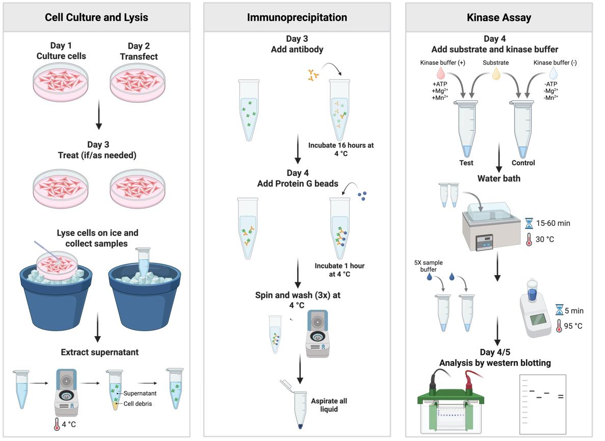
Graphical overview
Background
Protein kinase B, commonly referred to as Akt, is a serine/threonine kinase that serves as a central integrator of signaling pathways in mammalian cells. Akt regulates a broad spectrum of cellular functions, including cell growth, survival, migration, and metabolism, through phosphorylation of more than 100 identified substrates [1]. Activation of Akt is initiated by growth factor–stimulated phosphoinositide 3-kinase–mediated production of phosphatidylinositol 3,4,5-trisphosphate (PIP3) at the plasma membrane [2]. Binding of PIP3 to the N-terminal pleckstrin homology (PH) domain of Akt promotes its translocation to the plasma membrane [3]. Once localized at the membrane, Akt is sequentially phosphorylated at two critical regulatory sites: a threonine residue within the kinase domain by phosphoinositide-dependent kinase 1 (PDK1) [4,5] and a serine residue within the C-terminal hydrophobic motif primarily by mammalian target of rapamycin complex 2 [6]. Dual phosphorylation at these sites is required for full enzymatic activation, enabling Akt to phosphorylate downstream effectors and propagate signaling responses [2,7]. In the absence of receptor stimulation, intramolecular interactions between the PH and kinase domains enforce AKT autoinhibition [8]; stabilization of this conformation has informed the development of multiple allosteric inhibitors [9].
In cellular studies, Akt activation is most commonly inferred by immunoblot detection of phosphorylation at the two regulatory residues, which serve as widely used surrogate markers of Akt activation. While this approach provides a convenient measure of upstream signaling, Akt phosphorylation status alone does not directly assess catalytic activity toward substrates and may not fully reflect functional output under all experimental conditions. Direct measurement of substrate phosphorylation is therefore necessary to evaluate Akt enzymatic activity more accurately.
While historically kinase activity assays have relied on radioactivity, more recently, assay kits employing colorimetric readouts have been introduced (e.g., Abcam, catalog number: ab139436). This approach is suited for high-throughput in vitro screening and identification of potential Akt substrates. In addition to endpoint assays, lentivirus-based real-time kinetic methods have been developed to monitor Akt activity dynamically in living cells (e.g., Sartorius, catalog number: BA-04868). Although these approaches provide valuable quantitative determination and/or temporal resolution, they are expensive and require specialized instrumentation and expertise, restricting their use to laboratories with dedicated infrastructure and limiting broader accessibility. Also, some of the current assay kits to detect substrate phosphorylation rely on Akt expressed and purified from insect cells, often following co-expression with PDK1 to generate a constitutively active enzyme (e.g., Promega, catalog number: V1911). As a result, these methods are limited in their ability to capture physiologically relevant regulation or to evaluate allosteric inhibitors that depend on intact regulatory interactions.
The protocol described here provides a nonradioactive, immunoprecipitation-based Akt kinase assay that can be readily implemented in laboratories equipped for standard cell biology and biochemistry techniques. By measuring Akt1 kinase activity from mammalian cells transiently expressing an epitope-tagged Akt1, this approach directly assesses substrate phosphorylation while preserving autoinhibitory control and endogenous regulatory mechanisms. Limitations of this assay are that it is low-throughput and semiquantitative, but it is well-suited for initial investigation and hypothesis testing, enabling assessment of Akt regulation, mutant function, and allosteric inhibitor responses under physiologically relevant conditions. In addition to measuring basal and stimulus-dependent Akt activity, this assay can be applied to compare isoform-specific activity among the three Akt isoforms [10] and to evaluate the functional impact of disease-associated Akt mutations. With appropriate optimization, this approach may also be adapted to study other protein kinases whose activity is governed by autoinhibition or complex regulatory interactions.
Materials and reagents
Biological materials
1. HEK293-T (ATCC, catalog number: CRL-3216)
2. NEB5α competent E. coli high efficiency (New England Biolabs, catalog number: C2987H)
Reagents
1. Dulbecco's high glucose modified eagles medium (DMEM) (Cytiva, catalog number: SH30022.02)
2. Dulbecco's phosphate-buffered saline (DPBS) (Cytiva, catalog number: SH30028.02)
3. 0.25% trypsin, 0.1% EDTA in HBSS w/o calcium, magnesium and sodium bicarbonate (Corning, catalog number: 25-053-CI)
4. Fetal bovine serum (FBS) characterized (Cytiva, catalog number: SH30071.03)
5. Penicillin/streptomycin/glutamine (PSG), 100× (Gibco, catalog number: 10378-016)
6. OPTI-MEM I (1×) reduced serum medium (Gibco, catalog number: 31985-062)
7. TransIT®-LT1 transfection reagent (Mirus Bio, catalog number: MIR 2300)
8. pcDNA3.1-HA-Akt1 WT construct (Addgene plasmid #78778) (a gift from Dr. Jaewhan Song)
9. pcDNA3.1-HA-Akt1 K179M construct (Addgene plasmid #73409) (a gift from Dr. Jie Chen)
10. ZymoPURE II Plasmid Midiprep kit (ZYMO RESEARCH, catalog number: D4200)
11. Dimethyl sulfoxide (DMSO) (Corning, catalog number: 25950CQC)
12. MK-2206 hydrochloride (Cayman Chemical Company, catalog number: 11593)
13. ATP solution (100 mM) (Thermo Scientific, catalog number: R0441)
14. PRAS40 recombinant protein antigen (Novus Biologicals, catalog number: NBP2-57165PEP)
15. Dithiothreitol (DTT) (Thermo Fisher Scientific, catalog number: R0861)
16. HEPES (Fisher Scientific, catalog number: BP310-1)
17. Manganese chloride tetrahydrate (Fisher Scientific, catalog number: M87-100)
18. Magnesium chloride hexahydrate (Fisher Scientific, catalog number: M33-500)
19. Phosphatase inhibitor cocktail I (Thermo Fisher Scientific, catalog number: J63907.AA)
20. Phosphatase inhibitor cocktail II (Thermo Fisher Scientific, catalog number: J61022.AA)
21. Protease inhibitor tablets (Thermo Fisher Scientific, catalog number: A32955)
22. Triton X-100 (TX-100) (Fisher Scientific, catalog number: BP151-500)
23. HA-Tag mouse monoclonal antibody (0.2 μg/μL) (Santa Cruz Biotechnology, catalog number: SC-7392)
24. Protein G PLUS-agarose (Santa Cruz Biotechnology, catalog number: SC-2002)
25. PRAS40 rabbit monoclonal antibody (Cell Signaling, catalog number: 2691)
26. Phospho-PRAS40 (Thr246) rabbit monoclonal antibody (Cell Signaling, catalog number: 2997)
27. IRDye® 680RD goat anti-mouse IgG (H+L), 0.1 mg (Li-COR Biosciences, catalog number: 925-68070)
28. IRDye® 680RD goat anti-rabbit IgG (H+L), 0.1 mg (Li-COR Biosciences, catalog number: 925-68071)
29. β-Mercaptoethanol (β-ME) (Millipore Sigma, catalog number: 444203)
30. Bromophenol blue (Fisher Scientific, catalog number: BP115-25)
31. Glycerol (Thermo Fisher Scientific, catalog number: J61059AP)
32. TRIS base (Fisher Scientific, catalog number: BP152-10)
33. HCl (Millipore Sigma, catalog number: HX06034)
34. Glycine (Fisher Scientific, catalog number: BP381-5)
35. Ammonium persulfate (APS) (Fisher Scientific, catalog number: BP179-100)
36. Sodium dodecyl sulfate (SDS) (Fisher Scientific, catalog number: BP166-500)
37. N,N,N',N'-Tetramethylethylenediamine (TEMED) (Thermo Fisher Scientific, catalog number: 17919)
38. Sodium phosphate dibasic anhydrous (Na2HPO4) (Fisher Scientific, catalog number: BP332-500)
39. Potassium phosphate monobasic (KH2PO4) (Fisher Scientific, catalog number: BP362-500)
40. Potassium chloride (KCl) (Fisher Scientific, catalog number: P217-500)
41. Sodium chloride (NaCl) (Fisher Scientific, catalog number: S271-10)
42. TWEEN-20 (Fisher Scientific, catalog number: BP337-500)
43. 30% Acrylamide/Bis solution 29:1 (Bio-Rad, catalog number: 1610156)
44. Precision Plus Protein Dual Color Standards (Bio-Rad, catalog number: 1610374)
45. Methanol (Fisher Scientific, catalog number: A412-4)
46. Non-fat dry milk (Lab Scientific, catalog number: M0841)
Solutions
1. Complete DMEM medium (see Recipes)
2. Lysis buffer (see Recipes)
3. Kinase assay buffer (+) (see Recipes)
4. Kinase assay buffer (-) (see Recipes)
5. 5× sample buffer (see Recipes)
6. 10× running buffer (see Recipes)
7. 1× running buffer (see Recipes)
8. 10× transfer buffer (see Recipes)
9. 1× transfer buffer (see Recipes)
10. 10× PBS (see Recipes)
11. 1× PBS (see Recipes)
12. 1× PBS with Tween 20 (PBS-T) (see Recipes)
13. SDS-PAGE resolving gel (see Recipes)
14. SDS-PAGE stacking gel (see Recipes)
15. 5% non-fat dry milk (see Recipes)
Recipes
1. Complete DMEM medium
| Reagent | Final concentration | Quantity or volume |
|---|---|---|
| DMEM | n/a | 445 mL |
| FBS | 10% (v/v) | 50 mL |
| 100× PSG | 1× | 5 mL |
Store at 4 °C.
2. Lysis buffer
| Reagent | Final concentration | Quantity or volume |
|---|---|---|
| 1 M HEPES, pH 7.4 | 25 mM | 162.5 μL |
| 1 M MgCl2 | 1 mM | 6.5 μL |
| 1 M NaCl | 150 mM | 975 μL |
| 20% Triton-X-100 | 1% | 325 μL |
| 1 M DTT | 1 mM | 6.5 μL |
| 50× Protease inhibitor | 1× | 130 μL |
| 50× Phosphatase inhibitor cocktail I | 1× | 130 μL |
| 100× Phosphatase inhibitor cocktail II | 1× | 60 μL |
| Autoclaved MilliQ water | - | 4704.5 μL |
| Total | N/A | 6.5 mL |
The calculations and volumes shown above are sufficient for 10 dishes. Make the lysis buffer fresh for same-day usage only and keep it on ice. The storage conditions for the stock solutions used to make lysis buffer are as follows: 1 M HEPES at 4 °C; autoclaved MilliQ water, 1 M MgCl2, 1 M NaCl, and 20% TX-100 at room temperature; 1 M DTT, 50× protease inhibitor cocktail, 50× phosphatase inhibitor cocktail I (PIC-I), and 100× phosphatase inhibitor cocktail II (PIC-II) at -20 °C. The stock solutions are stable at these storage conditions for at least 6 months.
3. Kinase assay buffer (+)
| Reagent | Final concentration | Quantity or volume |
|---|---|---|
| 1 M HEPES, pH 7.4 | 25 mM | 3.25 μL |
| 0.1 M MgCl2 | 5 mM | 6.5 μL |
| 0.1 M MnCl2 | 5 mM | 6.5 μL |
| 0.1 M DTT | 1 mM | 1.3 μL |
| 50× protease inhibitor | 1× | 2.6 μL |
| 50× phosphatase inhibitor cocktail I | 1× | 2.6 μL |
| 100× phosphatase inhibitor cocktail II | 1× | 1.3 μL |
| 100 mM ATP | 5 mM | 6.5 μL |
| PRAS40 (0.5 mg/mL) | 0.5 μg | 5.2 μL |
| Autoclaved MilliQ water | N/A | 94.25 μL |
| Total | N/A | 130 μL |
The calculations and volumes shown above are sufficient for five reactions. Make the kinase assay buffer fresh for same-day usage only and keep it on ice. The storage conditions for the solutions used to make the kinase assay buffer are as follows: 1 M HEPES at 4 °C; autoclaved MilliQ water, 0.1 M MgCl2, and 0.1 M MnCl2 at room temperature; 1 M DTT, 100 mM ATP, 50× protease inhibitor cocktail, 50× PIC-I, and 100× PIC-II at -20 °C. The stock solutions are stable at these storage conditions for at least 1 year. The PRAS40 antigen should be stored in small aliquots (10 μL) at -80 °C.
4. Kinase assay buffer (-)
| Reagent | Final concentration | Quantity or volume |
|---|---|---|
| 1 M HEPES, pH 7.4 | 25 mM | 3.25 μL |
| 0.1 M DTT | 1 mM | 1.3 μL |
| 50× protease inhibitor | 1× | 2.6 μL |
| 50× phosphatase inhibitor cocktail I | 1× | 2.6 μL |
| 100× phosphatase inhibitor cocktail II | 1× | 1.3 μL |
| PRAS40 (0.5 μg/μL) | 0.5 μg | 5.2 μL |
| Autoclaved MilliQ water | N/A | 113.75 μL |
| Total | N/A | 60 μL |
The calculations and volumes shown above are sufficient for five reactions. Make the kinase assay buffer fresh for same-day usage only and keep it on ice. The storage conditions for the solutions used to make the kinase assay buffer are as follows: 1 M HEPES at 4 °C; autoclaved MilliQ water at room temperature; 1 M DTT, 50× protease inhibitor cocktail, 50× PIC-I, and 100× PIC-II at -20 °C. The stock solutions are stable at these storage conditions for at least 1 year. The PRAS40 antigen should be stored in small aliquots (10 μL) at -80 °C.
5. 5× sample buffer
| Reagent | Final concentration | Quantity or volume |
|---|---|---|
| 1 M Tris pH 6.8 | 312.5 mM | 3.125 mL |
| Glycerol | 50% (v/v) | 5 mL |
| SDS | 0.35 M | 1 g |
| β-ME | 5% (v/v) | 0.5 mL |
| Bromophenol blue | 0.01% (w/v) | 1 mg |
| Autoclaved MilliQ water | N/A | ~0.375 mL |
| Total | N/A | 10 mL |
Save in aliquots (100–500 μL) at -20 °C. Thaw before use.
6. 10× running buffer (pH ~8.3)
| Reagent | Final concentration | Quantity or volume |
|---|---|---|
| Tris base | 0.25 M | 30 g |
| Glycine | 1.9 M | 144 g |
| SDS | 1% | 10 g |
| MilliQ water | N/A | ~816 mL |
| Total | N/A | 1 L |
Store at room temperature.
7. 1× running buffer (pH ~8.3)
| Reagent | Final concentration | Quantity or volume |
|---|---|---|
| 10× running buffer | 1× | 100 mL |
| MilliQ water | N/A | 900 mL |
| Total | N/A | 1 L |
Store at room temperature.
8. 10× transfer buffer (pH ~8.3)
| Reagent | Final concentration | Quantity or volume |
|---|---|---|
| Tris base | 0.25 M | 30 g |
| Glycine | 1.9 M | 144 g |
| MilliQ water | N/A | ~826 mL |
| Total | N/A | 1 L |
Store at room temperature.
9. 1× transfer buffer (pH ~8.3)
| Reagent | Final concentration | Quantity or volume |
|---|---|---|
| 10× transfer buffer | 1× | 100 mL |
| Methanol | 20% (v/v) | 200 mL |
| MilliQ water | N/A | 700 mL |
| Total | N/A | 1 L |
Store at 4 °C.
10. 10× PBS
| Reagent | Final concentration | Quantity or volume |
|---|---|---|
| NaCl | 1.4 M | 81.82 g |
| KCl | 2.68 mM | 199.8 mg |
| Na2HPO4 | 0.1 M | 14.2 g |
| KH2PO4 | 0.02 M | 2.72 g |
| MilliQ water | N/A | ~900 mL |
| Total | N/A | 1 L |
Store at room temperature.
11. 1× PBS
| Reagent | Final concentration | Quantity or volume |
|---|---|---|
| 10× PBS | 1× | 100 mL |
| MilliQ water | N/A | 900 mL |
| Total | N/A | 1 L |
Store at room temperature.
12. 1× PBS-T
| Reagent | Final concentration | Quantity or volume |
|---|---|---|
| 10× PBS | 1× | 100 mL |
| Tween 20 | 0.1% (v/v) | 0.1 mL |
| MilliQ water | N/A | 899.1 mL |
| Total | N/A | 1 L |
Store at room temperature.
13. SDS-PAGE resolving gel
| Reagent | Final concentration | Quantity or volume |
|---|---|---|
| 30% Acrylamide/Bis solution, 29:1 | 10% | 13.34 mL |
| 1.5 M TRIS pH 8.8 | 0.375 M | 10 mL |
| 10% SDS | 0.1% | 400 μL |
| 10% APS | 0.1% | 400 μL |
| TEMED | 0.1% | 40 μL |
| MilliQ water | N/A | 15.82 mL |
| Total | N/A | 40 mL |
Store the acrylamide solution, 1.5 M Tris pH 8.8, and 10% APS in aliquots at 4 °C. APS stock solution should be made fresh weekly. Store 10% SDS and TEMED at room temperature.
14. SDS-PAGE stacking gel
| Reagent | Final concentration | Quantity or volume |
|---|---|---|
| 30% Acrylamide/Bis solution, 29:1 | 5% | 3.34 mL |
| 1.0 M TRIS pH 6.8 | 0.127 M | 2.54 mL |
| 10% SDS | 0.1% | 200 μL |
| 10% APS | 0.1% | 200 μL |
| TEMED | 0.1% | 20 μL |
| MilliQ water | N/A | 13.7 mL |
| Total | N/A | 20 mL |
Store the acrylamide solution, 1.0 M Tris pH 6.8, and 10% APS in aliquots at 4 °C. APS stock solution should be made fresh weekly. Store 10%SDS and TEMED at room temperature.
15. 5% dry non-fat milk
| Reagent | Final concentration | Quantity or volume |
|---|---|---|
| Dry non-fat milk | 5% | 2.5 g |
| 1× PBS | n/a | 50 mL |
Make it fresh when needed. It should be ok to store at 4 °C for 1–2 days.
Laboratory supplies
1. 100 mm sterile tissue culture dish (Fisher Scientific, catalog number: FB012924)
2. 15 mL centrifuge tubes (Olympus, purchased from Genesee Scientific, catalog number: 28-103)
3. 1.7 mL microcentrifuge tubes (Olympus, purchased from Genesee Scientific, catalog number: 24-282LR)
4. 50 mL centrifuge tubes (Olympus, purchased from Genesee Scientific, catalog number: 28-108)
5. 10 μL barrier tips sterile (Olympus, purchased from Genesee Scientific, catalog number: 24-400)
6. 200 μL barrier tips sterile (Olympus, purchased from Genesee Scientific, catalog number: 26-412)
7. 1,000 μL barrier tips sterile (Olympus, purchased from Genesee Scientific, catalog number: 24-430)
8. 10 μL non-filter pipette tips (Fisherbrand SureOne, catalog number: 02-707-455)
9. 200 μL yellow non-filter pipette tips (Fisherbrand SureOne, catalog number: 02-707-451)
10. 1,000 μL non-filter pipette tips (Olympus, purchased from Genesee Scientific, catalog number: 23-615 R)
11. 200 μL wide-bore universal pipette tips (Axygen, catalog number: T205WBCRS)
12. Gel loading tips (1–200 μL) (Fisherbrand, catalog numer: 02-707-182)
13. 5 mL serological pipette sterile (Genclone, purchased from Genesee Scientific, catalog number: 12-102)
14. 10 mL serological pipette sterile (Genclone, purchased from Genesee Scientific, catalog number: 12-104)
15. 9” disposable Pasteur pipettes (Fisher Scientific, catalog number: 13-678-20D)
16. Cell scrapers (Fisher Scientific, catalog number: 08-100-241)
17. Kimwipes (Kimtech Science Brand, catalog number: 34155)
18. Short plates Mini-Protean (Bio-Rad, catalog number: 1653308)
19. Glass plates Mini-Protean (Bio-Rad, catalog number: 1653312)
20. Blotting paper 703 (VWR International, catalog number: 28298-020)
21. Immobilon FL transfer membrane (Merck Millipore, catalog number: IPFL00010)
22. Black western blot incubation boxes (Li-COR Bioscience, catalog number: 929-97201)
23. CountessTM cell counting chamber slides (Thermo Fisher Scientific, Invitrogen, catalog number: C10283)
24. Parafilm (Genesee Scientific, catalog number: 16-100)
25. Kirkland Stretch-Tite plastic wrap
26. Liquid nitrogen dewar
27. Tube racks
28. Lab coat
29. Gloves
30. Safety glasses
Equipment
1. Biosafety cabinet (The Baker Company, model: RND-BSC-001)
2. HERAcell VIOS 160i CO2 Incubator (Thermo Fisher Scientific, model: HERAcell VIOS 160i)
3. Isotemp GPD 10 Stone Bath (Fisher Scientific)
4. CountessTM II FL (Thermo Fisher Scientific) automated cell counter
5. Evos XL Core Microscope (Invitrogen)
6. Nanodrop One (Thermo Fisher Scientific)
7. Mini-PROTEAN Tetra Vertical Electrophoresis Cell (Bio-Rad)
8. PowerPacTM Basic Power Supply (Bio-Rad)
9. Refrigerated Centrifuge (Eppendorf, model: 5424 R)
10. Li-COR Odyssey XF (Li-COR Bioscience)
11. BenchRocker 2D Variable Speed 2-D Rocker (Genesee Scientific)
12. Basic AB315 Benchtop Laboratory pH/mV Meter (Fisher Scientific)
13. Stripettor Ultra Electric Pipette Controller (Corning)
14. Freezer (-80 °C)
15. Freezer (-20 °C)
16. Refrigerator (2–8 °C)
Procedure
A. Cell culture
Note: You will need 100 mm dishes of ~90% confluent HEK293-T cells (3–4 dishes are sufficient to pass HEK293-T cells into ten 100 mm plates ready for transfection the next day). See General notes 1 and 2.
1. Take the dishes containing HEK293-T cells out of the incubator and place them in a biosafety cabinet.
2. Aspirate spent medium using a sterile glass Pasteur pipette attached to the vacuum line.
3. Add 5 mL of DPBS gently to the side wall of the dishes using a pipette controller and a sterile serological pipette.
4. Gently swirl to make sure the entire surface is covered with DPBS.
5. Aspirate DPBS using vacuum, as in step A2.
6. Add 0.5 mL of trypsin-EDTA solution dropwise evenly across the dishes.
7. Incubate cells in a humidified 37 °C, 5% CO2 incubator for 2–3 min to detach cells from the dishes.
Note: Check for complete detachment using the Evos XL Core Microscope or any other microscope available in your lab.
8. While incubating, open ten new 100 mm cell culture–treated dishes in the biosafety cabinet.
9. Add 9 mL of complete DMEM medium to each plate and label them with the cell type, date, and passage number.
10. After incubation with trypsin-EDTA, return the dishes to the biosafety cabinet and add 3.5 mL of complete DMEM medium to each dish.
11. Using a 5 mL serological pipette, gently pipette up and down 5–6 times to evenly distribute cells in the suspension. Combine the cell suspensions into a single tube.
12. Count cells with Countess II FL automated cell counter.
Notes:
1. Detailed instructions can be found here:
2. A traditional hemocytometer and manual counting can be used here, e.g., Neubauer counting chamber (EMS #68052-14). Briefly, cells are mixed 1:1 with trypan blue and loaded onto the chamber; viable cells are counted in the grid to calculate cell concentration using standard methods.
13. Based on the cell count, plate 4.5 × 10⁶ cells into each new dish.
Note: We use this cell density to plate HEK293-T cells in our lab, so the cells are ready for transfection the next day. We recommend optimizing the number of cells needed for your cell line of interest.
14. Incubate cells at 37 °C, 5% CO2. Culture cells for 24 h or until ~75% confluent.
B. Transfection
1. Prepare transfection mixes by combining 600 μL of Opti-MEM medium, 15 μL of Mirus TransIT transfection reagent, and either no DNA (mock), 5 μg of pcDNA3.1-HA-Akt1 (WT) [11], or 5 μg of catalytically inactive pcDNA3.1-HA-Akt1 (K179M) [12] per plate in sterile microcentrifuge tubes.
Notes:
1. The number of plates prepared for each condition depends on the experimental design. In this study, one set of transfections (two plates each for mock, WT, and K179M) was used to demonstrate the specificity of the kinase assay (Figure 1A), and another (four plates with WT) was used to demonstrate inhibition of Akt1 activity by the allosteric inhibitor MK-2206 (Figure 1B).
2. We use NEB5α high-efficiency competent E. coli cells transformed with the pcDNA3.1-HA-Akt1 constructs (WT or K179M) to extract endotoxin-free, transfection-quality pDNA using the ZymoPURE II Plasmid Midiprep kit.
3. The amount of pDNA can be increased up to 10 μg for cell lines that are harder to transfect. The manufacturer-recommended pDNA:Mirus TransIT ratio is 1 μg:3 μL.
2. Incubate the transfection mix in the biosafety cabinet for 20 min.
3. Aspirate the medium from the dishes and add 5.5 mL of fresh complete DMEM medium.
4. After the 20-min incubation is over, add the transfection mix dropwise to each dish in the biosafety cabinet.
5. Incubate dishes in the humidified 37 °C, 5% CO2 incubator for 21 h.
Notes:
1. If not treating cells (Figure 1A), let the plates incubate for 24 h and skip section C.
2. HEK293-T cells show robust expression of transfected constructs; therefore, 24 h of transfection time is sufficient. The transfection time can be increased to 48 h for cell lines that show relatively lower expression of recombinant proteins.
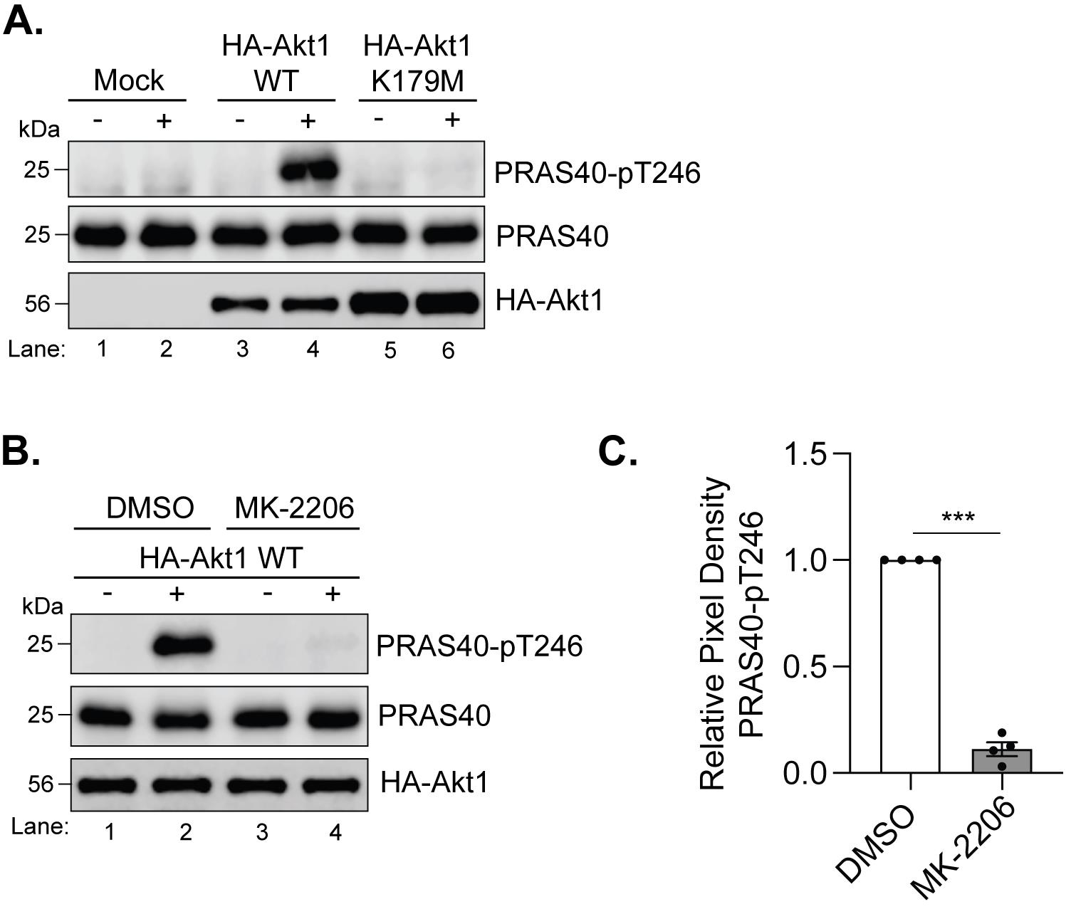
Figure 1. Immunoprecipitation-based Akt kinase assay. (A) Lysates of HEK293-T cells either mock-transfected or transfected with 5 μg of the pcDNA3.1-HA-Akt1 WT or K179M construct were immunoprecipitated with the mouse monoclonal anti-HA antibody. The immunoprecipitates were incubated with 0.5 μg of purified recombinant PRAS40 C-terminal fragment in kinase assay (+) buffer at 30 °C for 15 min. A parallel reaction was set up for each set in the kinase buffer (-) without ATP and the divalent metal cations. The reactions were analyzed by immunoblotting for PRAS40-pT246, PRAS40, and HA. (B) Lysates of HEK293-T cells transfected with 5 μg of the pcDNA3.1-HA-Akt1 WT and treated with either DMSO or 5 μM of MK-2206 (an Akt1 allosteric inhibitor) were processed for immunoprecipitation and immunoblotting as in panel A. (C) Band intensities for PRAS40-pT246 and PRAS40 from DMSO- and MK-2206-treated samples in panel B (lanes 2 and 4) were quantified from immunoblots using Image Studio Lite 6 software (Li-COR). For each experiment, the PRAS40-pT246 signal was normalized to the corresponding PRAS40 signal. Normalized values from MK-2206-treated samples were then expressed relative to the corresponding DMSO control, which was set to 1. Data from four independent experiments were analyzed, error bars represent the standard error of the mean, and statistical significance (***p < 0.001) was determined using a paired t-test in GraphPad Prism.
C. Treatment
1. Take two 50-cm sterile conical tubes and add 20 mL of fresh complete DMEM medium to each in the biosafety cabinet.
2. Add 5 μM MK-2206 to one tube and an equal volume of DMSO to the other. Label them accordingly.
Note: The 5 μM concentration of MK-2206 was selected based on prior dose-response experiments [10].
3. Take the dishes out (transfection set for Figure 1B), place them in the biosafety cabinet, and aspirate the spent medium using vacuum.
4. Label two plates as “MK-2206” and the other two as “DMSO.” Add 10 mL of the respective complete medium prepared in the 50 mL conical tubes in step C1 to the dishes.
5. Incubate dishes in a humidified 37 °C, 5% CO2 incubator for 3 h.
D. Lysis
1. With ~1 h remaining in the treatment period (section C), or at ~23 h post-transfection for untreated samples (section B), begin preparing for lysis.
a. Prepare the lysis buffer and keep it on ice.
b. Pre-chill the microcentrifuge to 4 °C.
c. Turn on the heating block to 95 °C.
2. Remove dishes from the incubator at the end of the treatment or incubation period and immediately place dishes flat on ice.
Note: From now on, the biosafety cabinet is not needed.
3. Aspirate the medium from each plate using vacuum.
4. Add 5 mL of cold DPBS per dish gently along the sides of the dishes, swirl gently, and aspirate DPBS using vacuum.
5. Tilt plates at a 45° angle on ice to collect any additional DPBS left behind. Gently aspirate the dishes a second time with vacuum.
6. Add 600 μL of cold lysis buffer to each dish.
7. Scrape cells from the dishes using a cell scraper, ensuring full coverage of the dish.
8. Collect the lysates and transfer to pre-labeled, sterile 1.7 mL microcentrifuge tubes on ice. Let the tubes incubate on ice for 10 min.
9. Place microcentrifuge tubes in the pre-chilled microcentrifuge and centrifuge at 21,130× g for 10 min at 4 °C.
10. Remove tubes carefully without disturbing the cell debris at the bottom and place them on ice.
11. Using a p1000 micropipette, transfer 550 μL of the supernatant (i.e., cleared lysate) to new, labeled, sterile 1.7 mL microcentrifuge tubes. These will be used to immunoprecipitate HA-Akt1, as described in section E.
Notes:
1. Make sure not to disturb the cell debris at the bottom when transferring supernatant.
2. Under the conditions described, 550 μL of cleared lysate from HEK293-T cells typically corresponds to ~2.5 μg/μL protein (~1.25–1.5 mg total protein input per immunoprecipitate).
12. Transfer the remaining 50 μL of the supernatant from each tube to fresh microcentrifuge tubes.
13. To these tubes, add 15 μL of 5× sample buffer, heat for 5 min at 95 °C, and store at -20 °C.
Note: These lysates can be used to check the expression level of HA-Akt1 in case troubleshooting is needed (see General notes and troubleshooting section).
E. Immunoprecipitation
1. Using a p10 micropipette, add 2 μL of the anti-HA mouse monoclonal antibody to each microcentrifuge tube containing 550 μL of the cleared lysate from step D11.
2. Seal the lids of the tubes with parafilm and incubate for ~16 h at 4 °C while gently rocking on a nutator.
3. The following day, place the tubes on ice and remove parafilm.
4. Centrifuge briefly (~30 s) at 21,130× g at 4 °C.
5. Use wide-bore pipette tips to add 20 μL of Protein G PLUS-agarose bead slurry to each sample.
Notes:
1. Protein G PLUS-agarose beads should be stored at 4 °C.
2. Make sure to mix the beads by gently pipetting up and down using a p1000 pipettor before adding 20 μL of the bead slurry to the samples.
3. Wide-bore pipette tips help ensure consistent transfer of Protein G beads, particularly in small volumes, and minimize bead loss during pipetting. If wide bore tips are not available, standard pipette tips can be cut or trimmed to widen the opening.
6. Seal the lids with parafilm again and incubate for 1 h at 4 °C while rocking gently on a nutator.
Notes:
1. During this incubation, prepare the lysis buffer for washes (you will need 15 mL for 10 tubes) and kinase assay buffers (+ and -) (prepare 130 μL of each). Keep these on ice.
2. Turn on the water bath at 30 °C and the heating block at 95 °C.
7. After 1 h, place samples on ice again and remove parafilm.
8. Centrifuge briefly (~30 s) at 21,130× g at 4 °C so the beads settle down at the bottom of the tube.
9. Attach a p200 tip to the vacuum line and very carefully aspirate, leaving behind ~50 μL of liquid above the beads.
10. Pipette 500 μL of cold lysis buffer to each tube, close, and invert the tubes 5–6 times.
11. Repeat steps E8–10 a total of three times.
12. After the final wash, switch the p200 tip with a loading tip and very carefully aspirate all remaining liquid off, taking care not to aspirate the beads.
F. Kinase assay
1. For each transfection condition (performed in duplicate plates), set up two reactions: one with 25 μL of kinase buffer (−) (no ATP, no divalent cations) and one with 25 μL of kinase buffer (+) containing ATP and divalent cations.
2. Gently tap to resuspend the beads.
3. Incubate the tubes in a 30 °C water bath for 15 min.
Note: The 15-min reaction time was selected based on our previously published time-course analysis [10], as it captures the early phase of the reaction and allows for more sensitive detection of differences in Akt activity prior to saturation (60–90 min). Comparable results were obtained at 60 min, indicating that the assay described here is robust across a 15–60 min time window in HEK293-T cells. Depending on the cell type and experimental conditions, reaction time may require optimization, and a time-course analysis is recommended when adapting the assay to new cell lines.
4. After incubation, add 6.25 μL of 5× sample buffer.
5. Heat samples at 95 °C for 5 min.
Pause point: Samples can be stored at -20 °C if not immediately running SDS-PAGE.
G. SDS-PAGE and western blotting
Note: SDS-PAGE and western blotting (i.e., immunoblotting) are well-established techniques. Detailed step-by-step protocols are provided in [13]. The steps below focus on those specific to the analysis here.
1. Make 10% resolving/5% stacking gels with a 1.5 mm spacer and 15-well combs.
Note: Pre-made gels can be purchased instead of handmade gels.
2. Electrophorese samples using 1× running buffer at constant voltage of 100 V until the samples enter the resolving gel, and then increase the voltage to 120 V until the dye front reaches the bottom of the gel.
Note: Make sure the 25-kDa marker of the protein ladder stays in the gel, as the PRAS40 antigen is ~26 kDa. See further explanation in step G5.
3. From the kinase assay samples, load the gels in two sets.
a. The first set, with 10 μL samples each in the following order (this set is used for detection of PRAS40-pT246 and HA):
Panel A: Ladder (1 μL), 1× sample buffer, mock (-), mock (+), WT (-), WT (+), K179 (-), K179 (+), 1× sample buffer, ladder (1 μL).
Panel B: Ladder (1 μL), 1× sample buffer, DMSO (-), DMSO (+), MK2206 (-), MK2206 (+), 1× sample buffer, ladder (1 μL).
b. The second set, with 2.5 μL samples each in the same order as above. This set is used for the detection of total PRAS40.
Note: Serial dilution experiments established that 10 μL of sample provides a signal within the linear range and is sufficient for reliable detection of PRAS40-pT246. For PRAS40, 2.5 μL is sufficient for detection due to its higher signal intensity, which helps avoid saturation. The remaining sample can be retained for any potential repeat immunoblotting analyses if needed due to technical issues.
4. Transfer the gel to an activated Polyvinylidene Fluoride Immobilon FL transfer membrane using cold 1× transfer buffer at 100 V for 60 min. Keep the transfer unit on ice.
5. After the transfer is complete, remove the membrane and place it on a plastic wrap on a cutting board. Using a ruler, cut the membrane for the first set of samples horizontally between protein markers 20–37 kDa and 37–100 kDa for PRAS40-pT246 and HA antibodies, respectively. Cut the membrane for the second set of samples horizontally between protein markers 20–37 kDa for PRAS40.
Note: The recombinant PRAS40 protein used in this assay is a carboxyl-terminal fragment (residues 182–256) that retains the Akt phosphorylation site, T246. This commercially available fragment provides a convenient and consistent substrate for the assay. The protein fragment has an apparent molecular mass of ~26 kDa, which should be considered when performing and interpreting western blots.
6. Label blots on the side and incubate in 5% dry non-fat milk in 1× PBS for 30 min at room temperature, shaking gently.
7. Perform three 5-min washes in 1× PBS-T at room temperature, shaking gently.
8. Incubate blots for 16–18 h while rocking at 4 °C in the appropriate primary antibody diluted in 1× PBS-T.
a. Anti-HA mouse monoclonal antibody, 1:500.
b. Anti-phospho-PRAS40 (Thr246) rabbit monoclonal antibody, 1:1,000.
c. Anti-PRAS40 rabbit monoclonal antibody, 1:1,000.
9. After incubation, perform three 5-min washes in 1× PBS-T at room temperature, shaking gently.
10. Incubate blots in appropriate secondary antibody and incubate for 1 h at room temperature while rocking gently. Use the light-protecting blot containers to avoid photobleaching of the fluorophores attached to the secondary antibodies.
a. IRDye® 680RD goat anti-mouse IgG, 1:5,000 (for the anti-mouse HA primary antibody blot).
b. IRDye® 680RD goat anti-rabbit IgG, 1:5,000 (for the anti-rabbit PRAS40 and PRAS40-pT246 primary antibody blots).
11. After incubation, perform three 5-min washes in 1× PBS-T at room temperature, shaking gently.
12. Perform a last wash for 5 min in 1× PBS before developing the blots using the Li-COR Odyssey XF system.
Validation of protocol
This protocol has been used and validated in the following research article(s):
• Palma et al. [10]. Substrate preference of protein kinase B isoforms can vary depending on the cell line. PLoS One (Figure 11).
General notes and troubleshooting
General notes
1. Always wear appropriate personal protective equipment (PPE)—lab coat, gloves, and safety glasses—while performing this protocol.
2. Perform all cell culture steps described in sections A–C in a certified biosafety cabinet. Use only cell culture–grade dishes and sterile, individually wrapped serological pipettes, and always use sterile, barrier pipette tips. Because complete DMEM and trypsin-EDTA are stored at 4 °C, prewarm them to 37 °C in a water bath or heating block for approximately 15 min before use. Prior to placement in the biosafety cabinet, spray all bottles, pipette aids, and pipettors with 70% ethanol.
3. To minimize potential changes in activity during sample processing, all lysis and immunoprecipitation steps must be performed under cold conditions using pre-chilled buffers and equipment.
4. This protocol can be adapted to and optimized for other cell lines.
5. Methanol is toxic and flammable. Handle carefully and dispose of methanol-containing waste according to institutional safety guidelines.
6. Unpolymerized acrylamide is a neurotoxin and a suspected carcinogen. Handle unpolymerized acrylamide solutions with care and avoid skin contact and inhalation. Dispose of acrylamide-containing waste according to institutional safety guidelines.
7. SDS is an irritant and is harmful if inhaled or in contact with skin or eyes. Handle SDS-containing solutions with care and avoid generating dust or aerosols when weighing it.
8. DTT is a strong reducing agent and is harmful if swallowed or in contact with skin or eyes.
9. β-ME is a strong reducing agent and has a strong odor. Add β-mercaptoethanol to the 5× sample buffer solutions in a chemical fume hood and avoid inhalation or skin contact.
10. This assay provides a semiquantitative readout of catalytic activity from transiently expressed Akt1 and should be interpreted within the context of defined experimental conditions. This assay has not been validated for the detection of endogenous Akt activity.
11. Reagent batch effects should be considered, as variability in antibody performance, serum composition, or other key reagents may impact Akt immunoprecipitation efficiency and downstream substrate phosphorylation. Where possible, consistent reagent lots should be used, and new batches should be empirically validated to ensure comparable performance.
12. Immunoblot-based detection may be subject to signal saturation; therefore, signal linearity should be verified, and appropriate sample volumes should be used.
13. Because the assay relies on immunoprecipitated kinase, co-immunoprecipitated proteins may contribute to substrate phosphorylation; inclusion of appropriate controls (e.g., mock-transfected and catalytically inactive construct) is essential to confirm specificity.
14. This assay uses a relatively high ATP concentration, consistent with standard nonradioactive kinase assays. As such, the assay is better suited for evaluating allosteric inhibitors than ATP-competitive inhibitors.
15. The use of Mn2+ in the kinase reaction is consistent with common in vitro kinase assay conditions, although divalent cation composition (Mn2+ vs. Mg2+) can influence kinase activity and thus should be kept consistent across experiments.
16. While absolute IP efficiency may vary between independent experiments, the inclusion of matched controls within each experimental set allows for reliable comparison of Akt activity under the conditions tested.
17. The assay can, in principle, be adapted to substrates other than PRAS40 that contain defined Akt phosphorylation sites and have suitable phospho-specific antibodies available for detection. However, substrate selection should consider specificity, as Akt consensus motifs are shared with other kinases. PRAS40 was selected here as a validated and readily available Akt substrate with a well-characterized phospho-specific antibody.
Troubleshooting
Problem 1: Uneven signal for HA-Akt1 in the immunoprecipitates across samples.
Possible causes: Uneven amount of Protein G beads added and/or loss of Protein G beads during wash steps. Another reason could be variable transfection efficiency across plates.
Solutions: Make sure to resuspend beads thoroughly before dispensing. Since the packed bead volume from a 20 μL slurry, even though sufficient to immunoprecipitate, can be difficult to see, you can add a higher amount of bead slurry (30 μL) per tube so it is easier to see the bead pellet. If substantial variation in HA-Akt1 levels is observed, comparisons of kinase activity across samples may be confounded by differences in immunoprecipitation efficiency and should not be considered reliable without further optimization. To determine whether variability arises from transfection efficiency, lysates saved in step D13 can be analyzed by western blotting for HA and a loading control (e.g., actin). Maintaining standardized cell seeding density, confluency at transfection, transfection duration, and consistent handling during lysis, immunoprecipitation, and wash steps promotes consistent HA-Akt1 levels across samples and reduces variability in immunoprecipitation efficiency.
Problem 2: Weak/absent signal in all blots.
Possible causes: Transfer orientation error, poor antibody performance.
Solutions: If not even the protein ladder shows up, it is likely a transfer orientation error. If the protein ladder shows up fine, make a fresh antibody dilution of secondary antibodies. Make sure the tubes/containers containing secondary antibody dilutions are either opaque or wrapped with aluminum foil to protect from light.
Acknowledgments
Conceptualization, D.B.; Investigation, A.P. and J.M.; Writing—Original Draft, A.P., J.M., and D.B.; Writing—Review & Editing, D.B.; Funding acquisition, D.B.; Supervision, D.B. This work was supported by the National Institute of General Medical Sciences (NIGMS) grant SC3GM139707 to D.B. A.P. is currently supported by the NIH-NIGMS Bridges to the Doctorate fellowship #T32GM1380175. The graphical abstract was created with Biorender.com. This protocol has been validated in and is adapted from Palma et al. [10].
Competing interests
The authors declare no conflicts of interest.
References
- Manning, B. D. and Toker, A. (2017). AKT/PKB Signaling: Navigating the Network. Cell. 169(3): 381–405. https://doi.org/10.1016/j.cell.2017.04.001
- Alessi, D. R., Andjelkovic, M., Caudwell, B., Cron, P., Morrice, N., Cohen, P. and Hemmings, B. A. (1996). Mechanism of activation of protein kinase B by insulin and IGF-1. EMBO J. 15(23): 6541–6551. https://doi.org/10.1002/j.1460-2075.1996.tb01045.x
- Frech, M., Andjelkovic, M., Ingley, E., Reddy, K. K., Falck, J. R. and Hemmings, B. A. (1997). High Affinity Binding of Inositol Phosphates and Phosphoinositides to the Pleckstrin Homology Domain of RAC/Protein Kinase B and Their Influence on Kinase Activity. J Biol Chem. 272(13): 8474–8481. https://doi.org/10.1074/jbc.272.13.8474
- Alessi, D. R., James, S. R., Downes, C., Holmes, A. B., Gaffney, P. R., Reese, C. B. and Cohen, P. (1997). Characterization of a 3-phosphoinositide-dependent protein kinase which phosphorylates and activates protein kinase Bα. Curr Biol. 7(4): 261–269. https://doi.org/10.1016/s0960-9822(06)00122-9
- Stokoe, D., Stephens, L. R., Copeland, T., Gaffney, P. R. J., Reese, C. B., Painter, G. F., Holmes, A. B., McCormick, F. and Hawkins, P. T. (1997). Dual Role of Phosphatidylinositol-3,4,5-trisphosphate in the Activation of Protein Kinase B. Science. 277(5325): 567–570. https://doi.org/10.1126/science.277.5325.567
- Sarbassov, D. D., Guertin, D. A., Ali, S. M. and Sabatini, D. M. (2005). Phosphorylation and Regulation of Akt/PKB by the Rictor-mTOR Complex. Science. 307(5712): 1098–1101. https://doi.org/10.1126/science.1106148
- Yang, J., Cron, P., Good, V. M., Thompson, V., Hemmings, B. A. and Barford, D. (2002). Crystal structure of an activated Akt/Protein Kinase B ternary complex with GSK3-peptide and AMP-PNP. Nat Struct Biol. 9(12): 940–944. https://doi.org/10.1038/nsb870
- Pervanidis, K. A., D’Angelo, G. D., Weisner, J., Brandherm, S. and Rauh, D. (2024). Akt Inhibitor Advancements: From Capivasertib Approval to Covalent-Allosteric Promises. J Med Chem. 67(8): 6052–6063. https://doi.org/10.1021/acs.jmedchem.4c00075
- Calleja, V., Laguerre, M., Parker, P. J., Larijani, B. (2009). Role of a novel PH‐kinase domain interface in PKB/Akt regulation: structural mechanism for allosteric inhibition. PLoS Biol. 7: e17. https://doi.org/10.1371/journal.pbio.1000017
- Palma, M. S., Perez, S. R., Husain, A., Bhandari, D. (2024). Substrate preference of protein kinase B isoforms can vary depending on the cell line. PLoS One. 19(3): e0298322. https://doi.org/10.1371/journal.pone.0298322
- Lee, M. S., Jeong, M. H., Lee, H. W., Han, H. J., Ko, A., Hewitt, S. M., Kim, J. H., Chun, K. H., Chung, J. Y., Lee, C., et al. (2015). PI3K/AKT activation induces PTEN ubiquitination and destabilization accelerating tumourigenesis. Nat Commun. 6(1): e1038/ncomms8769. https://doi.org/10.1038/ncomms8769
- Zeng, M., van der Donk, W. A. and Chen, J. (2014). Lanthionine synthetase C–like protein 2 (LanCL2) is a novel regulator of Akt. Mol Biol Cell. 25(24): 3954–3961. https://doi.org/10.1091/mbc.e14-01-0004
- Sambrook, J., Russell, D. W. (2001). Molecular Cloning: A Laboratory Manual. 3rd edition. Cold Spring Harbor, NY: Cold Spring Harbor Laboratory Press.
Article Information
Publication history
Received: Jan 19, 2026
Accepted: May 10, 2026
Available online: May 28, 2026
Published: Jul 5, 2026
Copyright
© 2026 The Author(s); This is an open access article under the CC BY-NC license (https://creativecommons.org/licenses/by-nc/4.0/).
How to cite
Peek, A., Mehta, J. N. and Bhandari, D. (2026). An Immunoprecipitation-Based Nonradioactive Kinase Assay to Measure Akt Kinase Activity in Mammalian Cell Lines. Bio-protocol 16(13): e5724. DOI: 10.21769/BioProtoc.5724.

 Download Citation in RIS Format
Category
Biochemistry > Protein > Activity
Biochemistry > Protein > Posttranslational modification
Cell Biology > Cell signaling > Phosphorylation
Do you have any questions about this protocol?
Post your question to gather feedback from the community. We will also invite the authors of this article to respond.
 Post a Question


