


- Protocols

- Articles and Issues
- For Authors
- About
- Become a Reviewer


NADH-Dependent Oxidoreductase Activity Assay of OsAIM1 Using a Microplate Reader

(*contributed equally to this work) Published: Vol 16, Iss 13, Jul 5, 2026 DOI: 10.21769/BioProtoc.5722 Views: 252
Reviewed by: Shweta PanchalFélix de CarpentierYue Xi
 PDF
PDF  Ask a question
Original research article
The authors used this protocol in:

Feb 2026
Advertisement


Protocol Collections
Comprehensive collections of detailed, peer-reviewed protocols focusing on specific topics




Abstract
Peroxisomal β-oxidation is a key step in jasmonic acid biosynthesis. Quantitative biochemical characterization of enzymes involved in the β-oxidation pathway is essential for validating their catalytic functions and comparing differences among genetic variants. Existing enzyme activity assays largely rely on chromatographic techniques to quantify substrate consumption or product formation, but these approaches are not well-suited for high-throughput or continuous kinetic measurements. Here, we describe a spectrophotometric assay based on a plate reader determining OsAIM1 enzymatic activity by monitoring the decrease in NADH absorbance at 340 nm. The method employs a 96-well plate reaction system, enabling real-time kinetic measurements and providing a standardized workflow for calculating reaction rates. Reaction components, protein concentration ranges, and data processing parameters were systematically optimized to ensure linearity, reproducibility, and quantitative accuracy. This assay is simple to perform, requires small reaction volumes, and offers relatively high throughput, making it suitable for functional characterization and kinetic analysis of NADH-dependent enzymes.
Key features
• Enzyme activity assay based on NADH absorbance changes, enabling real-time measurement.
• Conducted in a 96-well plate format with a low reaction volume and minimal protein requirement.
• Allows calculation of reaction rates and specific activity with good reproducibility.
• Applicable to NADH-dependent enzymes and suitable for comparison between different samples.
Keywords: Spectrophotometric assay
 Enzyme activity measurement
Plate reader assay
NADH oxidation
High-throughput analysis
Enzyme activity measurement
Plate reader assay
NADH oxidation
High-throughput analysis
Graphical overview
Background
β-oxidation, an enzymatic process in which fatty acids are sequentially converted into acetyl-CoA, plays a key role in the biosynthesis of jasmonic acid (JA) and salicylic acid (SA) [1]. JA and SA play important roles in plant growth, development, and stress responses. Therefore, quantitative biochemical characterization of enzymes in the β-oxidation pathway is essential for linking genetic findings to catalytic function.
OsAIM1 (ABNORMAL INFLORESCENCE MERISTEM1) encodes a peroxisome-localized multifunctional enzyme that functions in the β-oxidation pathway. For recombinant expression, OsAIM1 was cloned into a pGEX-4T-2 vector and expressed in Escherichia coli as a GST (glutathione S-transferase) fusion protein, followed by affinity purification. It catalyzes the hydratase–dehydrogenase steps of acyl-CoA intermediates, thereby contributing to chain-shortening reactions in peroxisomes. In plants, this pathway is involved not only in fatty acid turnover but also in the production of benzoic acid–derived metabolites such as SA [2–3]. During catalysis, enzymes of this class are typically coupled to the conversion of NADH to NAD+. Because NADH exhibits a characteristic absorbance peak at 340 nm, whereas NAD+ shows minimal absorbance at this wavelength, enzymatic reaction rates can be continuously monitored by measuring the decrease in absorbance at 340 nm [4].
At present, activity assays for enzymes involved in β-oxidation commonly rely on high-performance liquid chromatography or mass spectrometry to quantify substrate consumption or product formation [5–8]. Despite their high sensitivity, these methods are inherently discontinuous, as they require intermittent sampling, reaction quenching, and offline analysis. Such procedures interrupt the reaction process and limit the ability to capture rapid changes over time, thereby preventing continuous, real-time monitoring of enzyme kinetics. In addition, the relatively low throughput and labor-intensive workflows further restrict their applicability in dynamic or high-throughput kinetic analyses. In contrast, spectrophotometric assays based on changes in NADH absorbance can be performed on a plate reader platform, enabling real-time detection in small reaction volumes and parallel measurement of multiple samples.
In this study, we describe an NADH-dependent enzyme activity assay using a plate reader and provide a detailed description of the reaction setup and the calculation of enzymatic reaction rates to improve reproducibility and quantitative accuracy. This method can be extended to the functional analysis of other NADH-dependent enzymes, such as 3-hydroxyacyl-CoA dehydrogenase and alcohol dehydrogenase [9–11].
Materials and reagents
Biological materials
1. DH5α chemically competent Escherichia coli cells (Tsingke Biotechnology Co., Ltd., catalog number: TSC-C14)
2. BL21 (DE3) Escherichia coli cells (Tiangen Biotech, catalog number: CB105-02)
3. pGEX-4T-2 vector
Reagents
1. Ampicillin (Aladdin, catalog number: A408319)
2. Yeast extract (Biomed, catalog number: LP0021)
3. Tryptone (Biomed, catalog number: LP0042)
4. Sodium chloride (NaCl) (Sigma-Aldrich, catalog number: S9625)
5. Agar powder (Solarbio, catalog number: A8190)
6. Isopropyl-β-D-thiogalactopyranoside (IPTG) (Sigma-Aldrich, catalog number: 367-93-1)
7. Phosphate-buffered saline (powder) buffer (PBS) (Pytbio, catalog number: FZ1055)
8. Triton X-100 (Sigma-Aldrich, catalog number: X-100)
9. L-Glutathione reduced (Sigma-Aldrich, Abcam, catalog number: 70-18-8)
10. Tris (Solarbio, catalog number: T8060)
11. Hydrochloric acid (HCl) (CaymanChemical, catalog number: 401137-50)
12. Bovine serum albumin (BSA) (Sigma-Aldrich, catalog number: A7906)
13. Nicotinamide adenine dinucleotide (reduced form) (NADH) (MCE, catalog number: 606-68-8)
14. Potassium dihydrogen phosphate (KH2PO4) (Aladdin, catalog number: P113041)
15. Potassium hydroxide (KOH) (Sigma-Aldrich, catalog number: 306568)
16. Acetoacetyl-CoA sodium salt (MCE, catalog number: HY-N7392A)
17. GST magnetic agarose (Solarbio, catalog number: M2320)
18. Ampicillin (MCE, catalog number: HY-B0522)
19. Protein loading buffer (SDS, 4×) (MREDA, catalog number: M212252-5ml)
Solutions
1. LB culture medium (see Recipes)
2. 50 mM Tris-HCl (see Recipes)
3. 20 mg/mL BSA (see Recipes)
4. 1 M KH2PO4, pH 7.0 (see Recipes)
5. 5.45 mM acetoacetyl-CoA (see Recipes)
6. 10 mM NADH (see Recipes)
7. Reaction buffer (see Recipes)
Recipes
1. LB culture medium
| Reagent | Quantity or volume |
|---|---|
| Yeast extract | 0.5 g |
| Tryptone | 1 g |
| NaCl | 1 g |
| ddH2O | To 100 mL |
For solid medium, add 1.5 g of agar. Sterilize at 121 °C for 20 min and store at 4 °C.
2. 50 mM Tris-HCl
| Reagent | Quantity or volume |
|---|---|
| Tris | 6.06 g |
| ddH2O | To 1 L |
Adjust the pH to 8.0 with concentrated HCl at room temperature (25 °C). Store at 4 °C.
3. 20 mg/mL BSA
| Reagent | Quantity or volume |
|---|---|
| BSA | 20 mg |
| ddH2O | To 1 mL |
Store at -20 °C.
4. 1 M KH2PO4, pH 7.0
| Reagent | Quantity or volume |
|---|---|
| KH2PO4 | 0.136 g |
| ddH2O | To 1 mL |
Adjust the pH to 7.0 with concentrated KOH at room temperature. Store at 4 °C.
5. 5.45 mM acetoacetyl-CoA
| Reagent | Quantity or volume |
|---|---|
| Acetoacetyl-CoA sodium salt | 0.005 g |
| ddH2O | To 1 mL |
Store at -20 °C.
6. 10 mM NADH
| Reagent | Quantity or volume |
|---|---|
| Nicotinamide adenine dinucleotide | 0.007 g |
| ddH2O | To 1 mL |
Store at -20 °C protected from light.
7. Reaction buffer
| Reagent | Final concentration | Quantity or volume |
|---|---|---|
| 20 mg/mL BSA | 0.2 mg/mL | 1 μL |
| 1 M KH2PO4 | 0.2 M | 20 μL |
| 5.45 mM acetoacetyl-CoA | 30 μM | 0.55 μL |
| ddH2O | n/a | 78.45 μL |
| Total | n/a | 100 μL |
Prepare fresh immediately before use and do not store.
Laboratory supplies
1. 1.5, 2.0, and 50 mL tubes
2. Magnetic rack (Aikerui Bioengineering Co., Ltd., catalog number: AG21301)
3. 96-well EIA/RIA plate (Corning, catalog number: 3590)
4. Erlenmeyer flask (Sigma-Aldrich, catalog number: Z567868)
5. Petri dish (Gadro, catalog number: GH10078)
Equipment
1. Low-temperature ultra-high-pressure continuous-flow cell disruptor (JNBIO, model: JN-Mini pro)
2. Microplate reader (Molecular Devices, LLC, model: Spectra M3)
3. NanoDrop (Thermo Fisher Scientific Inc, model: 840-317500)
4. Thermal cycler (Sigma-Aldrich, model: Z742467)
5. Water bath (POMEX, model: BM14099)
6. Autoclave
7. Laminar flow hood
8. Shaking incubator
Procedure
A. Expression and purification of recombinant OsAIM1 in E. coli
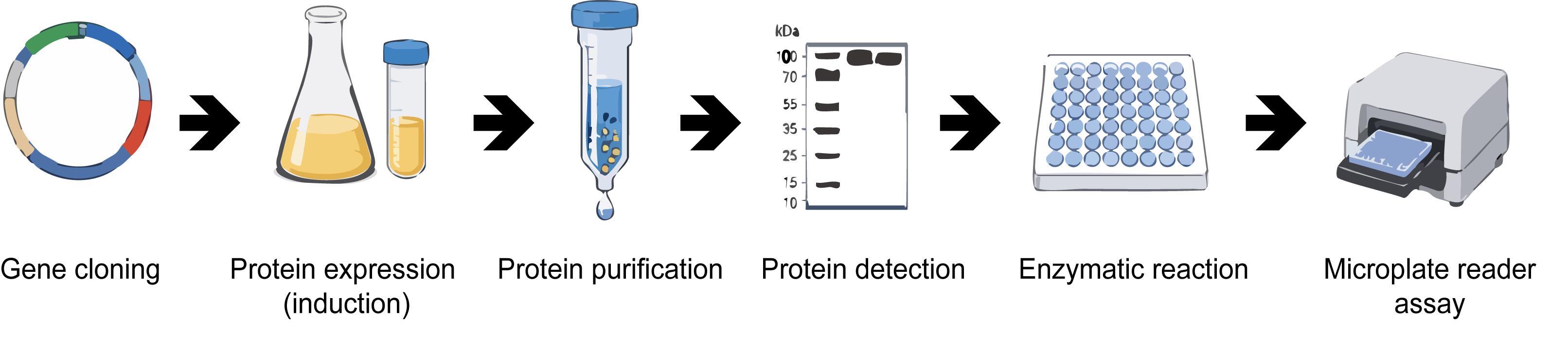
1. Clone the coding sequence of OsAIM1 into the pGEX-4T-2 vector for expression as an N-terminal GST fusion protein (Figure 1A), which facilitates soluble expression and enables efficient one-step affinity purification.
2. Confirm the integrity of the recombinant plasmid by Sanger sequencing using vector-specific primers (PGEX-5′) (Figure 1B).
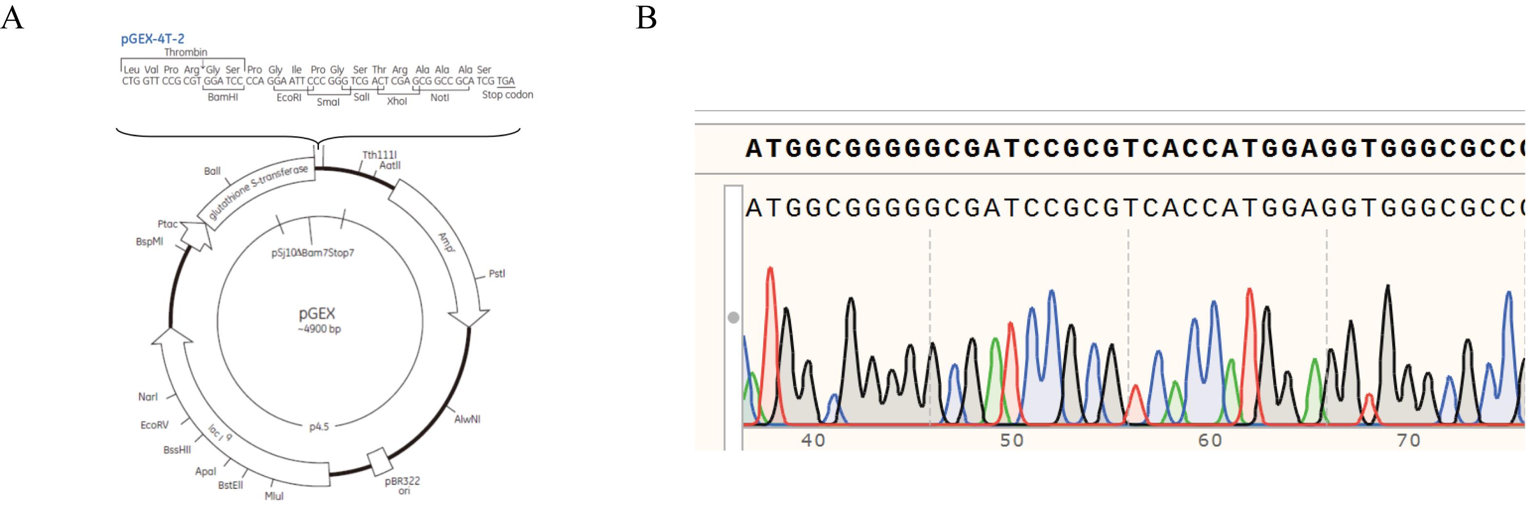
Figure 1. Construction and validation of the GST-OsAIM1 expression vector. (A) Schematic representation of the pGEX-4T-2 vector used for generating the N-terminal GST (glutathione S-transferase) fusion construct. (B) Sanger sequencing chromatogram and sequence alignment confirming the correct in-frame insertion and integrity of the GST-OsAIM1 expression construct.
3. Transform the verified plasmid into E. coli BL21 (DE3) competent cells and plate on LB agar containing 100 μg/mL ampicillin. Incubate overnight at 37 °C.
4. Inoculate a single colony into 600 μL of LB medium in a sterile 1.5- or 2.0-mL microcentrifuge tube and culture overnight at 37 °C with shaking at 220 rpm.
5. Transfer 600 μL of the overnight culture into 100 mL of LB medium containing ampicillin (100 μg/mL). Incubate at 37 °C with shaking at 220 rpm until OD600 reaches 0.6–0.8.
6. Add IPTG to a final concentration of 0.3 mM to induce protein expression. Continue incubation at 16 °C with shaking at 110 rpm for 20 h.
7. Collect 1 mL of culture before and after induction, pellet cells, and resuspend in SDS loading buffer. Analyze protein expression by SDS-PAGE to confirm induction of the GST-OsAIM1 fusion protein (~expected molecular weight) (Figure 2A).
8. Harvest cells by centrifugation at 8,228× g for 5–10 min at 4 °C.
9. Discard the supernatant and resuspend the pellet in 30 mL of pre-chilled PBS. Vortex thoroughly to ensure complete resuspension.
10. Centrifuge again at 8,228× g for 5–10 min at 4 °C and resuspend the pellet in 30 mL of pre-chilled PBS.
11. Lyse the cells using a low-temperature ultra-high-pressure continuous-flow cell disruptor for approximately 5 min. Centrifuge the lysate at 10,414× g for 10 min at 4 °C and transfer the supernatant to a new 50-mL centrifuge tube.
12. Add 100 μL of GST magnetic beads into a 1.5- or 2.0-mL microcentrifuge tube and wash 3–5 times with 1 mL of pre-chilled PBS. Add the washed beads to the supernatant and incubate at 4 °C with rotation for 2–4 h.
13. Collect the beads using a magnetic rack and wash 5–10 times with 1 mL of pre-chilled PBS containing 0.3% Triton X-100.
14. Elute the bound protein by adding 300 μL of 10 mM reduced glutathione dissolved in 50 mM Tris-HCl buffer (pH 8.0).
15. Analyze eluted fractions by SDS-PAGE to assess protein purity. Optionally, perform western blotting using anti-GST antibodies to confirm protein identity (Figure 2B).
16. Measure protein concentration using a NanoDrop spectrophotometer and store the purified protein at -80 °C.
Note: The overall recovery after GST affinity purification was estimated to be in the range of 20%–50%.
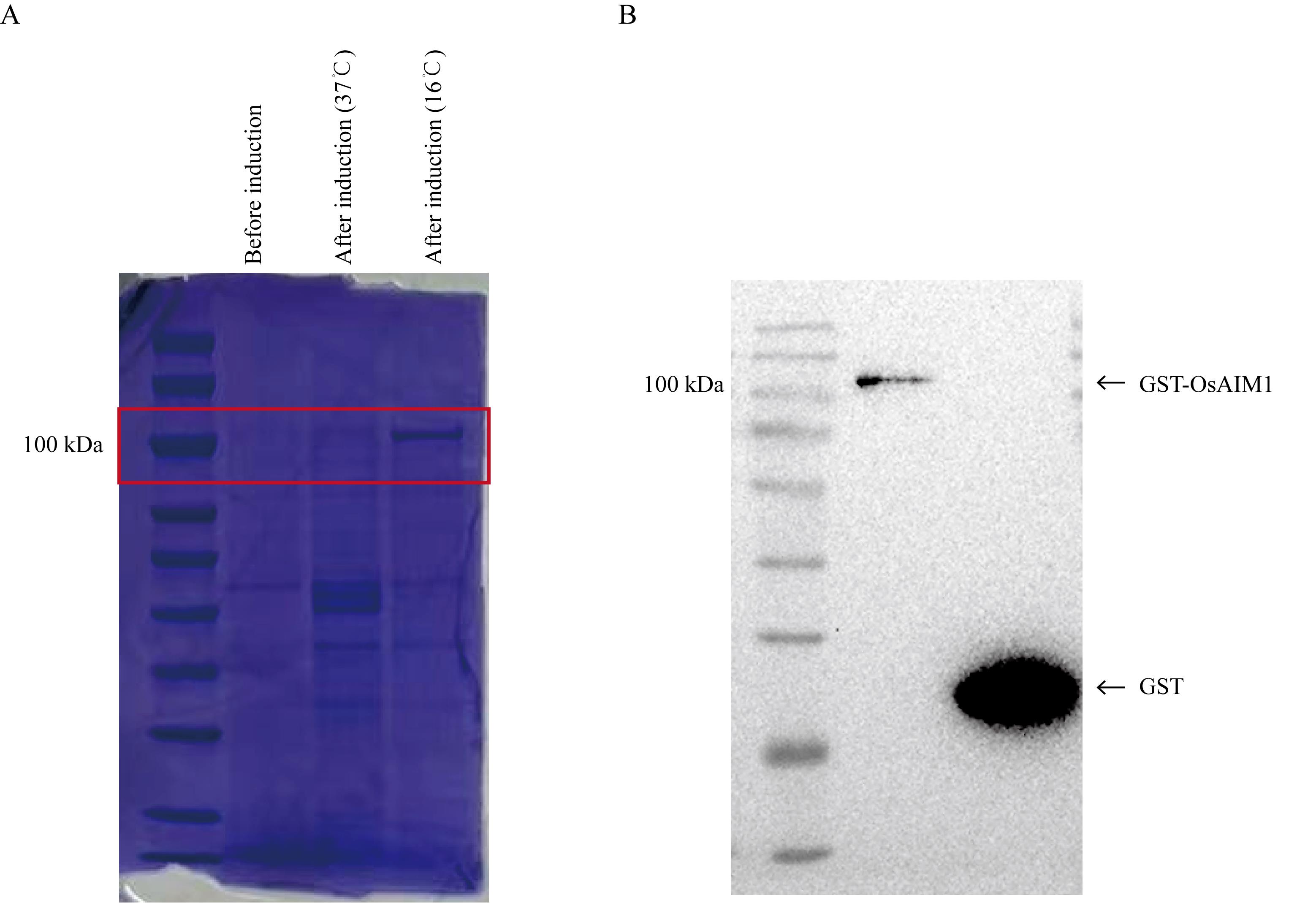
Figure 2. Protein expression and purification analysis. (A) SDS-PAGE analysis of the GST-OsAIM1 fusion protein before and after induction at two different temperatures. The red box indicates the position of GST-OsAIM1. (B) Western blot analysis of the purified GST-OsAIM1 fusion protein and the purified GST (glutathione S-transferase) control.
B. Enzymatic activity assay of 3-hydroxyacyl-CoA dehydrogenase
1. Add 100 μL of reaction buffer to each well of a 96-well microplate at room temperature (25 °C). The reaction buffer contains acetoacetyl-CoA (final concentration 30 μM) as the substrate. The assay is based on the reversible NADH-dependent reduction of acetoacetyl-CoA to 3-hydroxyacyl-CoA, accompanied by the oxidation of NADH to NAD+, consistent with the known activity of 3-hydroxyacyl-CoA dehydrogenases (EC 1.1.1.35) [12]. Subsequently, add 1 μL of a 10 mM NADH solution to each well to obtain a final NADH concentration of 0.1 mM in the reaction mixture. Gently mix the solution by pipetting up and down several times to ensure thorough homogenization while avoiding the introduction of air bubbles.
2. Initiate the reaction by adding 0.5 μg of purified OsAIM1 protein to each well containing the reaction mixture. Immediately mix gently by pipetting to ensure uniform distribution of the enzyme in the reaction solution. For the negative control, add 0.5 μg of purified GST protein to parallel wells under identical conditions and add an equal volume of reaction buffer without enzyme to account for non-enzymatic NADH oxidation.
3. Because the enzymatic reaction begins immediately after enzyme addition and proceeds rapidly, the microplate should be placed into the microplate reader without delay after mixing.
4. Measure the absorbance at 340 nm, which corresponds to the characteristic absorption peak of NADH. Record absorbance values every 15 s for the duration of the assay. The enzymatic reaction rate is determined from the linear decrease in absorbance at 340 nm over time (Figure 3), reflecting NADH oxidation during the reaction.
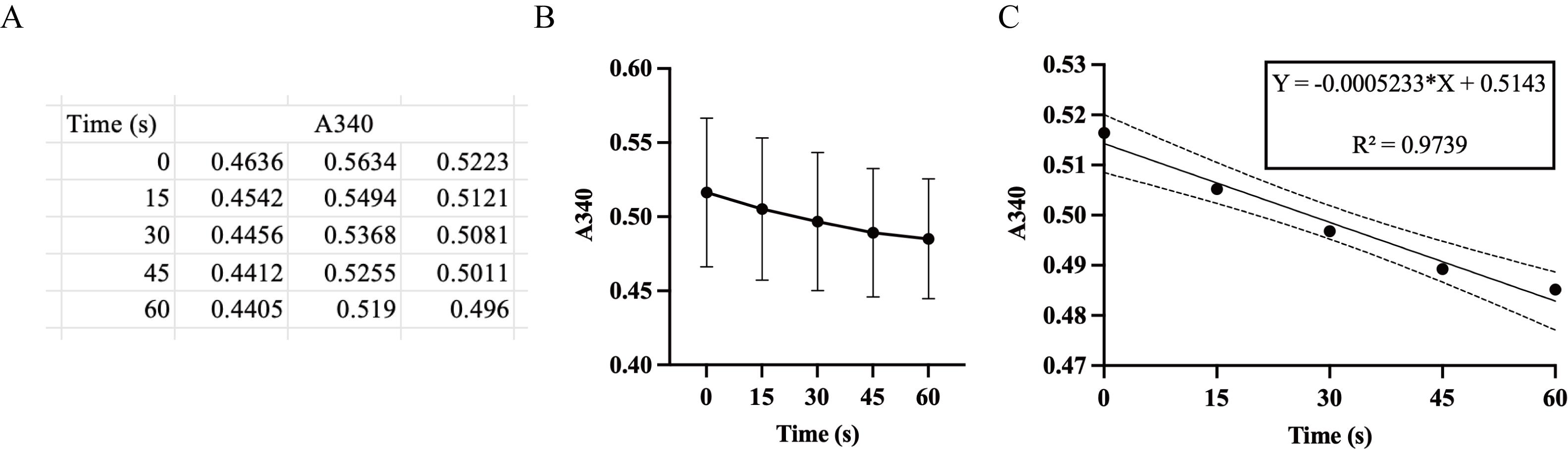
Figure 3. Measurements and calculation of the reaction rate. (A, B) Time-dependent changes in absorbance at 340 nm over a 60 s interval (A) and the corresponding kinetic curves (B), derived from three independent replicates. Values are presented as means ± SD (n = 3). (C) Linear regression analysis based on the mean values of the three replicates; dashed lines denote the 95% confidence interval of the fitted regression.
Data analysis
Absorbance values at 340 nm were exported from the plate reader. For each sample, the absorbance at the initial time point (t = 0) was defined as A0, and the absorbance at subsequent time points was defined as At. Reaction kinetics were visualized by plotting absorbance at 340 nm against time. A progressive decrease in absorbance reflects the oxidation of NADH during the enzymatic reaction.
The reaction rate was determined from the linear portion of the absorbance decrease curve. Specifically, the rate was calculated as the slope of the absorbance change over time:
Reaction rate (∆A340/min) = (A0 - At)/∆t
The product formation rate was calculated using the molar extinction coefficient of NADH:
Rate (μM·min-1) = (∆A340/min)/(ε × l) × 103
where ε is the molar extinction coefficient of NADH (6.22 mM-1·cm-1), and l is the effective path length (cm).
The initial reaction rate (v0) was obtained from the linear phase and expressed as follows:
v0 (μM·s-1) = Rate/60
The specific activity of the enzyme was calculated as follows:
Specific activity (μmol·min-1·mg-1) = (Rate × V)/protein
where V is the reaction volume (L), and protein is the amount of enzyme used in the assay (mg).
Validation of protocol
This protocol has been used and validated in the following research article:
Hu et al. [13]. The multifunctional protein OsAIM1 regulates floret opening and closure timing via jasmonic acid-mediated lodicule dynamics in rice. Plant physiology (Figure 8e)
General notes and troubleshooting
General notes
1. The enzyme concentration and reaction time should be optimized to ensure that the decrease in absorbance at 340 nm remains within the linear range (0.02–0.3). Excess enzymes may result in rapid NADH depletion and loss of linearity.
2. NADH is light-sensitive and prone to oxidation. Prepare fresh NADH solutions before use and minimize exposure to light during reaction setup.
3. Ensure that the plate reader is properly calibrated for 340 nm detection. If available, path length correction should be enabled or kept consistent between experiments to reduce variability.
4. Perform all reactions at the same temperature (25 °C) to avoid variability in enzymatic rates. Significant temperature fluctuations may affect reaction kinetics.
Troubleshooting
Problem 1: Absorbance decreases too rapidly, making slope calculation unreliable.
Possible causes: Excess enzyme concentration or NADH concentration too low.
Solutions: Reduce enzyme amount or increase the initial NADH concentration while ensuring a linear detection range.
Problem 2: High variability between replicate wells.
Possible causes: Inconsistent pipetting or delay between enzyme addition and measurement.
Solution: Use multichannel pipettes when possible and immediately place the plate into the reader after enzyme addition.
Acknowledgments
Conceptualization, J.W. and S.Z.; Investigation, Y.H., S.C., H.L., H.H., Z.X., B.H., L.C., L.Z., J.W., K.C., W.L., W.S.; Writing—Original Draft, Y.H.; Writing—Review & Editing, J.W. and S.Z.; Funding acquisition, J.W. and S.Z.; Supervision, J.W., S.Z., L.J., Z.Z., C.L., S.Z., Y.T., X.L., S.L., L.C. Funding support from the Biological Breeding-National Science and Technology Major Project (2024ZD04080), the Foundation of Biological Breeding Zhongshan Lab (ZSBBL-KY2023-07), the Jiangsu Provincial Key Research and Development Program (BE2023362), and the “JBGS” Project of Seed Industry Revitalization in Jiangsu Province [JBGS (2021) 007]. This protocol has been described and validated in the original research paper by Hu et al. [13].
Competing interests
The authors declare no conflicts of interest.
References
- Kao, Y. T., Gonzalez, K. L. and Bartel, B. (2017). Peroxisome Function, Biogenesis, and Dynamics in Plants. Plant Physiol. 176(1): 162–177. https://doi.org/10.1104/pp.17.01050
- Richmond, T. A. and Bleecker, A. B. (1999). A Defect in b-Oxidation Causes Abnormal Inflorescence Development in Arabidopsis. Plant Cell. 11(10): 1911. https://doi.org/10.2307/3871086
- Xu, L., Zhao, H., Ruan, W., Deng, M., Wang, F., Peng, J., Luo, J., Chen, Z. and Yi, K. (2017). ABNORMAL INFLORESCENCE MERISTEM1 Functions in Salicylic Acid Biosynthesis to Maintain Proper Reactive Oxygen Species Levels for Root Meristem Activity in Rice. Plant Cell. 29(3): 560–574. https://doi.org/10.1105/tpc.16.00665
- Binstock, J. F. and Schulz, H. (1981). Fatty acid oxidation complex from Escherichia coli. Meth Enzymol. 71: 403–411. https://doi.org/10.1016/0076-6879(81)71051-6
- Li, Y., Liu, Y. and Zolman, B. K. (2019). Metabolic Alterations in the Enoyl-CoA Hydratase 2 Mutant Disrupt Peroxisomal Pathways in Seedlings. Plant Physiol. 180(4): 1860–1876. https://doi.org/10.1104/pp.19.00300
- Gloerich, J., Denis, S., van Grunsven, E. G., Dacremont, G., Wanders, R. J. and Ferdinandusse, S. (2003). A novel HPLC-based method to diagnose peroxisomal D-bifunctional protein enoyl-CoA hydratase deficiency. J Lipid Res. 44(3): 640–644. https://doi.org/10.1194/jlr.d200039-jlr200
- Abdel-Mawgoud, A. M., Lépine, F. and Déziel, E. (2013). A chiral high-performance liquid chromatography–tandem mass spectrometry method for the stereospecific analysis of enoyl-coenzyme A hydratases/isomerases. J Chromatogr A. 1306: 37–43. https://doi.org/10.1016/j.chroma.2013.07.049
- Tsuchida, S., Kawamoto, K., Nunome, K., Hamaue, N., Yoshimura, T., Aoki, T. and Kurosawa, T. (2011). Analysis of Enoyl-Coenzyme A Hydratase Activity and Its Stereospecificity Using High-Performance Liquid Chromatography Equipped with Chiral Separation Column. J Oleo Sci. 60(5): 221–228. https://doi.org/10.5650/jos.60.221
- Jiang, L. L., Kurosawa, T., Sato, M., Suzuki, Y. and Hashimoto, T. (1997). Physiological Role of D-3-Hydroxyacyl-CoA Dehydratase/D-3-Hydroxyacyl-CoA Dehydrogenase Bifunctional Protein. J Biochem. 121(3): 506–513. https://doi.org/10.1093/oxfordjournals.jbchem.a021615
- Wanders, R. J. A., Vreken, P., Ferdinandusse, S., Jansen, G. A., Waterham, H. R., van Roermund, C. W. T. and Van Grunsven, E. G. (2001). Peroxisomal fatty acid α- and β-oxidation in humans: enzymology, peroxisomal metabolite transporters and peroxisomal diseases. Biochem Soc Trans. 29(2): 250–267. https://doi.org/10.1042/bst0290250
- Orywal, K. and Szmitkowski, M. (2016). Alcohol dehydrogenase and aldehyde dehydrogenase in malignant neoplasms. Clin Exp Med. 17(2): 131–139. https://doi.org/10.1007/s10238-016-0408-3
- Latruffe, N. (2020). Human Peroxisomal 3-Ketoacyl-CoA Thiolase: Tissue Expression and Metabolic Regulation. Adv Exp Med Biol. 1299: 161–167. https://doi.org/10.1007/978-3-030-60204-8_12
- Hu, Y., Li, H., Hou, H., Cui, S., Xu, Z., Hao, B., Cai, L., Zhu, L., Wang, J., Chang, K., et al. (2026). The multifunctional protein OsAIM1 regulates floret opening and closure timing via jasmonic acid-mediated lodicule dynamics in rice. Plant Physiol. 200(2): e1093/plphys/kiaf690. https://doi.org/10.1093/plphys/kiaf690
Article Information
Publication history
Received: Apr 3, 2026
Accepted: May 14, 2026
Available online: May 27, 2026
Published: Jul 5, 2026
Copyright
© 2026 The Author(s); This is an open access article under the CC BY-NC license (https://creativecommons.org/licenses/by-nc/4.0/).
How to cite
Readers should cite both the Bio-protocol article and the original research article where this protocol was used:
- Hu, Y., Cui, S., Li, H., Hou, H., Xu, Z., Hao, B., Cai, L., Zhu, L., Wang, J., Chang, K., Li, W., Shao, W., Zhu, S., Li, C., Zhao, Z., Jiang, L., Tian, Y., Liu, X., Liu, S., Chen, L., Zhou, S. and Wan, J. (2026). NADH-Dependent Oxidoreductase Activity Assay of OsAIM1 Using a Microplate Reader. Bio-protocol 16(13): e5722. DOI: 10.21769/BioProtoc.5722.
- Hu, Y., Li, H., Hou, H., Cui, S., Xu, Z., Hao, B., Cai, L., Zhu, L., Wang, J., Chang, K., et al. (2026). The multifunctional protein OsAIM1 regulates floret opening and closure timing via jasmonic acid-mediated lodicule dynamics in rice. Plant Physiol. 200(2): e1093/plphys/kiaf690. https://doi.org/10.1093/plphys/kiaf690

 Download Citation in RIS Format
Category
Biochemistry > Protein > Activity
Biochemistry > Other compound > NAD+/NADH
Plant Science > Plant biochemistry > Protein > Activity
Do you have any questions about this protocol?
Post your question to gather feedback from the community. We will also invite the authors of this article to respond.
 Post a Question


